|
1
|
Roberts S and Johnson WE: Analysis of
aging and degeneration of the human intervertebral disc. Spine
(Phila Pa 1976). 24:500–501. 1999.PubMed/NCBI
|
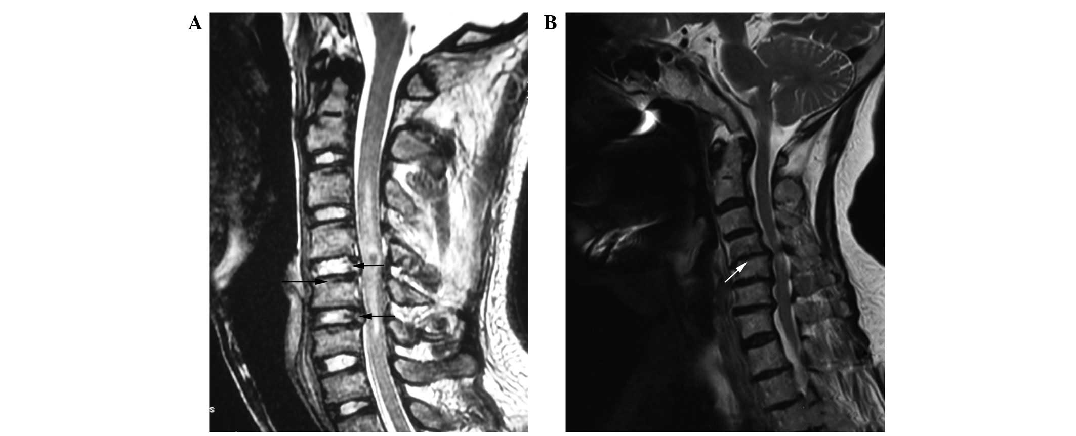
|
2
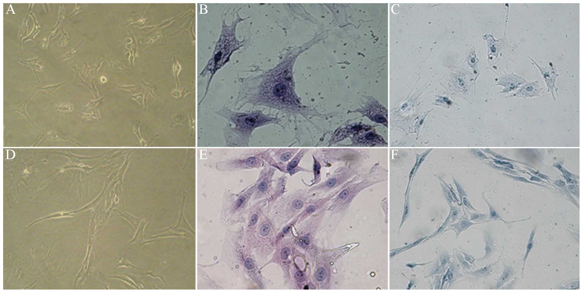
|
Jing L-B, Zhang X-L, Xu H-Z, et al:
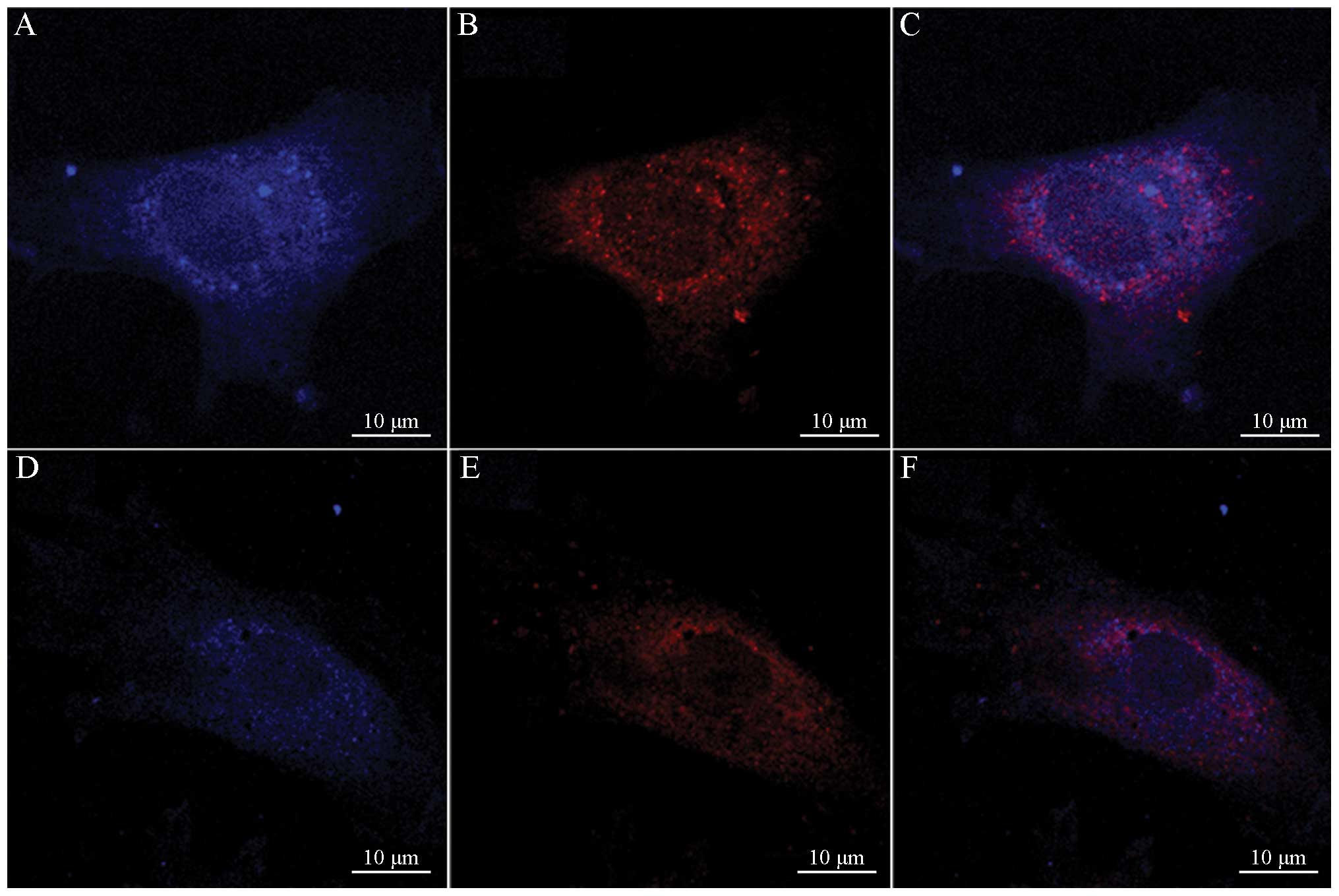
Protective effect of autophagy on nucleus pulposus cells under
starvation. Chin J Pathophysiol. 28:1302–1307. 2012.(In
Chinese).
|
|
3
|
Haschtmann D, Stoyanov JV, Gédet P and
Ferguson SJ: Vertebral endplate trauma induces disc cell apoptosis
and promotes organ degeneration in vitro. Eur Spine J. 17:289–299.
2008. View Article : Google Scholar : PubMed/NCBI
|
|
4
|
Reggiori F and Klionsky DJ: Autophagy in
the eukaryotic cell. Eukaryot cell. 1:11–21. 2002. View Article : Google Scholar : PubMed/NCBI
|
|
5
|
Chhangani D and Mishra A: Mahogunin ring
finger-1 (MGRN1) suppresses chaperone-associated misfolded protein
aggregation and toxicity. Sci Rep. 3:19722013. View Article : Google Scholar : PubMed/NCBI
|
|
6
|
Liu H, Cao W, Li Y, Feng M, Wu X, Yu K and
Liao M: Subgroup J avian leukosis virus infection inhibits
autophagy in DF-1 cells. Virol J. 10:1962013. View Article : Google Scholar : PubMed/NCBI
|
|
7
|
Miller G: The spine. Berquist TH: MRI of
the Musculoskeletal System. 2nd edition. Raven Press; New York, NY:
pp. 238–240. 1990
|
|
8
|
Thompson JP, Pearce RH, Schechter MT,
Adams ME, Tsang IK and Bishop PB: Preliminary evaluation of a
scheme for grading the gross morphology of the human intervertehral
disc. Spine (Phila Pa 1976). 15:411–415. 1990. View Article : Google Scholar : PubMed/NCBI
|
|
9
|
Xu HG, Peng HX, Cheng JF and Lü K:
Establishment and significance of an in vitro model of degeneration
of human cervical endplate chondrocytes. Zhonghua Yi Xue Za Zhi.
91:2912–2916. 2011.(In Chinese).
|
|
10
|
Le Maitre CL, Freemont AJ and Hoyland JA:
The role of interleukin-1 in the pathogenesis of human
intervertebral disc degeneration. Arthritis Res Ther. 7:R732–R745.
2005.
|
|
11
|
Biederbick A, Kern HF and Elsässer HP:
Monodansylcadaverine (MDC) is a specific in vivo marker for
autophagic vacuoles. Eur J cell Biol. 66:3–14. 1995.PubMed/NCBI
|
|
12
|
Shen C, Yan J, Jiang LS and Dai LY:
Autophagy in rat annulus fibrosus cells: evidence and possible
implications. Arthritis Res Ther. 13:R1322011. View Article : Google Scholar : PubMed/NCBI
|
|
13
|
Liang QQ, Cui XJ, Xi ZJ, et al: Prolonged
upright posture induces degenerative changes in intervertebral
discs of rat cervical spine. Spine (Phila Pa 1976). 36:E14–E19.
2011.PubMed/NCBI
|
|
14
|
Sowa G, Vadalà G, Studer R, et al:
Characterization of intervertebral disc aging: longitudinal
analysis of a rabbit model by magnetic resonance imaging,
histology, and gene expression. Spine (Phila Pa 1976).
33:1821–1828. 2008. View Article : Google Scholar
|
|
15
|
Shin BH, Lim Y, Oh HJ, et al:
Pharmacological activation of Sirt1 ameliorates
polyglutamine-induced toxicity through the regulation of autophagy.
PLoS One. 8:e649532013. View Article : Google Scholar : PubMed/NCBI
|
|
16
|
Caramés B, Taniguchi N, Otsuki S, Blanco
FJ and Lotz M: Autophagy is a protective mechanism in normal
cartilage, and its aging-related loss is linked with cell death and
osteoarthritis. Arthritis Rheum. 62:791–801. 2010.PubMed/NCBI
|
|
17
|
Sun K, Xie X, Liu Y, et al: Autophagy
lessens ischemic liver injury by reducing oxidative damage. Cell
Biosci. 3:262013. View Article : Google Scholar : PubMed/NCBI
|
|
18
|
Zhu X, Wu L, Qiao H, et al: Autophagy
stimulates apoptosis in HER2-overexpressing breast cancers treated
by lapatinib. J Cell Biochem. 114:2643–2653. 2013. View Article : Google Scholar : PubMed/NCBI
|
|
19
|
Bachar-Wikstrom E, Wikstrom JD, Ariav Y,
Tirosh B, Kaiser N, Cerasi E and Leibowitz G: Stimulation of
autophagy improves endoplasmic reticulum stress-induced diabetes.
Diabetes. 62:1227–1237. 2013. View Article : Google Scholar : PubMed/NCBI
|
|
20
|
Caramés B, Hasegawa A, Taniguchi N, Miyaki
S, Blanco FJ and Lotz M: Autophagy activation by rapamycin reduces
severity of experimental osteoarthritis. Ann Rheum Dis. 71:575–581.
2012.PubMed/NCBI
|
|
21
|
Mori K: Tripartite management of unfolded
proteins in the endoplasmic reticulum. Cell. 101:451–454. 2000.
View Article : Google Scholar : PubMed/NCBI
|
|
22
|
Kim HS, Montana V, Jang HJ, Parpura V and
Kim JA: Epigallocatechin gallate (EGCG) stimulates autophagy in
vascular endothelial cells: A potential role for reducing lipid
accumulation. J Biol Chem. 288:22693–22705. 2013. View Article : Google Scholar
|
|
23
|
Sanjuan MA, Dillon CP, Tait SW, et al:
Toll-like receptor signalling in macrophages links the autophagy
pathway to phagocytosis. Nature. 450:1253–1257. 2007. View Article : Google Scholar : PubMed/NCBI
|
|
24
|
Ye W, Xu K, Huang D, Liang A, Peng Y, Zhu
W and Li C: Age-related increases of macroautophagy and
chaperone-mediated autophagy in rat nucleus pulposus. Connect
Tissue Res. 52:472–478. 2011. View Article : Google Scholar : PubMed/NCBI
|
|
25
|
Zhu W-R, Ye W, Xu K, Lou Z-K, Ren J-K,
Chen J-H and Li C-H: Starvation-induced changes of LC-3 and
Beclin-1 expression in annulus fibrosus cells. Chin J Exp Sur.
28:983–985. 2011.(In Chinese).
|
|
26
|
Ye W, Chu Z, Xu K, et al: Expression and
significance of Beclin-1 and microtubule-associated protein 1 light
chain 3 in nucleus pulpous of rats during the aging process. Chin J
Exp Surg. 27:643–645. 2010.
|