Introduction
Death-associated protein kinase (DAPK) (1) is a pro-apoptotic protein identified
by scanning apoptosis-initiating genes and tumor-inhibiting genes
in the genome. It has been shown that DAPK participates in
apoptotic signal transduction pathways, resulting in the
suppression of tumor occurrence. Loss of the dynamic balance
between cell proliferation and apoptosis often leads to the
accumulation of gene mutations and the occurrence of tumors
(2–4).DAPK is located in the 9q34.1 site of
the human chromosome and has a molecular mass of 160 kDa (5). It is a serine/threonine protein
kinase regulated by calcium/calmodulin and participates in cell
survival, apoptosis and tumor suppression (6,7).
DAPK expression is absent from a variety of tumor cells and
tissues, and the loss of expression is closely correlated with CpG
methylation. DAPK consists of 7 domains, including a core kinase
domain, calcium/calmodulin-binding domain, ankyrin repeat region,
P-ring, cytoskeleton-binding domain, death domain and serine-rich
tail. The core kinase domain is located between the
calcium/calmodulin-binding domain and the N-terminus. It is
composed of an 11 serine/threonine structure with a conserved
lysine residue, which is associated with ATP binding. Mutation of
the residue eradicates the apoptotic effect of DAPK (8). Due to its apoptosis promoting and
cell adhesion and cell migration inhibiting effects, DAPK plays an
important role in tumor development and metastasis (9,10).
E-cadherin is a calcium-dependent glycoprotein that
is widely distributed in epithelial cells. It mediates homogeneous
cell adhesion, and maintains the integrity of the cell structure
and epithelial polarity. Reduced E-cadherin-mediated cell adhesion
is a significant cause of tumor invasion and metastasis. The
reduction or lack of E-cadherin expression leads to reduced mutual
adhesion between the cells. Therefore infiltrative growth and
metastasis occur (11). It has
been found that ezrin and E-cadherin are involved in the process of
esophageal cancer invasion and metastasis (12).
In the current study, in situ hybridization
and immunohistochemistry methods were used to detect the expression
of DAPK and E-cadherin in the surgically excised esophageal
squamous cell carcinoma (ESCC) tissue and the adjacent normal
tissues of patients. The study also aimed to further explore the
correlation between the expression levels of DAPK and E-cadherin
and the development of esophageal cancer in order to identify new
molecular markers of esophageal cancers. The immunoblot assay
indicated that the expression levels of DAPK and E-cadherin were
decreased significantly in the ESCC tissue when compared with the
adjacent normal tissues. The reverse transcription (RT)-PCR results
showed that the mRNA levels of DAPK and E-cadherin were also
reduced.
Materials and methods
Clinical data and specimens
Surgical specimens from 76 adult cases of esophageal
cancer, who were admitted to the Department of Thoracic Surgery,
Affiliated Hospital of Hebei Engineering University between July
2008 and July 2011, were selected and made into a tissue
microarray. There were 49 males and 27 females, corresponding to a
male to female ratio of ∼2:1. The patients were aged between 39 and
73 years old with average age of 61±5.1 years. The patients did not
receive preoperative radiotherapy, chemotherapy or
immunotherapy.
The pathological diagnoses were well-differentiated
squamous cell carcinoma (26 cases), undifferentiated carcinoma (33
cases) and poorly differentiated carcinoma (17 cases). There were
12 cases of T-I, 21 cases of T-II, 29 cases of T-III and 14 cases
of T-IV. There were 35 cases with lymph node metastasis and 41
cases without. All samples were fixed with 40 g/l poly formalin and
dehydrated conventionally. After embedding in paraffin, the samples
were serially sectioned to a thickness of 4–6 μm and stained
by H&E, immunohistochemical and in situ hybridization
staining.
Immunohistochemical and in situ
hybridization staining
Rabbit anti-human polyclonal antibody to DAPK was
purchased from Wuhan Boster Biological Technology, Ltd. (Wuhan,
China). The SP immunohistochemistry kit was purchased from Beijing
Zhongshan Golden Bridge Biotechnology Co., Ltd. (Beijing, China).
The SP method was performed using a 1:150 dilution of the DAPK
monoclonal antibody. The tissues were stained with DAB and
counterstained with hematoxylin. The staining procedure was carried
out strictly in accordance with the manufacturer’s instructions.
PBS was used as the negative control, instead of primary antibody.
The in situ pre-hybridization solution was purchased from
Wuhan Boster Biological Technology, Ltd. The 5′-end bio-labeling of
the complete phosphorothioate probe were performed by Beijing
AudioCodes Biotechnology Co., Ltd. (Beijing, China). The DAPK probe
sequence was CAGCTCGCCACCTGCAACGA. The specimens were dewaxed with
fresh xylene and dehydrated through an alcohol gradient. Endogenous
peroxidase was inactivated for 30 min by treatment with freshly
prepared 0.5% H2O2 at room temperature. The
DNA-binding protein in the specimens was digested with 3% fresh
citric acid protease (0.01 g/l) for 10 min at 37°C.
Pre-hybridization solution without probe (20 μl) was added
to each slide which was then maintained at 42°C for 4 h.
Hybridization solution with probe (1 ng/l) was hybridized in a
moist chamber at 42°C for 12 h. The samples were washed with 0.1X
standard sodium citrate (SSC) at 42°C and SA-Bio-AP was added.
After incubating at 37°C for 10 min and then rinsing, BCIP/NBT were
added and the samples were kept in the dark for 2–4 h to allow the
color reaction to occur. A sample without the probe was the
negative control.
Positive criteria of immunohistochemistry
and in situ hybridization staining
Positively stained DAPK protein is observed as pale
yellow or brown granules in the cytoplasm. Five fields in the high
magnification image were randomly selected and ≥200 cells were
observed in each field. The positive cell percentage and color
depth were determined (13). A
nine-point scoring system was adopted. The positive cell proportion
was determined and scored as follows: 1 point for <10% positive
cells, 2 points for 10–50% and 3 points for >50%. The color
depth was also scored: 0 point for negative staining, 1 point for
slight yellow color staining, 2 points for moderate yellow color
staining and 3 points for pale-brown color staining. Scores of 0–1,
2 and ≥3 are represented by (−), (+) and (++), respectively, with
(++) indicating normal expression and (−) and (+) indicating
missing or weak expression.
The positive expression of E-cadherin is shown as
yellow or brown-yellow stained fine particles in the cell membrane,
while negative expression is shown as expression in the cytoplasm
but not in the membrane. According to the criteria of Bajbouj
(14), (−) signifies negative
staining, (+) signifies a positive cell proportion of <75% and
(++) signifies a positive cell proportion of ≥75%; (++) indicates
normal expression, while (−) and (+) indicate missing or weak
expression.
Quantitative RT-PCR
The esophageal cancer tissues and the adjacent
normal mucosa from the surgical specimens of 76 cases were isolated
using the RNeasy FFPE kit (cat. #73504, Qiagen, Valencia, CA, USA)
and stored at −80°C for further analyses. Quantitative RT-PCR
analysis of DAPK and E-cadherin mRNA levels in the tissues were
performed. The RT-PCR experiments were repeated at least 3 times.
RNA was reverse transcribed into cDNA using random primers in a
Reverse Transcription II system (Promega, Madison, WI, USA)
according to the manufacturer’s instructions. Expression of DAPK
and E-cadherin mRNAs was quantified by quantitative PCR using an
ABI Prism Sequence Detection system (Applied Biosystems, Carlsbad,
CA, USA). The primers used in this study are listed in Table I. An assay reagent containing
premixed primers and a VIC-labeled probe (Applied Biosystems; cat.
#4310884E) was used to quantify the expression of endogenous GAPDH
mRNA. Template-negative and RT-negative conditions were used as
controls. Amplification of the endogenous GAPDH cDNA was monitored.
The levels (mean values) of DAPK and E-cadherin transcripts in the
patients were calculated.
 | Table I.Primers used for RT-PCR. |
Table I.
Primers used for RT-PCR.
| Gene | F/R | Primer sequence |
|---|
| GAPDH | F |
5′-GTGGGGCGCCCCAGGCACCA-3′ |
| GAPDH | R |
5′-CTCCTTAATGTCACGCACGATTT-3′ |
| DAPK | F |
5′-AGCATATCTACTAGATATCTGAT-3′ |
| DAPK | R |
5′-GGTACTCCACGTCGACGAGAC-3′ |
| E-cadherin | F | 5′-CAGTGAGCG
GAGATAGTGCC-3′ |
| E-cadherin | R | 5′-CAA
AAGTTGGAAAGCCCGTG-3′ |
Immunoblot assays
Total proteins were harvested from the esophageal
cancer tissues and the adjacent normal mucosa from the surgical
specimens of 76 cases. The proteins were separated by 10% SDS/PAGE
and then subjected to immunoblot analyses. The primary antibodies
against DAPK (∼160 kDa), E-cadherin (∼120 kDa) and β-actin were
purchased from Santa Cruz Biotechnology, Inc. (anti-DAPK, cat.
#sc-10805, 1:200; anti-E-cadherin, cat. #sc-7870, 1:200;
anti-β-actin, cat. #sc-130301, 1:10,000; Santa Cruz, CA, USA). The
secondary antibodies used in this study were goat anti-mouse
IgG-HRP (cat. #sc-2005, 1:10,000; Santa Cruz Biotechnology, Inc.)
and anti-rabbit IgG-HRP (cat. #31460, 1:5,000; Pierce
Biotechnology, Rockford, IL, USA). Bound antibodies were detected
using an ECL system (Pierce Biotechnology). The immunoblot
experiments were repeated at least 3 times. The mean normalized
optical density (OD) of DAPK and E-cadherin protein bands relative
to the OD of the β-actin band from the same individual was
calculated.
Statistical analysis
All statistical calculations were performed using
SPSS 13.0 statistical software. Continuous variables were
summarized as the mean values (mean ± standard error) and compared
using the independent sample t-test. P<0.05 was considered to
indicate a statistically significant result. In addition, the
χ2 test and Spearman’s rank correlation coefficient
analysis were used. α=0.05 was considered to indicate a
statistically significant difference.
Results
Expression of DAPK and E-cadherin in ESCC
and adjacent normal mucosa
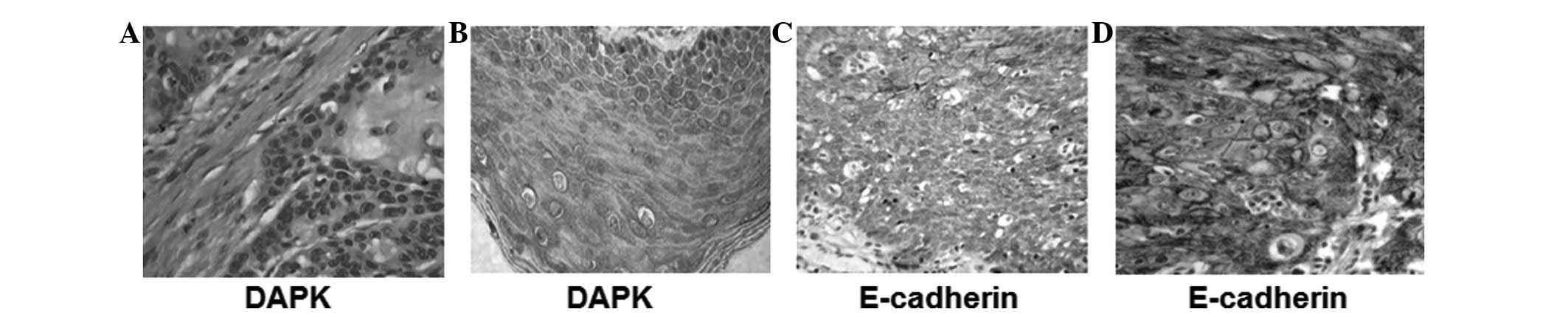
DAPK protein is localized in the cytoplasm and
presents as pale yellow or brown granules when stained. No positive
staining was observed in the control tissue (Fig. 1A and B). The positive expression of
E-cadherin is mainly localized in the cell membrane, and presents
as brownish-yellow granules when stained.
E-cadherin expression was observed in the epithelial
cells of the esophageal carcinoma cancer tissue and normal tissue
and was strongly expressed in the basal cells and spinous cell
layers in the squamous epithelial tissues of the normal esophagus
(Fig. 1C and D). There were
significantly differences in DAPK and E-cadherin expression between
the esophageal cancer tissue and the adjacent normal epithelium
tissue, as shown in Table II.
| Table II.Expression of DAPK and E-cadherin in
esophagus squamous carcinomatissue and adjacent normal tissue
(%). |
Table II.
Expression of DAPK and E-cadherin in
esophagus squamous carcinomatissue and adjacent normal tissue
(%).
| Category | DAPK(+) | DAPK(−) | P-value | E-cadherin(+) | E-cadherin(−) | P-value |
|---|
| Normal tissue | 65 (85.5) | 11 (14.5) | | 74 (97.36) | 2 (2.63) | |
| Cancer tissue | 27 (35.5) | 49 (64.5) | <0.001a | 21 (27.63) | 55 (72.36) | <0.001b |
Correlation between expression of DAPK
and E-cadherin and clinical and biological behavior of ESCC
No correlations were noted between DAPK expression
and the age, gender and tumor differentiation degree of the
patients. However, there were correlations between DAPK expression
and the tumor invasion depth and lymph node metastasis.
No correlations were observed between E-cadherin
expression and the age, gender and tumor invasion depth of the
patients. There were significant correlations between E-cadherin
expression and tumor differentiation and lymph node metastasis, as
shown in Table III.
| Table III.Correlation between expression of DAPK
and E-cadherin and clinical parameters of esophageal squamous
carcinoma. |
Table III.
Correlation between expression of DAPK
and E-cadherin and clinical parameters of esophageal squamous
carcinoma.
| | DAPK expression
| | E-cadherin expression
| |
|---|
| Clinical and
pathological factors | n | − | + | ++ | % | P-value | − | + | ++ | % | P-value |
|---|
| Gender | | | | | | | | | | | |
| Male | 49 | 21 | 10 | 18 | 36.7 | | 25 | 10 | 14 | 28.5 | |
| Female | 27 | 11 | 7 | 9 | 33.3 | | 17 | 3 | 7 | 25.9 | |
| Zc | | | −0.205 | | | 0.838 | | −0.383 | | | 0.452 |
| Age (years) | | | | | | | | | | | |
| <60 | 44 | 17 | 11 | 16 | 36.3 | | 27 | 4 | 13 | 29.5 | |
| ≥60 | 32 | 16 | 5 | 11 | 34.3 | | 15 | 9 | 8 | 25.0 | |
| Zc | | | −0.196 | | | 0.844 | | −0.698 | | | 0.485 |
| Tumor size | | | | | | | | | | | |
| <5 cm | 33 | 14 | 9 | 10 | 30.3 | | 16 | 8 | 9 | 27.2 | |
| ≥5 cm | 43 | 18 | 8 | 17 | 39.5 | | 26 | 5 | 12 | 27.9 | |
| Zc | | | −0.475 | | | 0.635 | | −0.695 | | | 0.487 |
| Degree of
differentiation | | | | | | | | | | | |
| High | 26 | 11 | 6 | 9 | 34.6 | | 8 | 6 | 12 | 46.2 | |
| Moderate | 33 | 14 | 7 | 12 | 36.3 | | 21 | 5 | 7 | 21.2 | |
| Low | 17 | 7 | 4 | 6 | 35.2 | | 13 | 2 | 2 | 11.7 | |
| χ2
value | | | 0.006 | | | 0.997 | | 10.148 | | | 0.006 |
| Infiltration
degree | | | | | | | | | | | |
| Did not
infiltrate outer membrane | 31 | 22 | 4 | 5 | 16.1 | | 16 | 5 | 10 | 32.2 | |
| Infiltrated outer
membrane and tissue | 45 | 10 | 13 | 22 | 48.9 | | 26 | 8 | 11 | 24.4 | |
|
Zc | | | −3.981 | | | 0.000 | | −0.775 | | | 0.450 |
| Lymph node
metastasis | | | | | | | | | | | |
| Yes | 41 | 13 | 8 | 20 | 48.7 | | 28 | 7 | 6 | 14.6 | |
| No | 35 | 19 | 9 | 7 | 20.0 | | 14 | 6 | 15 | 42.8 | |
|
Zc | | | −2.409 | | | 0.016 | | −2.758 | | | 0.006 |
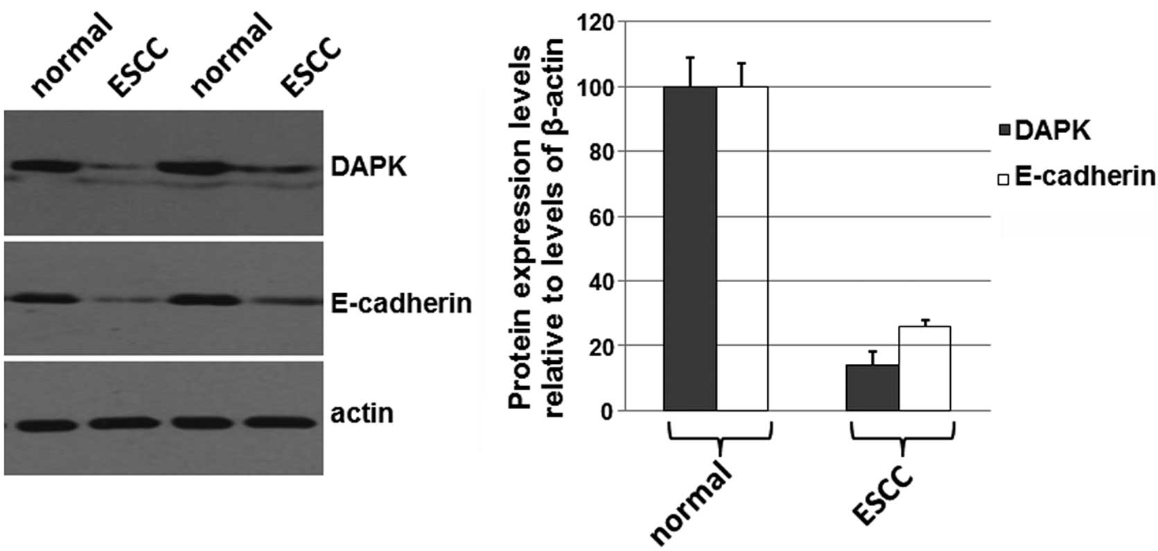
Expression levels of DAPK and E-cadherin
are decreased in ESCC tissues
To determine if the expression levels of DAPK and
E-cadherin were correlated with the development of carcinomas, the
total proteins were isolated from esophageal cancer tissues and the
adjacent normal mucosa from surgery specimens of the 76 cases.
Western blotting was performed. As shown in Fig. 2A, the expression levels of DAPK and
E-cadherin were decreased in ESCC compared with the levels in the
adjacent normal tissues. The levels of β-actin were used as a
loading control. The mean normalized OD of DAPK and E-cadherin
protein bands relative to the OD of β-actin bands from each of
patients was calculated (Fig. 2B).
The results shown in Fig. 2
suggest that expression levels of DAPK and E-cadherin are decreased
in ESCC tissues, which may be related to the development of
ESCC.
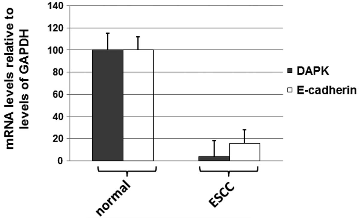
Levels of DAPK and E-cadherin mRNA are
decreased in ESCC tissues
To determine if levels of DAPK and E-cadherin mRNAs
were decreased in ESCC tissues, real-time PCR was performed. As
shown in Fig. 3, the expression
levels of DAPK and E-cadherin mRNA were reduced in ESCC tissues
compared with the levels in the adjacent normal tissues. The
results shown in Fig. 3 suggest
that levels of DAPK and E-cadherin mRNA were decreased in ESCC
tissues.
Discussion
In the current study, it is shown that the DAPK gene
is expressed in esophageal cancer tissue and adjacent normal
esophageal tissues. However, the expression levels of these
proteins are significantly reduced in cancer tissues, as revealed
by the results of the immunoblot assay and RT-PCR. The results also
indicate that the abnormal DAPK gene expression is closely
correlated with the invasion depth of esophageal squamous cell
carcinoma and lymph node metastasis, which suggests that the
abnormal expression of DAPK may be an indicator of prognosis.
E-cadherin expression is associated with the degree of esophageal
cancer differentiation and lymph node metastases, but there is no
significant correlation with age, gender, tumor length or the depth
of invasion. It was also identified in this study that the
E-cadherin expression rate in cancer tissues with lymph node
metastasis was significantly lower than that in cancer tissues
without lymph node metastasis (P<0.05). It was also observed
that the E-cadherin expression rate in poorly differentiated cancer
tissue was significantly lower than that in well-differentiated
cancer tissues (P<0.05), indicating that the reduction of
E-cadherin expression is associated with the differentiation of
esophageal squamous cell carcinoma and lymph node metastasis.
However, no significant difference between the E-cadherin
expression with or without fiber membrane infiltration was
identified, suggesting that E-cadherin is not correlated with the
invasion depth of esophageal squamous cell carcinoma. Further
analysis of in situ hybridization and immunohistochemical
detection showed consistent DAPK and E-cadherin positive expression
in esophageal carcinoma. The DAPK protein expression rate and
E-cadherin expression were positively correlated (P<0.01),
suggesting the correlation of DAPK and E-cadherin in tumor invasion
and metastasis.
The invasion and metastasis of tumor cells are
processes involving multiple stages, steps and factors. They are
associated with the main characteristics of the tumor cells,
overall host immune status and the characteristics of transformed
local tissues. DAPK and E-cadherin expression correlate with
esophageal squamous cell carcinoma and metastasis, suggesting that
DAPK and E-cadherin may have a synergistic effect. In situ
hybridization and immunohistochemical detection were used to
analyze the expression of DAPK and E-cadherin in esophageal
squamous cell carcinoma, which initially showed the reduction or
lack of DAPK expression may lead to the development of esophageal
cancer. The combined methods contribute to the comprehensive
diagnosis of ESCC, the degree of malignancy, metastatic potential
and prognosis.
Acknowledgements
This study was supported by the
Science and Technology Department of Hebei Province: Hebei
Technology Support Program (Grant #112061176D).
References
|
1.
|
Deiss LP, Feinstein E, Berissi H, Cohen O
and Kimchi A: Identification of a novel serine/threonine kinase and
a novel 15-kD protein as potential mediators ofthe gamma
interferon-induced cell death. Genes Dev. 9:15–30. 1995. View Article : Google Scholar
|
|
2.
|
Brabender J, Arbab D, Huan X, et al:
Death-associated protein kinase (DAPK) promoter methylation and
response to neoadjuvant radiochemotherapy in esophageal cancer. Ann
Surg Oncol. 16:1378–1383. 2009. View Article : Google Scholar : PubMed/NCBI
|
|
3.
|
Tong A, Lynn G, Ngo V, et al: Negative
regulation of Caenorhabditis elegans epidermal damage
responses by death-associated protein kinase. Proc Natl Acad Sci
USA. 106:1457–1461. 2009.
|
|
4.
|
Li H, Ray G, Yoo BH, Erdogan M and Rosen
KV: Down-regulation of death-associated protein kinase-2 is
required for beta-catenin-induced anoikis resistance of malignant
epithelial cells. Biol Chem. 284:2012–2022. 2009. View Article : Google Scholar : PubMed/NCBI
|
|
5.
|
Zalckvar E, Berissi H, Eisenstein M and
Kimchi A: Phosphorylation of Beclin 1 by DAP-kinase promotes
autophagy by weakening its interactions with Bcl-2 and Bcl-XL.
Autophagy. 5:720–722. 2009. View Article : Google Scholar : PubMed/NCBI
|
|
6.
|
Yanagawa N, Osakabe M, Hayashi M, Tamura G
and Motoyama T: Detection of HPV-DNA, p53 alterations, and
methylation in penile squamous cell carcinoma in Japanese men.
Pathol Int. 58:477–482. 2008. View Article : Google Scholar : PubMed/NCBI
|
|
7.
|
Hoffmann AC, Vallböhmer D, Prenzel K, et
al: Methylated DAPK and APC promoter DNA detection in peripheral
blood is significantly associated with apparent residual tumor and
outcome. J Cancer Res Clin Oncol. 135:1231–1237. 2009. View Article : Google Scholar : PubMed/NCBI
|
|
8.
|
Fendri A, Masmoudi A, Khabir A, et al:
Inactivation of RASSF1A, RARbeta2 and DAP-kinase by promoter
methylation correlates with lymph node metastasis in nasopharyngeal
carcinoma. Cancer Biol Ther. 8:444–451. 2009. View Article : Google Scholar : PubMed/NCBI
|
|
9.
|
Santos-García A, Abad-Hernández MM,
Fonseca-Sánchez E, et al: E-cadherin, laminin and collagen IV
expression in the evolution from dysplasia to oral squamous cell
carcinoma. Med Oral Patol Oral Cir Bucal. 11:E100–E105. 2006.
|
|
10.
|
Zhai JW, Yang XG, Yang FS, Hu JG and Hua
WX: Expression and clinical significance of Ezrin and E-cadherin in
esophageal squamous cell carcinoma. Chin J Cancer. 29:317–320.
2010. View Article : Google Scholar : PubMed/NCBI
|
|
11
|
Gao DL, Li SL, Chen KS, Zhao ZH, Zhao QM,
Liu ZW and Zhang YH: The inhibition of metalloproteinase in RECK
expression in esophageal squamous cell carcinoma and its biological
significance. World J Gastroenterol. 16:1634–1638. 2008.
|
|
12.
|
Gonzalez MA, Pinder SE, Wencyk PM, et al:
An immunohistochemical examination of the expression of E-cadherin,
alpha- and beta/gamma-catenins, and alpha2- and betal-integrins in
invasive breast cancer. J Pathol. 187:523–529. 1999. View Article : Google Scholar
|
|
13.
|
Lou C, Yang B, Gao YT, Wang YJ, Nie FH,
Yuan Q, Zhang CL and Du Z: Aberrant methylation of multiple genes
and its clinical implication in hepatocellularcarcinoma. Zhonghua
Zhong Liu Za Zhi. 30:831–836. 2008.(In Chinese).
|
|
14.
|
Bajbouj K, Poehlmann A, Kuester D, et al:
Identification of phosphorylated p38 as a novel DAPK-interacting
partner during TNFalpha-induced apoptosis in colorectal tumor
cells. Am J Pathol. 175:557–570. 2009. View Article : Google Scholar : PubMed/NCBI
|