Introduction
Malignant bile duct obstruction may develop into
clinically serious diseases, including obstructive jaundice and
cholangitis. Malignant bile duct obstruction is a condition that is
difficult to treat and has a high mortality rate. Secondary hepatic
inadequacy as well as digestion and absorption hypofunction
following bile duct obstruction-induced cholestasis are the primary
causes of accelerated mortalities due to obstructive jaundice.
Malignant hilar obstructive jaundice has long been a challenge in
clinical practice due to a low resective rate, insufficient
drainage and numerous complications. In the treatment of biliary
obstructive jaundice, endoscopic retrograde
cholangio-pancreatography (ERCP) combined with endoscopic biliary
stenting has been extensively applied as a minimally invasive
palliative therapy (1,2). Currently, percutaneous biliary stent
implantation, due to a higher success rate, the ability to meet
physiological requirements and a negligible influence on the
quality of life, has become another minimally invasive palliative
therapy for malignant hilar obstructive jaundice. In addition,
under conditions of unsuccessful surgery or drainage insufficiency,
this technique has gradually become an alternative to failed
biliary stent implantation under ERCP in 33 to 88% of cases
(3–10). One-channel double stent
implantation has the advantages of little trauma, few complications
and sufficient drainage, and has been increasingly widely used for
the treatment of partial hilar biliary obstruction. However,
studies on this therapy, particularly its combined use with
antitumor interventional techniques, such as arterial
chemoembolization, are rare.
Between September 2004 and September 2009, a total
of 8 patients with hilar obstruction were subjected to percutaneous
one-channel double stent implantation combined with
chemoembolization or I125 seed implantation for
antitumor treatment at the Interventional Department, Henan
Provincial People’s Hospital (Zhengzhou, China). The aims of the
study were to evaluate the effect, safety and clinical value of
percutaneous one-channel double stent implantation combined with
anti-tumor treatment.
Materials and methods
Clinical data
A total of 8 patients with hilar biliary
obstruction, including 5 males and 3 females, were enrolled in the
present study. Their ages ranged from 31 to 78 years with an
average of 50.2±11.4 years. Among these patients, four had bile
duct cancer, two had hilar hepatic carcinoma, one had metastatic
carcinoma and one had pancreatic head carcinoma. Their clinical
manifestations included jaundice, itching and rock-like stools.
Preoperative computed tomography (CT) or magnetic resonance imaging
(MRI) revealed hilar lesions which caused bilateral hepatic duct
and/or common hepatic duct obstruction but without surgical
indications. The surgical instruments used included 18–22 G
trocars, balloons, nitinol mesh biliary stents with diameters of
8–10 mm and lengths of 50–80 mm (Nanjing Micro-tech Co., Ltd,
Nanjing, China), 7–9 F stent conveyors and 7–8.5 F biliary drainage
tubes (Bard Co., Murray Hill, NJ, USA). The study was conducted in
accordance with the Declaration of Helsinki and with approval from
the Ethics Committee of Henan Provincial People’s Hospital. Written
informed consent was obtained from all patients.
One-channel double stent
implantation
Surgery was performed under fluoroscopic monitoring.
The right hepatic duct was punctured through the right midaxillary
line. The route of the biliary system was observed by angiography.
A guidewire was inserted through the right hepatic duct and the
potential lacune of the obstruction segment between the left and
right hepatic ducts was sought. The guidewire entered the left
hepatic duct through the obstruction segment in the right hepatic
duct. A stent was implanted at the obstruction segment to allow the
bile from the left hepatic duct to flow into the right hepatic
duct. The guidewire was then sent into the common bile duct through
the right hepatic duct and passed down to the duodenum. Another
stent was implanted between the right hepatic duct and the common
bile duct. Both stents were released after the obstructed segment
was sufficiently dilated by a balloon catheter. The two stents were
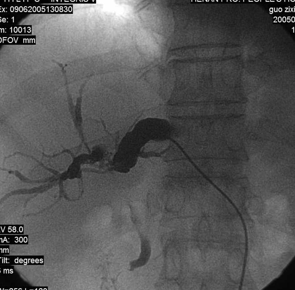
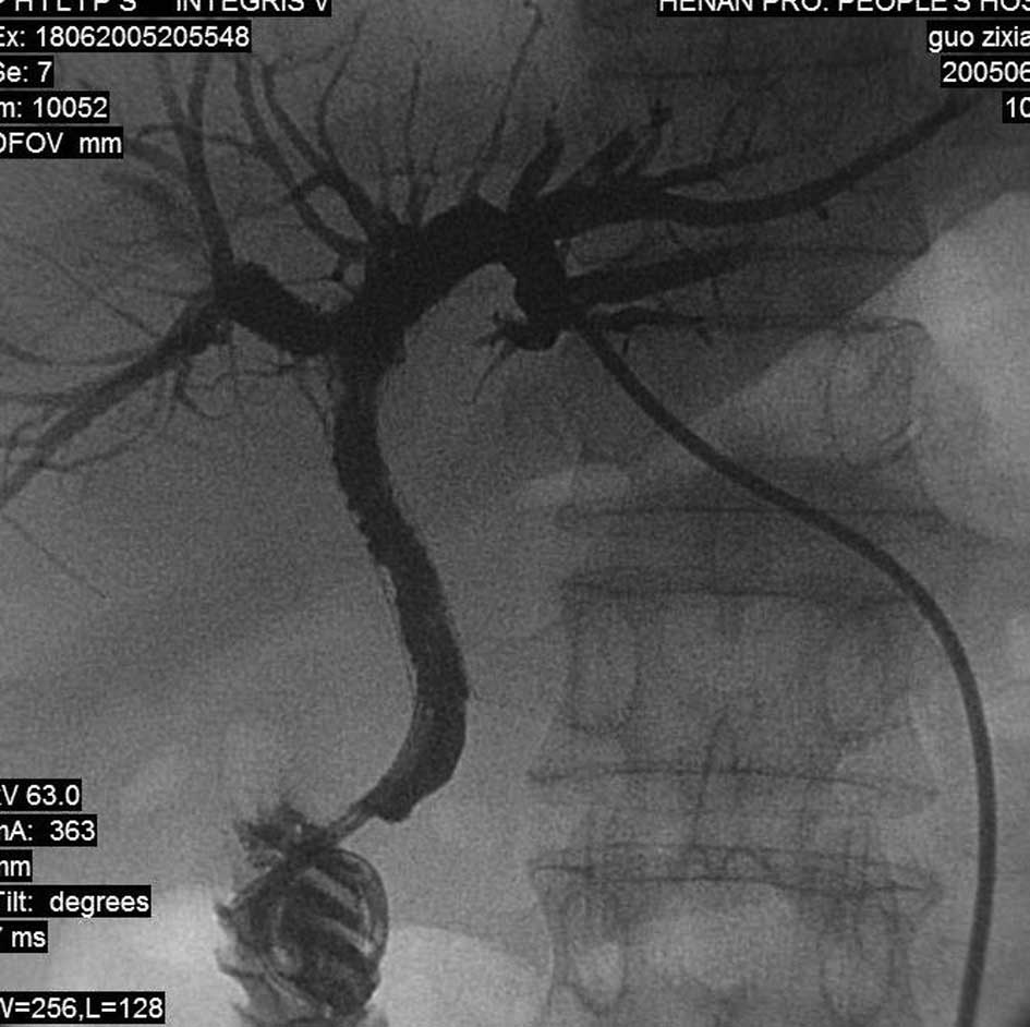
in a reversed ‘7’-shaped arrangement. However, if the right
approach was not suitable or the patient showed more noticeable
dilation of the left hepatic duct, a left approach slightly
deviated from the subxiphoid was adopted. The remainder of the
procedure was as described previously, with two stents in a ‘7’
arrangement (Figs. 1–4). If the patient was weak and had
numerous diseases, or the obstruction was so severe that it was
difficult for the guidewire to pass, the guidewire was adjusted as
much as possible to enter the opposite intrahepatic bile duct. An
external biliary drainage tube was introduced into the opposite
bile duct along the guidewire for simultaneous external drainage of
the intrahepatic bile ducts. Stent implantation was performed when
jaundice improved 3–10 days later.
Following implantation, stent morphology and
locations were observed via transcatheter angiography. The puncture
tract was sealed off with a gelatine sponge. Under conditions of
bile duct hemorrhage, incomplete stent opening or preoperative
diagnosis of cholangitis, external biliary drainage was maintained.
At 3–7 days, a follow-up angiography was performed. When a contrast
agent was confirmed to enter the duodenum smoothly, the drainage
tube was removed.
Combined antitumor treatment
Percutaneous I125 seed implantation into
cancer with insufficient blood supply was performed for six
patients, including 4 patients with bile duct cancer, one patient
with metastatic carcinoma and one patient with pancreatic head
carcinoma, and chemoembolization was performed for hepatocarcinoma
with a rich blood supply was performed for two patients. The
commonly used drugs were pirarubicin (40 mg), mitomycin (20 mg) and
embolization agents (between 10 and 20 ml). The time between
interventions was approximately 1 month over a course of 1–3
treatments.
Results
Surgery and postoperative effect
A total of 16 stents were successfully implanted
into 8 patients. A right approach was adopted for 6 patients, with
the drainage between stents in a reversed ‘7’ shape. A left
approach was adopted for 2 patients, with the drainage between
stents in a ‘7’ shape. Immediate stent implantation following
puncture was performed for 7 patients. One patient received early
external drainage of both hepatic ducts and the biliary duct stents
following the improvement of jaundice. Immediate postoperative
sealing of the puncture tract was implemented in 7 patients. The
external biliary drainage of one patient was arrested due to a
small amount of bile duct hemorrhage. At 3 days, the patient’s
drainage fluid turned yellowish and an angiographic review showed
satisfactory stent locations and morphology, as well as smooth bile
duct drainage. The external drainage tube was then extracted. The
preoperative total bilirubin concentrations in the patients ranged
from 138.2 to 796.9 μmol/l with an average of 267.1±154.7
μmol/l. This range decreased to 40.1–256.3 μmol/l
with an average of 61.2±13.4 μmol/l at 2 weeks after
surgery. The patients were followed up for 1–12 months. One
mortality occurred within 1 month and five mortalities within 2–6
months. Two patients survived for >6 months.
Complication management
No severe complications or mortalities associated
with the implanted stents occurred. One patient exhibited a small
volume of bile duct hemorrhage but had stable vital signs.
Following external drainage and the application of hemostatics,
hemorrhaging stopped 2 days later. Another patient showed
persistent low-grade fever (∼38°C) but without rigors or
convulsions. The fever improved following an anti-infective
therapy. The long-term complications in this study mainly took the
form of in-stent restenosis. During follow-ups, one patient
exhibited in-stent restenosis/obstruction. After external drainage,
the jaundice improved.
Discussion
Hilar biliary obstruction refers to a medical
condition in which primary bile duct carcinoma at the confluence of
the liver, common bile duct and both hepatic ducts, or other
malignancies near the site, encroaches or compresses the bile
ducts, causing obstruction at any site of the bile ducts,
particularly bile duct carcinoma jaundice. Hilar biliary
obstruction does not have specific clinical manifestations. When
diagnosed, the majority of patients have already progressed to late
stage disease and have therefore missed the optimal surgical
opportunity. In addition, the porta hepatis has a
complicated anatomical position, which contributes to a 5-year
survival rate of <10% and a resective rate of <20%, with
>80% of patients receiving palliative treatment (11). The biliary-intestinal anastomosis
and ‘T’ tube drainage commonly used in surgery may cause extensive
trauma and are likely to induce restenosis or obstruction. As
endoscopic and interventional techniques develop, biliary stent
drainage under ERCP and percutaneous transhepatic choleductus
drainage have gradually replaced the previous surgical methods
(3,12). Endoscopic and percutaneous methods
of biliary decompression offer relief from malignant biliary
obstruction, and each method has its own performance
characteristics depending on the obstruction site (13). Compared with ERCP, percutaneous
transhepatic choleductus drainage is safer, more effective and
advantageous to survival due to the applications of
ultrasound-guided devices and self-expandable metallic stents
(1–10). However, patients with Klatskin
tumors or malignant hilar obstruction do not tolerate drainage
failure well (13). In addition, a
significant increase has been observed in the survival of patients
with initial successful drainage compared with those having a
failed first attempt but subsequent success (8.7 months vs. 1.8
months; P<0.001) (14).
Previously, interventional therapies focused only on
dominant bile duct single-stent drainage. However, partial
drainage, particularly the failure of drainage of the bile duct
obstruction that may be observed by percutaneous transhepatic
chlangiography (15), causes liver
function damage to the undrained bile duct area and may lead to
cholangitis and hepatophyma (16),
thus further influencing the antitumor treatment efficacy and
patient prognosis. Deviere et al reported the serious
negative impact on patient outcomes of sepsis in undrained segments
(17). Life-threatening septic
complications may occur and prolonged sepsis may delay or even
disqualify patients from the intended treatment. Therefore,
effective bile duct drainage not only improves quality of life and
extends survival but also provides an opportunity for radiotherapy,
chemotherapy or even radical surgery for certain patients (1,18–20).
Multi-stent implantation may enlarge drained segments and thus
achieve comprehensive and complete internal bile drainage of the
obstructed bile ducts. If it is possible to establish improved
internal drainage in the three main bile ducts (the right anterior,
right posterior and left lobes), internal drainage should be
attempted where possible. Multi-stent implantation should be
adopted to achieve complete internal static bile drainage,
particularly in patients with hilar biliary obstruction who are not
able to receive surgical treatment.
To date, multi-stent implantation in clinical
practice mainly takes the forms of double-channel and one-channel
double stent implantation. The former is performed by puncturing
bilateral hepatic ducts through the right midaxillary line and the
subxiphoid and then implanting two stents for drainage with the
stents passing through the stenotic segment and running in the
common bile duct in a ‘kissing’ or ‘Y’-shaped arrangement. The
latter is performed through a single puncture tract to establish a
stent internal drainage channel between the left and right hepatic
ducts, and then an internal drainage channel between the right
hepatic duct and the common bile duct for complete and sufficient
bile drainage, with the two stents in a ‘7’- or reversed ‘7’-shaped
arrangement. Compared with the former, the latter requires fewer
punctures, thereby alleviating pain and reducing the risks of
puncture-associated complications, including hemorrhage, biliary
fistula and biliary tract infection. However, it requires a higher
level of surgical skill.
Prior to surgery, the dilation of the intrahepatic
bile ducts and the included angle between the left and right
hepatic ducts shown by CT or MRI should be well known. Based on
these observations, the site and direction of puncture are
determined. Normally, the midaxillary line is the site for a
puncture in the right hepatic duct. However, if such a puncture is
not appropriate, or the dilation of the left hepatic duct is
greater, a left approach should be adopted in which a puncture in
the left hepatic duct is implemented at a site slightly deviated
from the subxiphoid. Puncturing should be monitored
fluoroscopically. After successful puncturing, angiography is
performed, and the direction of needle insertion is adjusted to
obtain the appropriate angle. Normally, a second- or third-grade
bile duct branch is used as the puncture entry point as it is
comparatively distant from the confluence of the left and right
hepatic ducts and thus allows greater room for a catheter, as well
as a large space for stent or internal drainage tube release at the
proximal end, thereby increasing the drainage achievement rate and
surgical safety. However, since patients with obstructive jaundice
have thin bile duct walls, puncturing may easily damage the
opposite bile duct wall. In addition, both an inappropriate
direction of the guidwire through the puncture site and an
excessive use of force may cause bile duct perforation. Therefore,
a ‘J’-shaped super-smooth guidewire should be used. During
insertion, the guidewire should be softly twirled with a
slightly-strengthened pushing force to avoid false passage,
perforation, or even biliary fistula or biloma. After the guidewire
passes through the obstructed segment an angiographic catheter is
sent along the guidewire. Balloon dilation may be performed after
angiography shows that the catheter is located anterior to the bile
duct. For patients with a serious obstruction where it is difficult
to pass the guidewire through the stenotic segment, 3–10 days of
external drainage may be performed. After such a treatment, icteric
indices decrease, liver function recovers, constitutional symptoms
improve, intra-bile duct pressure and tension reduce and bile duct
wall edema regresses. This contributes to the easy passage of the
guidewire through an obstructed segment and the recanalization of
certain originally obstructed bile ducts. Since a percutaneous
transhepatic puncture channel has been established by this time,
stent implantation tends to be easier to perform with less
hemorrhage or pain.
However, postoperative intra-stent obstruction
remains an urgent clinical challenge influencing the middle- and
long-term treatment effect. Once stenosis or obstruction recurs,
another puncture drainage or stent implantation is required. This
phenomenon inevitably increases the surgical difficulty and causes
rehospitalization, resulting in further complications and increased
medical care costs (21,22). Primary hilar bile duct and
pancreatic cancer metastasis or metastatic lymph nodes compressing
the porta hepatis develop into noninfiltrating hilar
carcinoma (23). Under these
conditions, the rate of tumor growth inside the lumen of the stent
is rather slow. However, for infiltrating hilar carcinoma,
including gallbladder carcinoma and hepatocarcinoma, its constant
infiltrative growth through the mesh screens of a stent or
longitudinal development passing over the top of a stent tends to
cause stent obstruction. In addition, as the tumor grows rapidly,
intrahepatic metastases encroaching on bile ducts often occur,
leading to intrahepatic and extrahepatic malignant strictures. In
such conditions, the treatment of primary tumors appears to be
critical. Since the tumors are not able to be resected,
interventional therapy provides a satisfactory choice. Hepatic
arterial chemoembolization has a curative effect on tumors with
sufficient blood supply. In the present study, all patients
receiving chemoembolization exhibited reduced tumor volumes and
improved lipiodol deposition one month after treatment according to
CT images. For bile duct carcinoma or metastatic carcinoma with
insufficient blood supply, percutaneous I125 seed
implantation may be applied. Due to the persistent release of
radiation, this technique is able to kill tumors, inhibit tumor
growth, and extend the duration of unobstructed biliary stents.
However, due to the small number of patients receiving such
treatment in this study, as well as a lack of a randomized control
trial, its curative effect remains unclear.
In summary, to reduce the incidence rates of
puncture-associated injury, hemorrhage and infection,
single-channel double stent implantation should be considered a
priority among interventional therapies for patients with hilar
biliary obstruction involving both hepatic ducts. However, since a
small included angle between the right and left bile duct and
serious obstruction may lead to treatment failure in clinical
practice, single-channel or double-channel double stent
implantation should be selected according to the morphology and
angles of the bile duct branches. For these patients, external
drainage and antitumor treatment should also be used when necessary
to further improve the comprehensive curative effect. This study
may provide a new insight for the treatment of malignant hilar
single-duct obstruction in clinical practice. With the continual
development of the interventional technique and of interventional
equipment, and the possible emergence of new intra-bile duct
angle-adjusting puncture instruments and excellent biliary duct
stents, improvements in the middle- and long-term curative effects
appear to be likely.
References
|
1
|
Brown KT and Covey AM: Management of
malignant biliary obstruction. Tech Vasc Interv Radiol. 11:43–50.
2008. View Article : Google Scholar : PubMed/NCBI
|
|
2
|
Saluja SS, Gulati M, Garg PK, et al:
Endoscopic or percutaneous biliary drainage for gallbladder cancer:
a randomized trial and quality of life assessment. Clin
Gastroenterol Hepatol. 6:944–950. 2008. View Article : Google Scholar : PubMed/NCBI
|
|
3
|
Kloek JJ, van der Gaag NA, Aziz Y, et al:
Endoscopic and percutaneous preoperative biliary drainage in
patients with suspected hilar cholangiocarcinoma. J Gastrointest
Surg. 14:119–125. 2010. View Article : Google Scholar : PubMed/NCBI
|
|
4
|
Piñol V, Castells A, Bordas JM, et al:
Percutaneous self-expanding metal stents versus endoscopic
polyethylene endoprostheses for treating malignant biliary
obstruction: randomized clinical trial. Radiology. 225:27–34.
2002.
|
|
5
|
Laméris JS, Obertop H and Jeekel J:
Biliary drainage by ultrasound-guided puncture of the left hepatic
duct. Clin Radiol. 36:269–274. 1985.PubMed/NCBI
|
|
6
|
Lammer J, Hausegger KA, Flückiger F, et
al: Common bile duct obstruction due to malignancy: treatment with
plastic versus metal stents. Radiology. 201:167–172. 1996.
View Article : Google Scholar : PubMed/NCBI
|
|
7
|
Knyrim K, Wagner HJ, Pausch J and Vakil N:
A prospective, randomized, controlled trial of metal stents for
malignant obstruction of the common bile duct. Endoscopy.
25:207–212. 1993. View Article : Google Scholar : PubMed/NCBI
|
|
8
|
Lee SH, Park JK, Yoon WJ, et al: Optimal
biliary drainage for inoperable Klatskin’s tumor based on Bismuth
type. World J Gastroenterol. 13:3948–3955. 2007.PubMed/NCBI
|
|
9
|
Silva MA, Tekin K, Aytekin F, Bramhall SR,
Buckels JA and Mirza DF: Surgery for hilar cholangiocarcinoma; a 10
year experience of a tertiary referral centre in the UK. Eur J Surg
Oncol. 31:533–539. 2005.PubMed/NCBI
|
|
10
|
Mansfield SD, Barakat O, Charnley RM, et
al: Management of hilar cholangiocarcinoma in the North of England:
pathology, treatment, and outcome. World J Gastroenterol.
11:7625–7630. 2005.PubMed/NCBI
|
|
11
|
Singhal D, van Gulik TM and Gouma DJ:
Palliative management of hilar cholangiocarcinoma. Surg Oncol.
14:59–74. 2005. View Article : Google Scholar : PubMed/NCBI
|
|
12
|
Larssen L, Medhus AW, Hjermstad MJ, et al:
Patient-reported outcomes in palliative gastrointestinal stenting:
a Norwegian multicenter study. Surg Endosc. 25:3162–3169. 2011.
View Article : Google Scholar : PubMed/NCBI
|
|
13
|
Ho CS and Warkentin AE: Evidence-based
decompression in malignant biliary obstruction. Korean J Radiol.
13:S56–S61. 2012. View Article : Google Scholar : PubMed/NCBI
|
|
14
|
Paik WH, Park YS, Hwang JH, et al:
Palliative treatment with self-expandable metallic stents in
patients with advanced type III or IV hilar cholangiocarcinoma: a
percutaneous versus endoscopic approach. Gastrointest Endosc.
69:55–62. 2009. View Article : Google Scholar : PubMed/NCBI
|
|
15
|
Chang WH, Kortan P and Haber GB: Outcome
in patients with bifurcation tumors who undergo unilateral versus
bilateral hepatic duct drainage. Gastrointest Endosc. 47:354–362.
1998. View Article : Google Scholar : PubMed/NCBI
|
|
16
|
Costamagna G, Tringali A, Petruzziello L
and Spada C: Hilar tumours. Can J Gastroenterol. 18:451–454.
2004.
|
|
17
|
Deviere J, Baize M, de Toeuf J and Cremer
M: Long-term follow-up of patients with hilar malignant stricture
treated by endoscopic internal biliary drainage. Gastrointest
Endosc. 34:95–101. 1988. View Article : Google Scholar : PubMed/NCBI
|
|
18
|
Lee BH, Choe DH, Lee JH, Kim KH and Chin
SY: Metallic stents in malignant biliary obstruction, prospective
long-term clinical results. AJR Am J Roentgenol. 168:741–745. 1997.
View Article : Google Scholar : PubMed/NCBI
|
|
19
|
Shitara K, lshinguro A, Munakata M, Sakata
Y, Mizuno Y and Wada R: A case of advanced gastric cancer with
obstructive jaundice that responded to TS-1/CPT-11 combination
therapy after percutaneous transhepatic cholaugio drainage. Gan To
Kagaku Ryoho. 32:1465–1468. 2005.(In Japanese).
|
|
20
|
Matsui T, Kojima H, Kato J, et al: A case
report of unrectable gastrie cancer that responded to 5-FU plus
paelitaxel (FT) therapy. Gan To Kagaku Ryoho. 31:939–942. 2004.(In
Japanese).
|
|
21
|
van Delden OM and Laméris JS: Percutaneous
drainage and stenting for palliation of malignant bile duct
obstruction. Eur Radiol. 18:448–456. 2008.PubMed/NCBI
|
|
22
|
Dy SM, Harman SM, Braun UK, Howie LJ,
Harris PF and Jayes RL: To stent or not to stent: an evidence-based
approach to palliative procedures at the end of life. J Pain
Symptom Manage. 43:795–801. 2012. View Article : Google Scholar : PubMed/NCBI
|
|
23
|
Gong B, Pan Y and Hu B: Interventional
treatment of patients with hiar obstruction. J Hepatobiliary Surg.
11:323–324. 2003.
|