Introduction
A number of findings in both mouse models of cancer
and humans with cancer provide compelling evidence that, as a
primary defense against cancer, the immune system specifically
identifies and eliminates tumor cells on the basis of their
expression of tumor-specific antigens or molecules induced by
cellular stress (1). Nevertheless,
various attempts to perform specific and nonspecific immunotherapy
for human cancer in clinical trials have shown limited or no
success (2). The immune
suppression directly or indirectly induced by tumor cells may
account in part for the reasons. Cancer cells display multiple
immunosuppressive mechanisms to evade T-cell responses, either to
avoid immune recognition or to disable effector T-cells (3). They produce and release factors to
suppress the function or induce the apoptosis of immune cells
(4). B16F10 cells as a melanoma
cell line derived from C57BL mice may also produce and release the
immune suppressing factors. The culture supernatant of B16F10 cells
(B16F10-CS) may, therefore, contain the immune suppressing factors
and thereby have the effects of suppression on lymphocyte
activation. Antagonism of the suppression of lymphocyte activation
may benefit tumor control, and Ganoderma lucidum
polysaccharides (Gl-PS), with its multiple bioactivities may
have this potential.
Gl-PS is the critical bioactive component of
Ganoderma lucidum (Gl), a type of fungus commonly
known as Lingzhi or Reishi mushroom which has been widely used as
medicine to promote health and longevity in China for thousands of
years (5,6). It has been shown that many natural
products have immunomodulatory and antitumor effects (7–15),
as well as Gl-PS. The multiple biological activities of
Gl-PS include improvement of host immune function (16), prevention of oxidative damage
(17), protection of liver and
reduction of serum glucose levels, along with a lack of toxicity
(18,19), modification of biological response
and potentiation of immune effectiveness (20). Previous studies demonstrated the
effects of Gl-PS on acceleration of wound repair in
intestinal epithelial cells (21),
antagonism against the tumor-induced immunosuppression (22), promotion of B16F10 cells to
activate lymphocytes (23),
induction of stronger cytotoxicity in cytotoxic lymphocytes (CTLs)
with granzyme B and porforin by action on B16F10 cells (24), and enhancement of major
histocompatability complex (MHC) class I and costimulatory
molecules on B16F10 cells to induce stronger anti-B16F10
cytotoxicity in lymphocytes (25).
Nevertheless, the effects of Gl-PS on the counteraction
against the suppression of CD71 and FasL expression upon lymphocyte
activation induced by B16F10-CS remain to be confirmed. In the
present study, a mouse melanoma cell line, the B16F10 cell, was
used to evaluate the role of Gl-PS on the counteraction
against the suppression induced by B16F10-CS of CD71 and FasL
expression upon lymphocyte activation.
Materials and methods
Animals and drugs
Inbred strain C57BL/6 (H-2b) mice were
purchased from the Department of Experimental Animals, Health
Science Centre, Peking University, Beijing, China. The use of mice
was approved by the ethics committee of The Affiliated Hospital of
Chengde Medical College (Chengde, China). As previously described
(26), Gl-PS was isolated
from the boiling water extract of the fruit bodies of Gl by
ethanol precipitation and dialysis, followed by deproteination with
Sevag. The molecular weight of the Gl-PS was 584,900 Da,
with a ratio of polysaccharides to peptides of 93.61:6.49. The
polysaccharides were composed of D-rhamnose, D-xylose, D-fructose,
D-galactose, D-mannose, D-glucose and uronic acid. The Gl-PS
as water-soluble powder was dissolved in the B16F10-CS, filtered
through a 0.22 μm filter and stored at 4°C before use.
Preparation of the B16F10-CS
As described previously, the B16F10-CS was prepared
with B16F10 melanoma cells. Mouse B16F10 cells were grown at 37°C
in a humidified atmosphere containing 5% CO2 in
RPMI-1640 medium supplemented with 10% fetal bovine serum (FBS),
penicillin (100 IU/ml) and streptomycin (100 μg/ml). The
B16F10 cells were cultured in 6-well culture plates
(1×10⁵ cells/well at the start). The RPMI-1640 medium
was replaced with a fresh solution when 80% confluency was reached,
and incubated for a further 8 h. The supernatants of the cultures
(B16F10-CS) were harvested, filtered through a 0.22-μm
filter and stored at 4°C.
Preparation of splenic mononuclear
lymphocytes
As described previously (22), mouse mononuclear lymphocytes were
isolated from splenocytes of C57BL/6 mice in a Ficoll-Urografin
density gradient and counted by light microscopy with few cells
nonviable. Cells were placed into the wells of 96-well
flat-bottomed microplates at 1×10⁶ cells/well, with the
B16F10-CS containing different concentrations of Gl-PS (0.2,
0.8, 3.2 and 12.8 μg/ml) and 20 μg/well of
phytohemagglutinin (PHA). The total volume of each well was 200
μl. Two controls were RPMI-1640 medium alone (containing
neither B16F10-CS nor Gl-PS) and B16F10-CS alone (without
Gl-PS). The cells were cultured for 48 h for detection of
CD71 (transferrin receptor) expression and 72 h for detection of
cell proliferation as well as FasL expression.
Assay of splenic mononuclear lymphocyte
proliferation
Cell proliferation was measured by
3-[4,5-dimethylthiazol-2-yl]-2,5-diphenyltetrazolium bromide (MTT)
assay after 72 h incubation (27).
A total of 20 μl (5 mg/ml) MTT (Sigma, St. Louis, MO, USA)
solution was added to each well 4 h before termination. After 4 h
incubation, the cells were lysed and the purple formazan crystals
were solubilized for detection of the optical density (OD) at 490
nm.
Determination of CD71 expression by
immunofluorescence staining and flow cytometry
The immunofluorescence staining was performed as
described previously (23). After
48 h incubation, splenic mononuclear lymphocytes were washed twice
with washing buffer [2% FBS in phosphate-buffered saline, (PBS)].
Serum of mice and purified rat anti-mouse CD16/CD32 antibody (BD
Pharmingen, San Jose, CA, USA) were used to block non-specific
antibody binding. PE-conjugated anti-mouse CD71 (Beckman Coulter,
Inc., Fullerton, CA, USA) antibody was added to the cells and left
at 4°C for 45 min in the dark. The cells were then mounted on
slides, and examined and photographed under a fluorescence
microscope. Replacement of primary antibody with PBS was used as a
negative control. Cells were also determined by flow cytometry
analysis. Fluorescence profiles were generated on a FACSCalibur
flow cytometer (Becton Dickinson, San Jose, CA, USA). Histogram and
density plots were produced by the CellQuest software package
(Becton Dickinson). Dead cells and debris were gated out.
Determination of FasL production by
immunocytochemistry assay
The immunocytochemistry assay was performed as
described previously (22). After
24 h incubation, splenic mono-nuclear lymphocytes were smeared on
slides and fixed with cold acetone for 5 min. The endogenous
peroxidase activity was quenched with 3% hydrogen peroxide. After
blocking with 10% normal serum, goat primary antibody against FasL
(Santa Cruz Biotechnology, Inc., Santa Cruz, CA, USA) was used at a
1:50 dilution and incubated overnight at 4°C. The next day, the
horseradish peroxidase (HRP)-labeled secondary antibody was applied
for 1 h and staining was finalized with a diaminobenzidine (DAB)
solution to detect the target antigen. Slides were extensively
washed with PBS between the different stages and counterstained
with hematoxylin before mounting. Slides were examined and
photographed under a light microscope. Replacement of primary
antibody with PBS was used as a negative control.
SDS-polyacrylamide electrophoresis and
western blot analysis
The protein levels of FasL in splenic mononuclear
lymphocytes were determined by western blot analysis. The levels of
total protein extracted from the splenic mononuclear lymphocytes
were determined with the Bradford assay. Equal amounts of protein
(50 μg) were subjected to SDS-PAGE and transferred to PVDF
membranes. The membranes were subsequently pre-blocked in TBS
containing 5% non-fat milk powder and then incubated with mouse
polyclonal anti-FasL antibody (Santa Cruz Biotechnology, Inc.) at a
dilution of 1:100 followed by peroxidase-conjugated secondary
antibody for detection. The antigen-antibody complex was visualized
with western blot analysis luminol reagent (Santa Cruz
Biotechnology, Inc.). The bands were quantified with a Gel Doc 2000
system and Quantity One software (Bio-Rad, Hercules, CA, USA) and
expressed as a ratio (FasL vs. β-actin) followed by
standardization.
Statistical analysis
The results, except immunofluorescence and
immunocytochemistry, are expressed as the mean (± SD) of triplicate
experiments in western blot analysis, or octuplus experiments in
cell proliferation assay with MTT, and statistical comparison
between the experimental groups versus the control was performed
using one-way ANOVA, followed by the Dunnett’s t-test. P<0.05
was considered to indicate a statistically significant result.
Results
Suppression by B16F10-CS and antagonism
by Gl-PS on CD71 expression in splenic mononuclear lymphocytes
induced by PHA
During the activation of lymphocytes, some cluster
of differentiation (CD) appears which may serve as activation
markers, including early (CD69 and CD71) and late (CD25, CD26,
HLA//DR) activation markers (28).
Therefore, in addition to CD69, CD71 is an early activation marker
which is useful in evaluating lymphocyte activation responses to
antigens or mitogens. The suppression of lymphocyte activation may
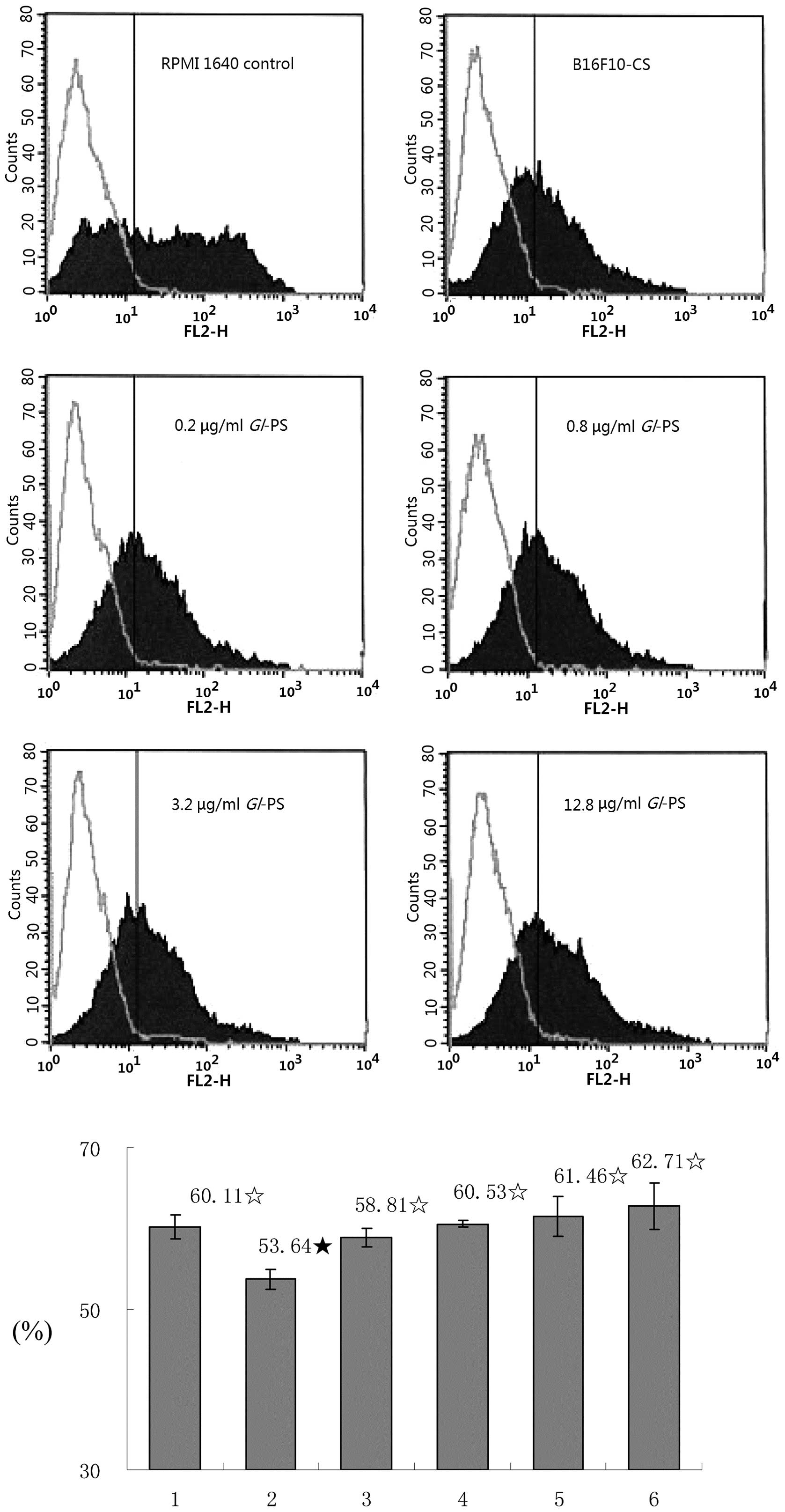
result in the suppression of CD71 expression. CD71 expression was
detected on lymphocytes. The lymphocytes were stimulated with PHA
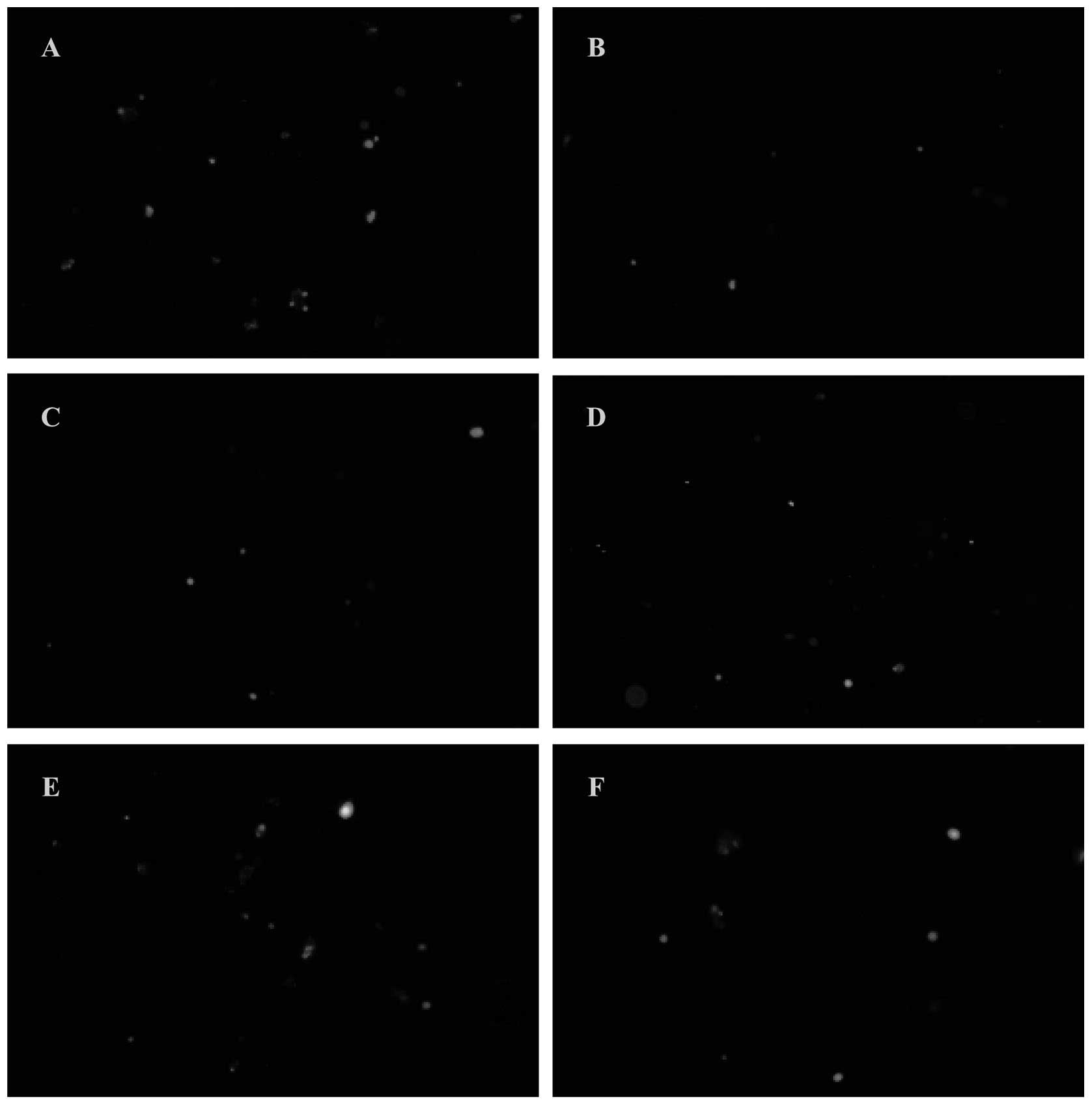
and cultured with B16F10-CS and Gl-PS for 48 h. It was shown
by immunofluorescence that the CD71 expression in mononuclear
lymphocytes was reduced after 48 h induction by PHA in B16F10-CS
wells (without Gl-PS), when compared with the RPMI-1640
medium control, while Gl-PS in the wells antagonized the
reduction of the CD71 expression (Fig.
1). It was also shown by flow cytometry that the expression
rate of CD71 in the lymphocytes was much lower in B16F10-CS wells
(without Gl-PS) than that in RPMI-1640 medium control wells
(P<0.05). The expression rate was higher in the wells with
B16F10-CS and any amount of Gl-PS used in this study than
that in wells with B16F10-CS without Gl-PS (all P<0.05,
Fig. 2), showing the antagonism of
Gl-PS on the reduction by B16F10-CS on the CD71 expression
in lymphocytes after induction by PHA.
Suppression by B16F10-CS and antagonism
by Gl-PS of splenic mononuclear lymphocyte proliferation induced by
PHA
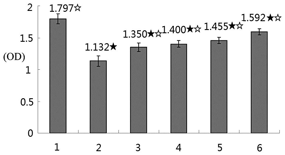
The activation of the lymphocytes can be disclosed
by proliferation induced by mitogens such as PHA. Therefore,
suppression of cell proliferation may reflect the suppression of
lymphocyte activation. The lymphocyte proliferation by MTT assay
after PHA stimulation was also detected. Compared with the
RPMI-1640 medium control, 72 h after induction with PHA, the OD in
B16F10-CS wells (without Gl-PS) was markedly reduced
(P<0.05), while in the wells containing different concentrations
of Gl-PS, the reduction of the OD was antagonized
significantly (P<0.05, Fig.
3).
Suppression by B16F10-CS and antagonism
by Gl-PS of FasL expression in splenic mononuclear lymphocytes
induced by PHA
FasL expression appears upon lymphocyte activation
for the maintenance of immune homeostasis as well as a pathway of
killing target cells. The FasL expression in splenic mononuclear
lymphocytes induced by mitogen such as PHA is also closely
associated with the activation of the lymphocytes to kill target
cells. The suppression of lymphocyte activation may, therefore,
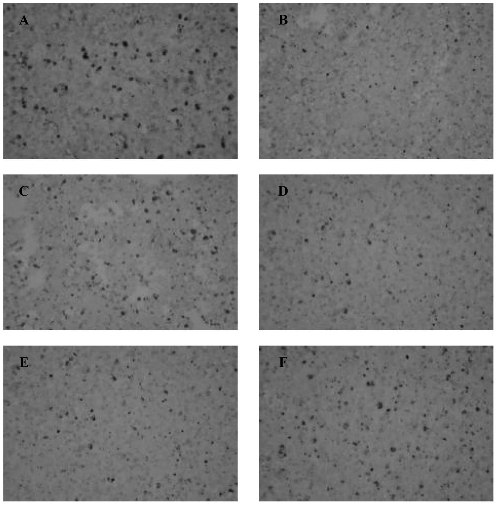
lead to the suppression of FasL expression. The FasL expression in
the lymphocytes was detected. Immunocytochemistry showed that FasL
expression in mononuclear lymphocytes was markedly reduced after 24
h incubation with PHA in B16F10-CS wells (without Gl-PS),
when compared with the RPMI-1640 medium control. Gl-PS in
the wells antagonized the reduction of the FasL expression
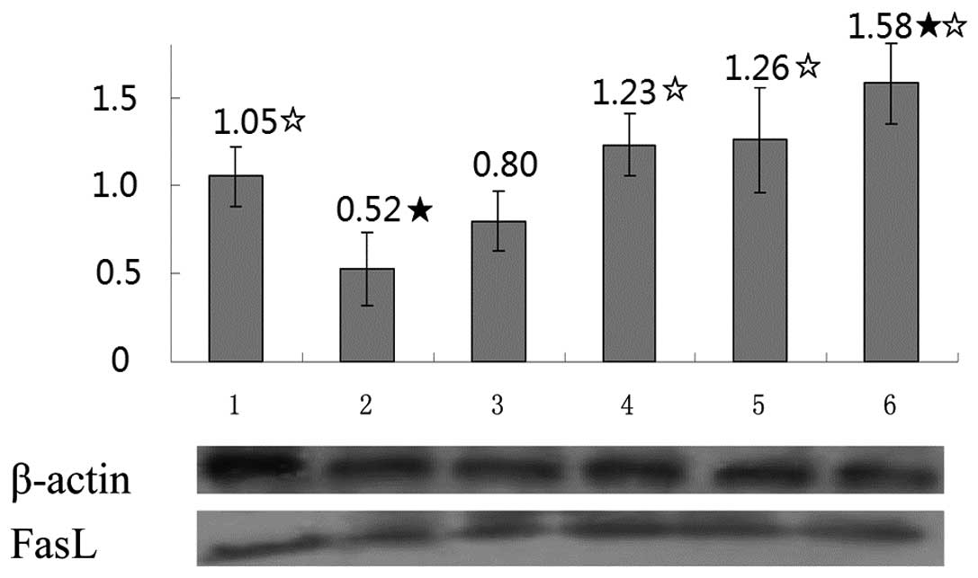
(Fig. 4). Western blot analysis
showed that FasL expression in mononuclear lymphocytes induced by
PHA was markedly reduced after 72 h in B16F10-CS wells (without
Gl-PS), when compared with the RPMI-1640 medium control
(P<0.05), while 0.8, 3.2 and 12.8 μg/ml Gl-PS
statistically antagonized the reduction of FasL expression
(P<0.05, Fig. 5).
Discussion
Immune responses to tumor-associated antigens (TAs)
are often detectable in tumor-bearing hosts, but they fail to
eliminate malignant cells or prevent the development of metastases
(29). Human tumors use many
different mechanisms of immuno-suppression to prevent immune cells
from exercising their antitumor activity, which enables the tumor
to escape from the host’s immune system and progress (30). The production and secretion of
soluble factors suppressing the functions of immune cells or
inducing the apoptosis of immune cells comprise part of the
immunosuppressive mechanisms (31). The IL-10, TGF-β, VEGF and
prostaglandins (PGs) are common immunosuppressive factors produced
by tumors which may directly or indirectly suppress the immune
response and obstruct immunotherapy (32). One of the most important aims of
tumor immunotherapy is, therefore, to counteract tumor-induced
immunosuppression. B16F10 cells are melanoma cells derived from
C57BL mice, which may produce immunosuppressive factors as well so
that the B16F10-CS may have the immunosuppressive effects (22).
Gl-PS, the critical component of Gl,
has multiple bio activities. In this study, the effects of
Gl-PS on the counteraction against the suppression induced
by B16F10-CS of CD71 and FasL expression upon lymphocyte activation
was examined. The proliferation, CD71 expression and FasL
expression were detected in the lymphocytes which were activated by
PHA to determine the suppression induced by B16F10-CS and the
counteraction by Gl-PS against the suppression.
The expression of activation markers on the cell
surface is useful in evaluating the lymphocyte response to antigens
or mitogens. CD71 is a lymphocyte activation marker, as are CD69
and CD25. CD71 expression on the cell surface represents the state
of metabolic activity in the cell and must be increased in order to
internalize the iron required for T-cell DNA synthesis and
proliferation (33). It was shown
in this study that CD71 expression in lymphocytes after activation
by PHA was suppressed by B16F10-CS, while the suppression was
antagonized by Gl-PS. CD71 is a transferrin receptor, a
membrane receptor involved in the control of iron supply to the
cell through the binding of transferrin, the major iron-carrier
protein. This receptor plays a key role in the control of cell
proliferation since iron is essential for sustaining
ribonucleo-tide reductase activity, and is the only enzyme that
catalyzes the conversion of ribonucleotides to deoxyribonucleotides
(34). Suppression of the CD71
expression in lymphocytes may, therefore, lead to suppression of
the proliferation and function of lymphocytes, while counteraction
of this suppression by Gl-PS may benefit the lymphocyte
activation and function.
T-cell activation is a very tightly regulated
process resulting in the production of cytokines as well as clonal
expansion and differentiation of effector T lymphocytes (35). Upon activation, T-cells undergo
complex structural and cytoskeletal changes leading to cell cycle
progression (36). The end result
is the activation and expansion of a pool of effector cells that
participate in cancer destruction. Proliferation is, therefore, a
common feature of lymphocyte activation. It was shown in this study
that after activation by PHA the proliferation of lymphocytes was
suppressed by the B16F10-CS, while the suppression was counteracted
by Gl-PS.
FasL is an inductive molecule expressed in T-cells,
and weighs 40 kDa (37). It is
homologous to the cytokine tumor necrosis factor (TNF), and is a
member of the TNFR-II type family (38). FasL expression depends on the
transcription factor levels. The positive regulators are NFAT,
Egr2/Egr3, NFκB, AP-1, c-myc SP1 and B1/Cdk1, while the negative
regulators are c-Fos and CIITA (39–41).
Binding of Fas with FasL causes trimerization and recruitment of
Fas-associated death domain (FADD) proteins through homotypic death
domain interactions. In turn, trimerized FADD recruits either
procaspase 8 or 10, which then becomes an activated caspase through
a process of autoproteolysis (42). Assembly of these components results
in the formation of a death-inducing signaling complex (DISC),
which is pivotal in the receptor-dependent apoptosis (42). Caspase 8 interacts with procaspases
3, 6, or 7 and, after a process of transproteolysis, they become
activated caspases. Finally, these effector caspases cleave DNA.
Caspase 8 can also hydrolyze Bid, which causes damage to the
mitochondrial outer membrane and triggers cytochrome-C release
(43,44). FasL expression is, therefore, also
an event in lymphocyte activation. In addition to maintaining
immune homeostasis, FasL expressed on activated lymphocytes
contributes to killing target cells by providing death signals.
FasL crosslinks Fas, a death receptor expressed on the target
cells, which causes the apoptosis of the target cells by triggering
the apoptosis pathway (45). It
was shown in this study that the expression of FasL in the
lymphocytes after activation by PHA was suppressed by the
B16F10-CS, while the suppression was antagonized by Gl-PS,
especially at higher concentrations, indicating the suppression of
lymphocyte activation by the B16F10-CS as well as the counteraction
by Gl-PS.
In conclusion, counteraction against the suppression
by tumor cells on CD71 expression, cell proliferation, FasL
expression in lymphocytes after induction with PHA denotes a
counteraction against the suppression by tumor cells on the
function of lymphocytes, which is important to facilitate cancer
immunotherapy, therefore, Gl-PS showing the effects of
counteraction against the suppression induced by B16F10-CS on CD71
expression, cell proliferation, as well as FasL expression after
induction with PHA, fully or partially, indicated the potential of
Gl-PS to facilitate cancer immunotherapy.
Acknowledgements
The authors thank Professor Shuqian
Lin of the Fuzhou Institute of Green Valley Bio-Pharm Technology
for providing the Gl-PS.
References
|
1
|
Swann JB and Smyth MJ: Immune surveillance
of tumors. J Clin Invest. 117:1137–1146. 2007. View Article : Google Scholar : PubMed/NCBI
|
|
2
|
Gross S and Walden P: Immunosuppressive
mechanisms in human tumors: why we still cannot cure cancer.
Immunol Lett. 116:7–14. 2008. View Article : Google Scholar : PubMed/NCBI
|
|
3
|
Rabinovich GA, Gabrilovich D and Sotomayor
EM: Immunosuppressive strategies that are mediated by tumor cells.
Annu Rev Immunol. 25:267–296. 2007. View Article : Google Scholar : PubMed/NCBI
|
|
4
|
Whiteside TL: Immune suppression in
cancer: effects on immune cells, mechanisms and future therapeutic
intervention. Semin Cancer Biol. 16:3–15. 2006. View Article : Google Scholar : PubMed/NCBI
|
|
5
|
Shao BM, Dai H, Xu W, Lin ZB and Gao XM:
Immune receptors for polysaccharides from Ganoderma lucidum.
Biochem Biophys Res Commun. 323:133–141. 2004. View Article : Google Scholar : PubMed/NCBI
|
|
6
|
Chan WK, Cheung CC, Law HK, Lau YL and
Chan GC: Ganoderma lucidum polysaccharides can induce human
monocytic leukemia cells into dendritic cells with
immunostimulatory function. J Hematol Oncol. 1:92008. View Article : Google Scholar
|
|
7
|
Amarante MK, Watanabe MA, Conchon-Costa I,
Fiori LL, Oda JM, Búfalo MC and Sforcin JM: The effects of propolis
on CCL5 and IFN-γ expression by peripheral blood mononuclear cells
from leishmaniasis patients. J Pharm Pharmacol. 64:154–160.
2012.
|
|
8
|
Kour K, Sangwan PL, Khan I, Koul S, Sharma
SN, Kitchlu S and Bani S: Alcoholic extract of Cicer microphyllum
augments Th1 immune response in normal and chronically stressed
Swiss albino mice. J Pharm Pharmacol. 63:267–277. 2011. View Article : Google Scholar : PubMed/NCBI
|
|
9
|
Sliva D: Medicinal mushroom Phellinus
linteus as an alternative cancer therapy. Exp Ther Med.
1:407–411. 2010.
|
|
10
|
Vermorken AJ, Zhu J, Van de Ven WJ, Cui Y
and Fryns JP: Curcumin for the prevention of progression in
monoclonal gammopathy of undetermined significance: A word of
caution. Exp Ther Med. 1:265–269. 2010. View Article : Google Scholar : PubMed/NCBI
|
|
11
|
Li QQ, Wang G, Reed E, Huang L and Cuff
CF: Evaluation of cisplatin in combination with β-elemene as a
regimen for prostate cancer chemotherapy. Basic Clin Pharmacol
Toxicol. 107:868–876. 2010.
|
|
12
|
Mishra N, Tiwari S, Vaidya B, Agrawal GP
and Vyas SP: Lectin anchored PLGA nanoparticles for oral mucosal
immunization against hepatitis B. J Drug Target. 19:67–78. 2011.
View Article : Google Scholar : PubMed/NCBI
|
|
13
|
Zhong Z, Dong Z, Yang L, Chen X and Gong
Z: Inhibition of proliferation of human lung cancer cells by green
tea catechins is mediated by upregulation of let-7. Exp Ther Med.
4:267–272. 2012.PubMed/NCBI
|
|
14
|
Yang Y, Jin C, Li H, He Y, Liu Z, Bai L
and Dou K: Improved radiosensitizing effect of the combination of
etanidazole and paclitaxel for hepatocellular carcinoma in vivo.
Exp Ther Med. 3:299–303. 2012.PubMed/NCBI
|
|
15
|
Yang X, Hu W, Zhang Q, Wang Y and Sun L:
Puerarin inhibits C-reactive protein expression via suppression of
nuclear factor kappaB activation in lipopolysaccharide-induced
peripheral blood mononuclear cells of patients with stable angina
pectoris. Basic Clin Pharmacol Toxicol. 107:637–42. 2010.
View Article : Google Scholar
|
|
16
|
Lin ZB and Zhang HN: Anti-tumor and
immunoregulatory activities of Ganoderma lucidum and its
possible mechanisms. Acta Pharmacol Sin. 25:1387–1395.
2004.PubMed/NCBI
|
|
17
|
You YH and Lin ZB: Protective effects of
Ganoderma lucidum polysaccharides peptide on injury of
macrophages induced by reactive oxygen species. Acta Pharmacol Sin.
23:787–791. 2002.
|
|
18
|
Zhang GL, Wang YH, Ni W, Teng HL and Lin
ZB: Hepatoprotective role of Ganoderma lucidum
polysaccharide against BCG-induced immune liver injury in mice.
World J Gastroenterol. 8:728–733. 2002.
|
|
19
|
Zhang HN, He JH, Yuan L and Lin ZB: In
vitro and in vivo protective effect of Ganoderma lucidum
polysaccharides on alloxan-induced pancreatic islets damage. Life
Sci. 73:2307–2319. 2003.PubMed/NCBI
|
|
20
|
Zhu XL and Lin ZB: Modulation of cytokines
production, granzyme B and perforin in murine CIK cells by
Ganoderma lucidum polysaccharides. Carbohydrate Polymers.
63:188–197. 2006. View Article : Google Scholar
|
|
21
|
Sun LX, Chen LH, Lin ZB, Qin Y, Zhang JQ,
Yang J, Ma J, Ye T and Li WD: Effects of Ganoderma lucidum
polysaccharides on IEC-6 cell proliferation, migration and
morphology of differentiation benefiting intestinal epithelium
healing in vitro. J Pharm Pharmacol. 63:1595–1603. 2011.
|
|
22
|
Sun LX, Lin ZB, Duan XS, Lu J, Ge ZH, Li
XJ, Li M, Xing EH, Jia J, Lan TF and Li WD: Ganoderma
lucidum polysaccharides antagonize the suppression on
lymphocytes induced by culture supernatants of B16F10 melanoma
cells. J Pharm Pharmacol. 63:725–35. 2011. View Article : Google Scholar
|
|
23
|
Sun LX, Lin ZB, Li XJ, Li M, Lu J, Duan
XS, Ge ZH, Song YX, Xing EH and Li WD: Promoting effects of
Ganoderma lucidum polysaccharides on B16F10 cells to
activate lymphocytes. Basic Clin Pharmacol Toxicol. 108:149–154.
2011.
|
|
24
|
Sun LX, Lin ZB, Duan XS, Lu J, Ge ZH, Song
YX, Li XJ, Li M, Xing EH, Yang N and Li WD: Stronger cytotoxicity
in CTLs with granzyme B and porforin was induced by Ganoderma
lucidum polysaccharides acting on B16F10 cells. Biomed Prev
Nutr. 2:113–118. 2012. View Article : Google Scholar
|
|
25
|
Sun LX, Lin ZB, Duan XS, Lu J, Ge ZH, Li
XF, Li XJ, Li M, Xing EH, Song YX, Jia J and Li WD: Enhanced MHC
class I and costimulatory molecules on B16F10 cells by Ganoderma
lucidum polysaccharides. J Drug Target. 20:582–592. 2012.
View Article : Google Scholar : PubMed/NCBI
|
|
26
|
Cao LZ and Lin ZB: Regulation on
maturation and function of dendritic cells by Ganoderma
lucidum polysaccharides. Immunol Lett. 83:163–169. 2002.
View Article : Google Scholar : PubMed/NCBI
|
|
27
|
Wang YY, Khoo KH, Chen ST, Lin CC, Wong CH
and Lin CH: Studies on the immuno-modulating and antitumor
activities of Ganoderma lucidum (Reishi) polysaccharides:
functional and proteomic analyses of a fucose-containing
glycoprotein fraction responsible for the activities. Bioorg Med
Chem. 10:1057–1062. 2002.PubMed/NCBI
|
|
28
|
Reddy M, Eirikis E, Davis C, Davis HM and
Prabhakar U: Comparative analysis of lymphocyte activation marker
expression and cytokine secretion profile in stimulated human
peripheral blood mononuclear cell cultures: an in vitro model to
monitor cellular immune function. J Immunol Methods. 293:127–142.
2004. View Article : Google Scholar
|
|
29
|
Whiteside TL: Immune responses to
malignancies. J Allergy Clin Immunol. 125:S272–S283. 2010.
View Article : Google Scholar : PubMed/NCBI
|
|
30
|
Whiteside TL, Mandapathil M, Szczepanski M
and Szajnik M: Mechanisms of tumor escape from the immune system:
adenosine-producing Treg, exosomes and tumor-associated TLRs. Bull
Cancer. 98:E25–E31. 2011.PubMed/NCBI
|
|
31
|
Wongkajornsilp A, Luankosolchai RA,
Huabprasert S, Chanyavanich V, Tisavipat N and Watanapa P: The
observation of immunosuppressor(s) derived from cultured tumor
cells and its partial neutralization with OK-432. J Med Assoc Thai.
84:212–122. 2001.PubMed/NCBI
|
|
32
|
Frumento G, Piazza T, Di Carlo E and
Ferrini S: Targeting tumor-related immunosuppression for cancer
immunotherapy. Endocr Metab Immune Disord Drug Targets. 6:233–237.
2006. View Article : Google Scholar : PubMed/NCBI
|
|
33
|
Cortés-Barberena E, González-Márquez H,
Gómez-Olivares JL and Ortiz-Muñiz R: Effects of moderate and severe
malnutrition in rats on splenic T lymphocyte subsets and activation
assessed by flow cytometry. Clin Exp Immunol. 152:585–592.
2008.PubMed/NCBI
|
|
34
|
Testa U, Pelosi E and Peschle C: The
transferrin receptor. Crit Rev Oncog. 4:241–276. 1993.
|
|
35
|
Goral S: The three-signal hypothesis of
lymphocyte activation/targets for immunosuppression. Dial
Transplant. 40:14–16. 2011. View Article : Google Scholar
|
|
36
|
Reicher B and Barda-Saad M: Multiple
pathways leading from the T-cell antigen receptor to the actin
cytoskeleton network. FEBS Lett. 584:4858–4864. 2010. View Article : Google Scholar : PubMed/NCBI
|
|
37
|
Chávez-Galán L, Arenas-Del Angel MC,
Zenteno E, Chávez R and Lascurain R: Cell death mechanisms induced
by cytotoxic lymphocytes. Cell Mol Immunol. 6:15–25. 2009.
|
|
38
|
Starling GC, Bajorath J, Emswiler J,
Ledbetter JA, Aruffo A and Kiener PA: Identification of amino acid
residues important for ligand binding to Fas. J Exp Med.
185:1487–1492. 1997. View Article : Google Scholar : PubMed/NCBI
|
|
39
|
Sun M, Ames KT, Suzuki I and Fink PJ: The
cytoplasmic domain of Fas ligand costimulates TCR signals. J
Immunol. 177:1481–1491. 2006. View Article : Google Scholar : PubMed/NCBI
|
|
40
|
Dzialo-Hatton R, Milbrandt J, Hockett RD
Jr and Weaver CT: Differential expression of Fas ligand in Th1 and
Th2 cells is regulated by early growth response gene and NF-AT
family members. J Immunol. 166:4534–4542. 2001. View Article : Google Scholar : PubMed/NCBI
|
|
41
|
Gourley TS and Chang CH: Cutting edge: the
class II transactivator prevents activation-induced cell death by
inhibiting Fas ligand gene expression. J Immunol. 166:2917–2921.
2001. View Article : Google Scholar : PubMed/NCBI
|
|
42
|
Carrington PE, Sandu C, Wei Y, Hill JM,
Morisawa G, Huang T, Gavathiotis E, Wei Y and Werner MH: The
structure of FADD and its mode of interaction with procaspase-8.
Mol Cell. 22:599–610. 2006. View Article : Google Scholar : PubMed/NCBI
|
|
43
|
Luo X, Budihardjo I, Zou H, Slaughter C
and Wang X: Bid, a Bcl2 interacting protein, mediates cytochrome c
release from mitochondria in response to activation of cell surface
death receptors. Cell. 94:481–490. 1998. View Article : Google Scholar : PubMed/NCBI
|
|
44
|
Li H, Zhu H, Xu CJ and Yuan J: Cleavage of
BID by caspase 8 mediates the mitochondrial damage in the Fas
pathway of apoptosis. Cell. 94:491–501. 1998. View Article : Google Scholar : PubMed/NCBI
|
|
45
|
de Vries JF, von dem Borne PA, van
Luxemburg-Heijs SA, Heemskerk MH, Willemze R, Falkenburg JH and
Barge RM: Differential activation of the death receptor pathway in
human target cells induced by cytotoxic T lymphocytes showing
different kinetics of killing. Haematologica. 92:1671–1678.
2007.PubMed/NCBI
|