Introduction
Breast cancer is a malignant tumor with a high
incidence in females from more developed western countries.
Although historically the incidence of breast cancer in women in
China was low, it has been on the increase in the past 20 years. In
large cities, such as Beijing and Shanghai, breast cancer is the
most common type of malignant tumor diagnosed in women. As the
country with the highest population in the world, there are a large
total number of cases annually. Anthracycline chemotherapeutic
agents, including Adriamycin, are among the main agents used for
breast cancer chemotherapy. Adriamycin has been essential in breast
cancer therapy, particularly in prolonging the survival of patients
with advanced and metastatic breast cancer. However, due to the
dose-limiting cardiotoxicity of Adriamycin, it is not suitable for
use in patients with cardiac disorders. The administration of
Adriamycin may cause varying degrees of myocardial toxicity,
seriously affecting the patients’ quality of life, and in certain
cases has caused toxicity-related mortality (1–3).
In recent years, studies of myocardial toxicity have
been reported, but no drug has been satisfactorily applied in a
clinical environment (4–6). With developments in the treatment of
malignant tumors, tumor reduction and disease relief in the short
term are no longer sufficient and steps to improve quality of life
and prolong the survival of patients are drawing an increasing
amount of attention. There has been a strong clinical demand for
cardioprotective drugs for use in tumor patients (7). There is a need for a drug that is not
only helpful in the treatment of breast cancer, but also alleviates
Adriamycin-induced cardiotoxicity.
Melatonin is an endocrine hormone naturally present
in the human body, which has a complicated and important
physiological role. Studies have confirmed that melatonin has an
inhibitory effect on multiple malignant tumors; the antitumor
effects in patients with ER+ breast cancer have been
reported (8–11). The inhibitory effect on breast
cancer cells was confirmed again at the cellular level in our
previous research (12,13). Notably, the resistance reversal
effect and sensitizing mechanism of Adriamycin have been observed.
Additionally, melatonin, currently the strongest known antioxidant
(14–17), has been confirmed to have a
significant multi-organ protective effect via a complex mechanism
(18–22).
It would be of interest to determine whether
melatonin has a cardioprotective effect that would aid the
anticarcinogenic effect of Adriamycin and, if so, to understand the
mechanism. Melatonin, therefore, may be an excellent adjuvant drug
in breast cancer chemotherapy, which would be worthy of further
study. A rat model of ER+ breast cancer was established
in order to investigate melatonin’s effects. The condition of the
human body was simulated as closely as possible in order to observe
the cardiotoxicity and the quality of life in rats concurrently
treated with melatonin and Adriamycin.
Materials and methods
Animals
In this study, 140 healthy female Sprague-Dawley
(SD) rats, weighing ∼200–250 g, were purchased from the Animal
Center of Hebei Medical University. The rats were maintained in
light-dark (LD) conditions. LD involved 12 h of light (from 6:00 to
18:00) and 12 h of darkness (from 18:00 to 6:00). The rats had free
access to water and food, and were maintained in a room with a
temperature of 25±2°C and humidity of 45–50%. The rats entered the
trial after 3 weeks of synchronization. This study was carried out
in strict accordance with the recommendations in the Guide for the
Care and Use of Laboratory Animals of the National Institutes of
Health. The protocol for animal use was reviewed and approved by
the Institutional Animal Care and Use Committee (IACUC) of the
First Hospital of Shijiazhuang, Hebei.
Establishment of the rat model of breast
cancer
According to the Russo method (23), of the 140 female SD rats, 130 were
injected with N-nitroso-N-methylurea (MNU; Sigma-Aldrich, St Louis,
MO, USA) to induce and establish the breast cancer models, and the
remaining 10 rats underwent an intraperitoneal injection with a
solvent which is used to dissolve MNU.
According to the Dagar method (24), the rats were weighed and numbered.
The rats were intraperitoneally injected with a 50 mg/kg dose of
MNU. After being weighed 2 weeks later, they were injected again
with the same dosage of MNU. The tumor growth in the rat breast was
observed on a weekly basis for the timely evaluation of successful
cases.
Animal groups
The rats were divided into the blank control group
(Blank); the solvent control group [Diss; dehydrated alcohol:
physiological saline (1:9)]; the melatonin group (MLT) who were
injected with a 10 mg/kg dose of melatonin (Sigma-Aldrich) once a
day for a total of 15 times; the Adriamycin group (ADM) who were
injected with a 2.5 mg/ml dose of Adriamycin every other day for a
total of 7 times; and the melatonin + Adriamycin (M+A) group. From
the first day in the M+A group the rats were treated with a 10
mg/kg dose of MLT once a day for 15 days, and from the third day
the rats were also treated with a 2.5 mg/ml dose of ADM every other
day. MLT was injected intraperitoneally prior to ADM. On the 18th
day, a number of each group of rats were sacrificed and taken for
analysis; the remaining rats continued to be observed to ascertain
the survival rates.
Detection of oxidative indices
Each group of rats underwent an intraperitoneal
injection of 30 mg/kg pentobarbital sodium as an anesthetic, prior
to opening the chest and the quick removal of the heart, which was
flushed with physiological saline three times and dried with filter
paper. After being weighed, the heart was prepared as a 10%
homogenate with a 0.2 M/l phosphate buffer solution (pH 8.6) in an
ice water bath to determine the concentration of lipid peroxide
(LPO) and the activities of superoxide dismutase (SOD) and
glutathione peroxidase (GSH-Px) in the myocardial tissue.
Preparation of myocardial tissue samples
for light microscopy
The rats underwent anesthesia as described above,
followed by the removal of the heart. The interventricular septum
was dissected and the myocardial tissues were removed from the left
ventricle. The myocardial tissues were cut into 8–10 blocks of 1
mm3 along with the striation of muscle fiber and fixed
in 10% paraformaldehyde phosphate buffer solution for pathological
sectioning and hematoxylin and eosin (H&E) staining. The
tissues were observed and photographic images captured under a
light microscope using low and high magnification.
Preparation of samples for electron
microscopy
The steps for processing the myocardial tissue
samples for electron microscopy included: sampling, pre-fixation,
washing, post-fixation, washing, dehydration, saturation,
embedding, polymerization, ultrathin sectioning, observation and
photographic image capture.
Statistical analysis
The data were analyzed with the statistical software
SPSS 13.0 (SPSS, Inc., Chicago, IL, USA). The oxidative indices
were compared using t-tests and the 1-month survival rate was
compared with the Chi-square test. P<0.05 was considered to
indicate a statistically significant difference
Results
Tumors and groups
In total, 140 female SD rats were used in this
study. Of the 10 rats in the MNU Diss group, there appeared to be
one death without an identifiable cause and the other rats were in
a good condition, without the generation of any tumors. Among the
130 rats who underwent MNU injection, four rats died during the
injection and three rats who had generated tumors died before they
were placed into groups. In total, seven rats did not generate
tumors and 116 rats did generate tumors and were randomly placed
into groups. The tumor initiation rate was 91.5% (119/130). In each
group, eight rats were randomly selected and dissected to remove
the tumors and hearts and then sacrificed, and the remaining rats
(24 rats in the blank group, 24 in the solvent group, 22 in the MLT
group, 24 in the ADM group, 22 in the M+A group) were observed for
six months in order to determine mortality rates.
Oxidative indices
The oxidative indices included LPO, SOD and GSH-Px
concentrations in the myocardial tissues. There were no marked
differences in each index among the Blank, Diss and MLT groups. The
concentration of LPO in the myocardial tissues in the ADM group was
significantly higher when compared with that in the M+A group
(P<0.05). The concentrations of SOD and GSH-Px in the ADM group
were significantly lower when compared with those in the M+A group
(P<0.05, Table I).
 | Table IComparison of LPO, SOD and GSH-Px
concentrations in the various groups. |
Table I
Comparison of LPO, SOD and GSH-Px
concentrations in the various groups.
| Groups | LPO (nmol/mgPr) | SOD
(μg/mgPr) | GSH-Px
(μ/mgPr) |
|---|
| Blank | 6.68±0.91 | 26.67±2.13 | 17.33±2.17 |
| Diss | 7.15±0.44 | 25.33±4.49 | 16.54±1.91 |
| MLT | 5.59±1.77 | 26.55±2.07 | 18.02±0.75 |
| ADM | 9.88±1.50 | 10.19±0.78 | 8.97±0.57 |
| M+A | 6.79±0.48a | 19.99±1.91b | 13.81±1.52c |
General characteristics of the
hearts
The shapes, sizes and weight of the hearts in the
MLT, Blank and Diss groups were close to normal. The hearts of the
rats in the ADM group appeared to be markedly congested and
swollen, with increased volumes and visible petechiae on the
pericardium. The hearts in the M+A group were essentially normal in
shape, with mild congestion, but without visible petechiae on the
pericardium.
Appearance of hearts under light and
electron microscopy
There were no marked anomalies in the Blank, Diss
and MLT groups, either under a light microscope with low
magnification or under an electron microscope for H&E-stained
myocardial sections.
In the ADM group, under a light microscope with a
low magnification, a large number of cardiac muscle bundle
fractures were observed, with mucus visible between the muscle
bundles (Fig. 1A). Under a light
microscope with a high magnification, it was observed that the
cardiac muscles appeared to be disordered with severe mucinous
degeneration (Fig. 1B). Under an
electron microscope, it was observed that the matrix in the nuclear
side appeared to have a marked edema, where the majority of the
ridges and a few of the mitochondrial membranes appeared to be
fused, indistinct or missing, some of the chromosomes appeared to
have undergone the cavitation phenomenon, glycogen granules were
significantly reduced under an electron microscope and parts of the
coxae were either arranged in an unorganized manner or missing
(Fig. 1C).
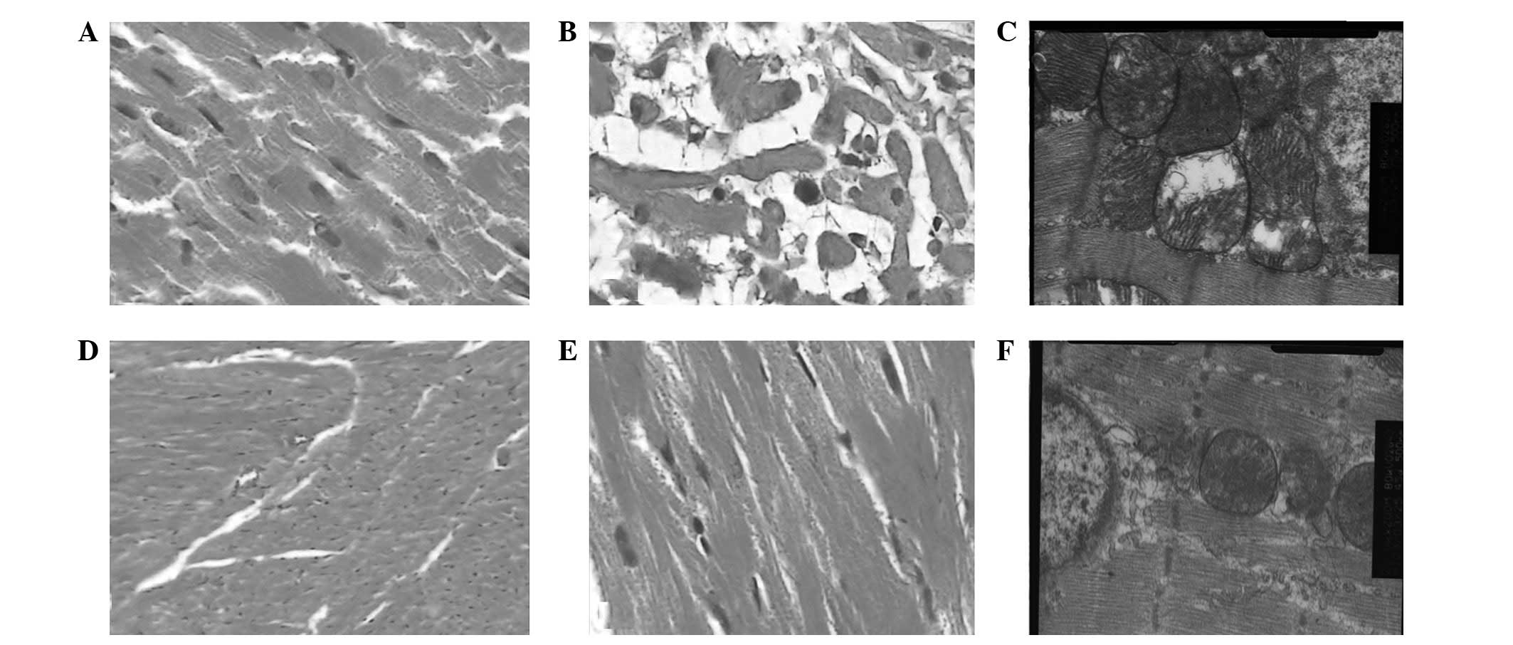 | Figure 1(A) Adriamycin-induced myocardial
injuries and muscle bundle fractures were visible under a light
microscope with low magnification in the ADM group (magnification,
×100); (B) Adriamycin-induced myocardial mucinous degeneration was
visible under a light microscope with high magnification in the ADM
group (magnification, ×400); (C) Adriamycin-induced organelle
injuries in myocardial cells and serious mitochondrial cavitation
were visible under a light microscope with high magnification in
the ADM group (magnification, ×20.0KX); (D) Essentially normal
cardiac muscles were visible under a light microscope with low
magnification in the M+A group (magnification, ×100); (E) Only mild
granular changes were visible under a light microscope with high
magnification in the M+A group (magnification, 400×); (F)
Adriamycin-induced organelle injuries in myocardial cells and
serious mitochondrial cavitation were visible under an electron
microscope with high magnification in the M+A group (magnification,
×20,000). Blank, no intervention; Diss, solvent intervention; MLT,
melatonin intervention; ADM, Adriamycin intervention; M+A,
melatonin + Adriamycin intervention. |
Under a light microscope with a low magnification,
it was observed that the cardiac muscle bundles were essentially
normal in the M+A group, but mild bundle fractures were present in
a few areas (Fig. 1D). Mild
granular degeneration was observed under a light microscope with
high magnification (Fig. 1E).
Under an electron microscope it was observed that a few sections of
the mitochondrial ridges appeared to be fused and indistinct and
the glycogen granules were reduced, but no cavitation phenomenon
was observed in the chromosomes (Fig.
1F).
Survival rates after 1-month
The 1-month survival rates of the remaining rats in
each group were analyzed and are shown in Table II. The 1-month survival rates were
4/16 in the Blank group and 6/16 in the Diss group, without a
statistically significant difference (P=0.35), 14/14 in the MLT
group, with a total survival rate significantly higher than in the
previous two groups (P=0.000), 5/16 in the ADM group and 11/14 in
the M+A group, with the latter higher than the former (P=0.012),
without a statistically significant difference between the MLT
group and the M+A group (P=0.11).
| Table IIComparison of 1-month survival rate
among groups. |
Table II
Comparison of 1-month survival rate
among groups.
| Groups | n | One month survival
(n) |
|---|
| Blank | 16 | 4 |
| Diss | 16 | 6 |
| MLT | 14 | 14 |
| ADM | 16 | 5 |
| M+A | 14 | 11 |
Discussion
Melatonin is an endocrine substance naturally
present in vivo that is secreted by the pinealocyte. It is
widely distributed in various organs of the human body and plays an
important and complicated biological function in vivo.
Studies have confirmed that melatonin has a multi-organ protective
effect, which indicates that melatonin may also play a role in
myocardial protection by promoting the anti-tumor effect of
Adriamycin (21,25–28).
In this study, rat models of ER+ breast cancer were
established in order to compare the cardiotoxicity in the ADM
group, with exposure to Adriamycin, and the M+A group, with
concurrent exposure to melatonin and Adriamycin. It was observed in
the general samples that the volumes of the hearts of the rats in
the ADM group were larger, with marked congestion and swelling, and
varying severities of petechiae on the pericardium. By contrast,
the hearts in the M+A group were almost normal in shape and color,
with visible congestion in severe cases, but without visible
petechiae on the pericardium. Under a light microscope with a low
magnification, it was observed that the myocardial tissues of the
ADM rats appeared to have a large number of cardiac muscle bundle
fractures, with mucus between the cardiac muscles (Fig. 1A). It was observed, under a light
microscope with high magnification, that the cardiac muscles were
arranged in a disorderly manner and appeared to have severe
mucinous degeneration (Fig. 1B).
It was also observed, under a light microscope with low
magnification, that the cardiac muscle bundles were essentially
normal in the M+T group, with no marked bundle fractures (Fig. 1D); mild granular degeneration was
visible under a light microscope with high magnification (Fig. 1E). Thereafter, changes in organelle
levels were observed using electron microscopy and it was
demonstrated that the organelles in the myocardial cells were badly
damaged in the ADM group and the majority of the mitochondria
appeared to undergo the cavitation phenomenon (Fig. 1C). There were only mild injuries to
the mitochondria in the M+A group (Fig. 1F). From different levels of
morphology, it was demonstrated that the myocardial injuries in the
M+A group, with intervention of melatonin, were alleviated compared
with those in the ADM group with a single application of
Adriamycin, which was consistent with our expectations, indicating
that melatonin has a protective effect against Adriamycin-induced
myocardial toxicity.
Some hypotheses concerning the mechanism of
anthracycline-induced myocardial injuries are as follows: i) the
oxidative stress effects produce a large number of free radicals,
causing myocardial injuries via oxidation; ii) calcium overload and
energy metabolism disorder may cause lipid peroxidation in
myocardial cells; iii) the tyrosine residing in myocardial cells
became nitrated by Adriamycin; iv) the myocardial cells appeared to
undergo direct apoptosis. Free radical damage and lipid
peroxidation in myocardial cells represented significant
anthracycline-induced myocardial injuries. Therefore, the
scavenging of free radicals is an important measure for preventing
myocardial injuries (29).
Superoxide dismutase (SOD) scavenges free radicals
in vivo. It catalyzes the conversion of superoxide radicals
into hydrogen peroxide and oxygen molecules and is key in resisting
cell damage caused by oxygen free radicals. Glutathione peroxidase
(GSH-Px) is an important peroxide-decomposing enzyme widely found
in vivo and is a detoxification enzyme that scavenges
hydrogen peroxide and other organic peroxides. It protects the
structure and the function of the cell membrane from peroxide
interference and damage through action as an antioxidant. LPO is
produced by the polyunsaturated fatty acids in the cell membrane
structure in vivo with the influence of oxygen free
radicals. The peroxidation of lipid membranes may damage the
membrane structure and cell function, causing a variety of
diseases. Floyd et al(30)
suggested that SOD, GSH-Px and LPO may be important indicators in
pharmacodynamic studies of Adriamycin-induced cardiotoxicity with
drug intervention, as they are closely related to the degree of
myocardial injury. In the current study, SOD and GSH-Px were
observed to be negatively correlated with Adriamycin-induced
myocardial injuries, while LPO was positively correlated (Table I), indicating that these three
indicators may be considered to be main outcome measures in the
evaluation of Adriamycin-induced cardiotoxicity. It was also
demonstrated that melatonin is significant for protecting against
oxidation and lipid peroxidation, which is consistent with the
results of previous studies. This may be one of the mechanisms by
which melatonin reduced Adriamycin-induced myocardial injury. This
is also consistent with the organ protective mechanism of melatonin
through antioxidation observed in previous studies (21,21,25,31,32).
In the current study, it was observed that all rats
treated with melatonin had a better overall quality of life. The
tumors shrank in the ADT group, but the rats in this group had the
worst quality of life and the shortest survival period (Table II). The tumors shrank slightly in
the M+A group compared with that in the ADM group for application
of melatonin, but the quality of life and survival period of the
rats in the M+A group were improved compared with those in the ADM
group. The treatment concept for advanced malignant tumors has
changed. Compared with the previous focus simply on tumor
shrinkage, at present, improvements to the quality of life and
extension of the survival period are considered to be more
important. It has been demonstrated in previous studies that
melatonin relieves toxicity and enhances the curative effect in
addition to improving the quality of life in the treatment of a
variety of tumors (33–35), which was confirmed again by the
current study, indicating that as an adjuvant drug of Adriamycin or
other chemotherapy drugs, melatonin may have a function concordant
with the current concept of tumor therapy. However, the mechanism
of melatonin is very complicated. Further studies into possible
mechanisms are required in order to make developments that are of
clinical value.
Acknowledgements
This study was supported by the
Science and Technology Support Program of Hebei Province
(No.11276178).
References
|
1.
|
Rock E and DeMichele A: Nutritional
approaches to late toxicities of adjuvant chemotherapy in breast
cancer survivors. J Nutr. 133:3785S–3793S. 2003.PubMed/NCBI
|
|
2.
|
Billingham ME, Mason JW, Bristow MR and
Daniels JR: Anthracycline cardiomyopathy monitored by morphologic
changes. Cancer Treat Rep. 62:865–872. 1978.PubMed/NCBI
|
|
3.
|
Bristow MR, Thompson PD, Martin RP, Mason
JW, Billingham ME and Harrison DC: Early anthracycline
cardiotoxicity. Am J Med. 65:823–832. 1978. View Article : Google Scholar
|
|
4.
|
Chicco AJ, Schneider CM and Hayward R:
Voluntary exercise protects against acute doxorubicin
cardiotoxicity in the isolated perfused rat heart. Am J Physiol
Regul Integr Comp Physiol. 289:R424–R431. 2005. View Article : Google Scholar : PubMed/NCBI
|
|
5.
|
Chicco AJ, Schneider CM and Hayward R:
Exercise training attenuates acute doxorubicin-induced cardiac
dysfunction. J Cardiovasc Pharmacol. 47:182–189. 2006. View Article : Google Scholar : PubMed/NCBI
|
|
6.
|
Hydock DS, Lien CY, Schneider CM and
Hayward R: Exercise preconditioning protects against
doxorubicin-induced cardiac dysfunction. Med Sci Sports Exerc.
40:808–817. 2008. View Article : Google Scholar : PubMed/NCBI
|
|
7.
|
Albini A, Pennesi G, Donatelli F,
Cammarota R, De Flora S and Noonan DM: Cardiotoxicity of anticancer
drugs: the need for cardio-oncology and cardio-oncological
prevention. J Natl Cancer Inst. 102:14–25. 2010. View Article : Google Scholar : PubMed/NCBI
|
|
8.
|
Lissoni P: Biochemotherapy with
immunomodulating pineal hormones other than melatonin:
5-methoxytryptamine as a new oncostatic pineal agent. Pathol Biol
(Paris). 55:198–200. 2007. View Article : Google Scholar : PubMed/NCBI
|
|
9.
|
Jawed S, Kim B, Ottenhof T, Brown GM,
Werstiuk ES and Niles LP: Human melatonin MT1 receptor induction by
valproic acid and its effects in combination with melatonin on
MCF-7 breast cancer cell proliferation. Eur J Pharmacol. 560:17–22.
2007. View Article : Google Scholar : PubMed/NCBI
|
|
10.
|
Lemus-Wilson A, Kelly PA and Blask DE:
Melatonin blocks the stimulation effects of prolactin and human
breast cancer cell growth in culture. Br J Cancer. 72:1435–1440.
1995. View Article : Google Scholar
|
|
11.
|
Sánchez-Barceló EJ, Cos S, Mediavilla D,
Martínez-Campa C, González A and Alonso-González C:
Melatonin-estrogen interactions in breast cancer. J Pineal Res.
38:217–222. 2005.
|
|
12.
|
Zhang Y, Zhu S and Liu J: The reverse
effect and mechanism of melatonin on breast carcinoma cell line
MCF-7/ADM resistant to Adriamycin. Chinese Journal of Clinical
Oncology. 36:291–295. 2009.(In Chinese).
|
|
13.
|
Zhang Y, Zhu S and Zhao W: The effect of
melatonin on the sensitivity of ER+breast carcinoma cell
line MCF-7 to Adriamycin and its mechanism. Chinese Journal of
Clinical Oncology. 36:1243–1247. 2009.(In Chinese).
|
|
14.
|
Reiter RJ, Tan DX, Manchester LC, Paredes
SD, Mayo JC and Sainz RM: Melatonin and reproduction revisited.
Biol Reprod. 81:445–456. 2009. View Article : Google Scholar : PubMed/NCBI
|
|
15.
|
Sener G, Jahovic N, Tosun O, Atasoy BM and
Yeğen BC: Melatonin ameliorates ionizing radiation-induced
oxidative organ damage in rats. Life Sci. 74:563–572. 2003.
View Article : Google Scholar : PubMed/NCBI
|
|
16.
|
Reiter RJ, Guerrero JM, Escames G,
Pappolla MA and Acuña-Castroviejo D: Prophylactic actions of
melatonin in oxidative neurotoxicity. Ann N Y Acad Sci. 825:70–78.
1997. View Article : Google Scholar : PubMed/NCBI
|
|
17.
|
Okatani Y, Wakatsuki A and Kaneda C:
Melatonin increases activities of glutathione peroxidase and
superoxide dismutase in fetal rat brain. J Pineal Res. 28:89–96.
2000. View Article : Google Scholar : PubMed/NCBI
|
|
18.
|
Reiter RJ, Acuña-Castroviejo D, Tan DX and
Burkhardt S: Free radical-mediated molecular damage. Mechanisms for
the protective actions of melatonin in the central nervous system.
Ann N Y Acad Sci. 939:200–215. 2001. View Article : Google Scholar : PubMed/NCBI
|
|
19.
|
Hardeland R, Pandi-Perumal SR and
Cardinali DP: Melatonin. Int J Biochem Cell Biol. 38:313–316. 2006.
View Article : Google Scholar
|
|
20.
|
Ganguly K, Kundu P, Banerjee A, Reiter RJ
and Swarnakar S: Hydrogen peroxide-mediated downregulation of
matrix metalloprotease-2 in indomethacin-induced acute gastric
ulceration is blocked by melatonin and other antioxidants. Free
Radic Biol Med. 41:911–925. 2006. View Article : Google Scholar
|
|
21.
|
Bruck R, Aeed H, Avni Y, et al: Melatonin
inhibits nuclear factor kappa B activation and oxidative stress and
protects against thioacetamide induced liver damage in rats. J
Hepatol. 40:86–93. 2004. View Article : Google Scholar : PubMed/NCBI
|
|
22.
|
Yurtcu E, Guney Y, Ergun MA, et al: Lack
of a time-dependent effect of melatonin on radiation-induced
apoptosis in cultured rat lymphocytes. Cell Biol Int. 31:1144–1149.
2007. View Article : Google Scholar : PubMed/NCBI
|
|
23.
|
Russo J, Tay LK and Russo IH:
Differentiation of the mammary gland and susceptibility to
carcinogenesis. Breast Cancer Res Treat. 2:5–73. 1982. View Article : Google Scholar : PubMed/NCBI
|
|
24.
|
Dagar S, Sekosan M, Rubinstein I and
Onyüksel H: Detection of VIP receptors in MNU-induced breast cancer
in rats: implications for breast cancer targeting. Breast Cancer
Res Treat. 65:49–54. 2001. View Article : Google Scholar : PubMed/NCBI
|
|
25.
|
Sener-Muratoğlu G, Paskaloğlu K, Arbak S,
Hürdağ C and Ayanoğlu-Dülger G: Protective effect of famotidine,
omeprazole, and melatonin against acetylsalicylic acid-induced
gastric damage in rats. Dig Dis Sci. 46:318–330. 2001.PubMed/NCBI
|
|
26.
|
Maestroni GJ: The immunotherapeutic
potential of melatonin. Expert Opin Investig Drugs. 10:467–476.
2001. View Article : Google Scholar : PubMed/NCBI
|
|
27.
|
Wang WZ, Fang XH, Stephenson LL, et al:
Microcirculatory effects of melatonin in rat skeletal muscle after
prolonged ischemia. J Pineal Res. 39:57–65. 2005. View Article : Google Scholar : PubMed/NCBI
|
|
28.
|
Topal T, Oter S, Korkmaz A, et al:
Exogenously administered and endogenously produced melatonin reduce
hyperbaric oxygen-induced oxidative stress in rat lung. Life Sci.
75:461–467. 2004. View Article : Google Scholar
|
|
29.
|
Fisher PW, Salloum F, Das A, et al:
Phosphodiesterase-5 inhibition with sildenafil attenuates
cardiomyocyte apoptosis and left ventricular dysfunction in a
chronic model of doxorubicin cardiotoxicity. Circulation.
111:1601–1610. 2005. View Article : Google Scholar : PubMed/NCBI
|
|
30.
|
Floyd JD, Nguyen DT, Lobins RL, et al:
Cardiotoxicity of cancer therapy. J Clin Oncol. 23:7685–7696. 2005.
View Article : Google Scholar : PubMed/NCBI
|
|
31.
|
Mirunalini S, Karthishwaran K, Dhamodharan
G and Shalini M: Melatonin attenuates lipid peroxidation and
enhances circulatory antioxidants during mammary carcinogenesis in
rats. J Biochem Tech. 2:171–174. 2010.
|
|
32.
|
Reiter RJ and Tan DX: Melatonin: a novel
protective agent against oxidative injury of the
ischemic/reperfused heart. Cardiovasc Res. 58:10–19. 2003.
View Article : Google Scholar : PubMed/NCBI
|
|
33.
|
Lissoni P, Barni S, Mandalá M, Ardizzoia
A, Paolorossi F, et al: Decreased toxicity and increased efficacy
of cancer chemotherapy using the pineal hormone melatonin in
metastatic solid tumour patients with poor clinical status. Eur J
Cancer. 35:1688–1692. 1999. View Article : Google Scholar
|
|
34.
|
Sánchez-Suárez P, Ostrosky-Wegman P,
Gallegos-Hernández F, et al: DNA damage in peripheral blood
lymphocytes in patients during combined chemotherapy for breast
cancer. Mutat Res. 640:8–15. 2008.PubMed/NCBI
|
|
35.
|
Lissoni P: Biochemotherapy with standard
chemotherapies plus the pineal hormone melatonin in the treatment
of advanced solid neoplasms. Pathol Biol (Paris). 55:201–204. 2007.
View Article : Google Scholar : PubMed/NCBI
|


