Introduction
Mechanical revascularization during percutaneous
coronary intervention (PCI) and coronary artery bypass grafting
(CABG) is largely effective in treating patients with myocardial
infarction; however, there are certain categories of patients in
which mechanical revascularization techniques are not able to be
applied, and this is affecting an increasing number of patients.
This is particularly true for patients with extensive ischemic
tissue and patients who have undergone multiple bypass and stenting
procedures (1,2). The new strategy of therapeutic
angiogenesis was developed for the treatment of such patients.
A number of growth factors have been demonstrated to
induce angiogenesis. Basic fibroblast growth factor (bFGF) is a
potent mitogen for cells of mesenchymal, neural and epithelial
origin, including all the cell types found in the vascular wall
(endothelial cells, smooth muscle cells and pericytes) (3). Preclinical studies have demonstrated
that the application of bFGF may lead to the development of
collateral circulation, accompanied by the restoration of
myocardial perfusion and an improvement in cardiac function
(4,5). The application of bFGF has been shown
to be safe and potentially efficacious for the treatment of
ischemic disease in a number of small clinical trials, including a
phase I randomized, double-blind, placebo-controlled trial
(6–8). Platelet-derived growth factor (PDGF),
which recruits smooth muscle cells and pericytes to promote
extracellular matrix deposition and stabilize neonatal vessels, has
been demonstrated to induce the formation of larger, more mature
vessels (9–12).
In our previous study, a novel multigenic vector
with two transcription units, allowing the combined expression of
two genes of interest from a single vector, was constructed
(13). In the study, the
synergistic effects of bFGF and PDGF were examined on therapeutic
angiogenesis in a rabbit model of hindlimb ischemia, using an
intramuscular injection of naked plasmid DNA. It was observed that
the transient combined delivery of bFGF and PDGF naked DNA resulted
in greater increases in capillary growth, collateral formation and
popliteal blood flow compared with those following the control and
single gene delivery (13). In the
present study, the therapeutic effect of this multigenic vector
encoding bFGF-2 and PDGF was examined in a rat model of acute
myocardial infarction (AMI), with the aim of verifying whether this
multigenic vector-based dual gene delivery of bFGF-2 and PDGF has a
potential application in the treatment of myocardial ischemic
disease.
Materials and methods
Animal model
Male Sprague-Dawley rats (weight, 200–250 g) were
obtained from the Laboratory Animal Center of Sichuan University
(Chengdu, China). All animals were handled in strict accordance
with good animal practice, as defined by the relevant national
and/or local animal welfare bodies, and in accordance with the
recommendations in the Guide for the Care and Use of Laboratory
Animals of the National Institutes of Health (8th edition, 2011).
The study was approved by the Ethics Review Board for Animal
Experiments of Sichuan University.
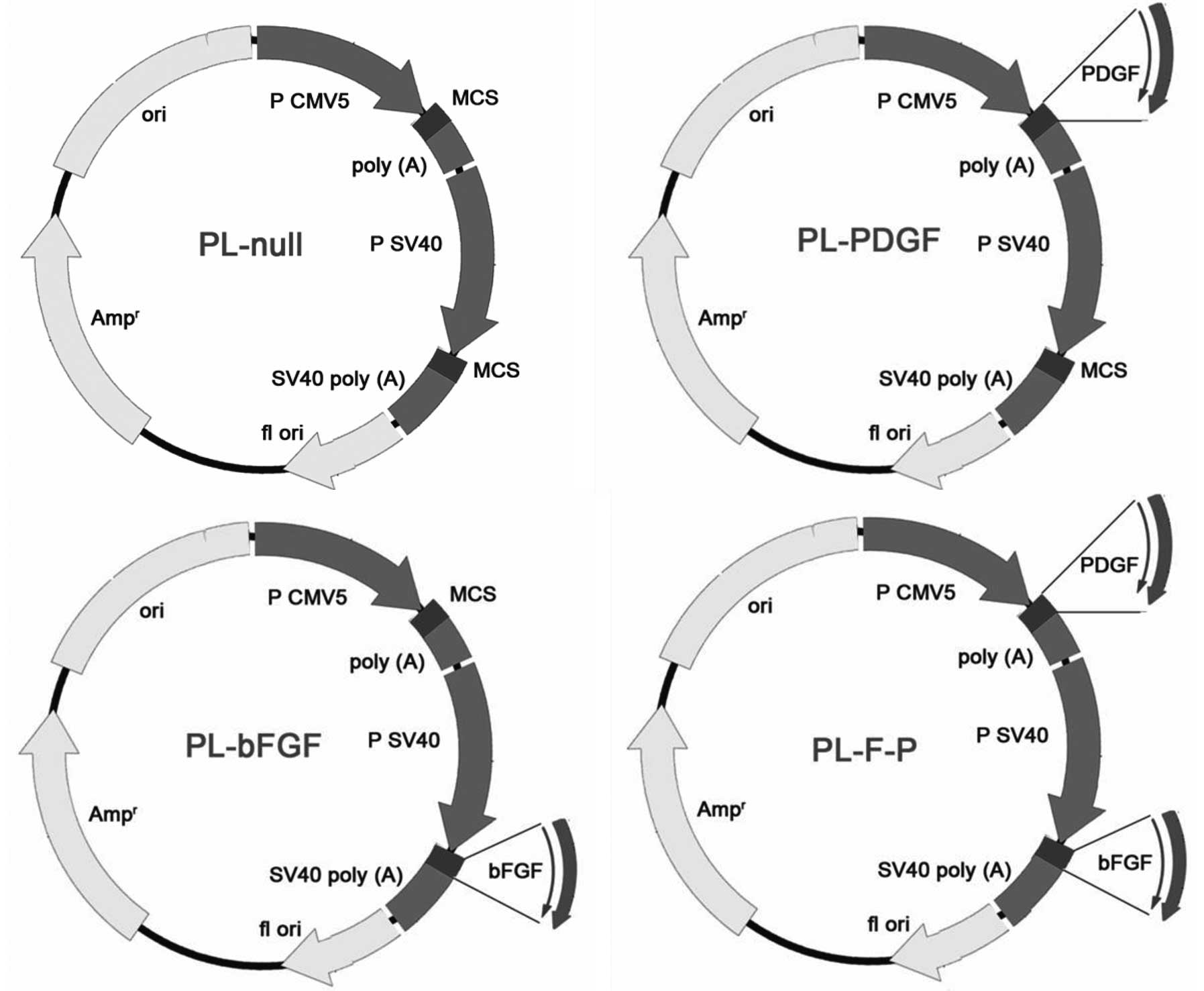
Construction of recombinant plasmids
A CMV5 promoter/enhancer and β-globin poly(A) region
from a pAdenovator-CMV5 vector were inserted upstream of the Simian
virus 40 (SV40) enhancer and early promoter sequence in the pSI
expression vector using multi-step polymerase chain reactions
(PCRs). The bFGF and PDGF genes were amplified from pBLAST45-hbFGF
and pBLAST49-hPDGF, respectively, and then cloned into the newly
constructed double-promoter plasmid (Fig. 1; Takara Tech., Dalian, China). The
resulting construct was identified using PCR, restriction
endonuclease digestion and sequence analysis.
Rat models of AMI and treatment
AMI was induced in the anterolateral wall of the
left ventricle by ligation of the left anterior descending coronary
artery. The rats were randomly assigned to one of four groups (n=10
rats per group): i) Rats treated with control plasmid (PL-Null);
ii) rats treated with plasmid encoding bFGF (PL-bFGF); iii) rats
treated with plasmid encoding PDGF (PL-PDGF); iv) rats treated with
plasmid encoding bFGF and PDGF (PL-F-P). The PL-Null group was
referred to as the control. Ten minutes subsequent to AMI, the
border zone of the infarct area was directly injected with plasmid
(100 μg in 100 μl) between the infarcted and normal tissue using a
1-ml syringe and a 25-gauge needle. All the rats were closely
monitored and were treated with antibiotics.
Functional echocardiography
assessment
Echocardiography was performed in all the
experimental subjects 28 days subsequent to gene transfer, under
controlled anesthesia and using a 3–8 MHz phased-array transducer
(Olympus Corporation, Tokyo, Japan) and Philips Sonos 7500
ultrasound system (Philips Healthcare, Andover, MA, USA). M-mode
tracing and 2D echocardiography images were recorded from
parasternal long- and short-axis views. The short-axis view was at
the level of the papillary muscles. Left ventricular (LV)
end-systolic and end-diastolic dimensions, as well as systolic and
diastolic wall thickness were measured from the M-mode tracings.
For each M-mode measurement, ≥3 consecutive cardiac cycles were
sampled. The average LV end-diastolic diameter (LVDd), LV
end-systolic diameter (LVDs), LV ejection fraction (LVEF) and LV
fractional shortening (FS) were measured. All measurements were
performed by two experienced echocardiographers who were blinded to
the treatment group.
Assessment of fibrosis using Masson’s
trichrome staining
The rats were sacrificed 28 days subsequent to gene
transfer. The hearts from each group were fixed in 4%
paraformaldehyde, embedded in paraffin, sectioned transversely and
stained with Masson’s trichrome. Sections from each heart were
evaluated in their entirety and quantified using a
computer-assisted automated image analyzer (Image ProPlus; Media
Cybernetics, Inc., Rockville, MD, USA). The extent of fibrosis was
assessed by measuring the collagen area as a proportion of the
total LV area. Morphometric studies were performed by two examiners
blinded to the treatment group.
Blood vessel measurements
The rats were sacrificed 28 days subsequent to gene
transfer. Heart muscle samples were fixed in 4% paraformaldehyde,
embedded in paraffin and sectioned transversely. Endothelial cells
were identified by the expression of von Willebrand factor (vWF),
while smooth muscle cells were identified by the expression of
smooth muscle actin (SMA). Immunohistochemistry was performed with
specific primary antibodies against vWF (1:50; Dako Denmark A/S,
Glostrup, Denmark) or SMA (1:2,000; Sigma, St. Louis, MO, USA).
Vessels from the infarction border zone were analyzed at ×200 and
×400 magnification for SMA and vWF immunostaining, respectively.
All vessel measurements were performed in a blinded manner in six
randomly selected fields on the infarction border zone from each
section. Blood vessel density was expressed as vessel
number/mm2.
Statistical analysis
Data are expressed as the mean ± standard error of
the mean (SEM) and were analyzed using SPSS version 11.0
statistical software (SPSS, Inc., Chicago, IL, USA). Differences
between groups were analyzed using analysis of variance (ANOVA) and
the least significant difference (LSD) method. P<0.05 was
considered to indicate a statistically significant difference.
Results
Changes in cardiac function following
gene transfer
Twenty-eight days subsequent to infarction, the
PL-F-P group had a significantly lower LVDd, and LVDs than the
PL-bFGF and PL-PDGF groups (P<0.01). Each of these three groups
had a significantly lower LVDd, and LVDs than the control group
(P<0.01). Conversely, the PL-F-P group showed a significantly
higher level of LVEF and FS than that of the control group
(P<0.01; Table I). These
results suggest that improved cardiac function was obtained
following the dual gene transfer of bFGF and PDGF.
 | Table ICardiac function 28 days subsequent to
infarction. |
Table I
Cardiac function 28 days subsequent to
infarction.
| Group | LVDd (mm) | LVDs (mm) | FS (%) | LVEF |
|---|
| Control | 5.83±0.02 | 3.34±0.01 | 44.54±0.11 | 0.82±0.01 |
| PL-bFGF | 5.81±0.02a,c | 2.56±0.02b,c | 56.78±0.13b,c | 0.91±0.01b,c |
| PL-PDGF | 5.52±0.02b,c | 2.56±0.02b,c | 54.50±0.16b,c | 0.92±0.01b,c |
| PL-F-P | 5.41±0.01b | 1.82±0.02b | 65.52±0.13b | 0.96±0.01b |
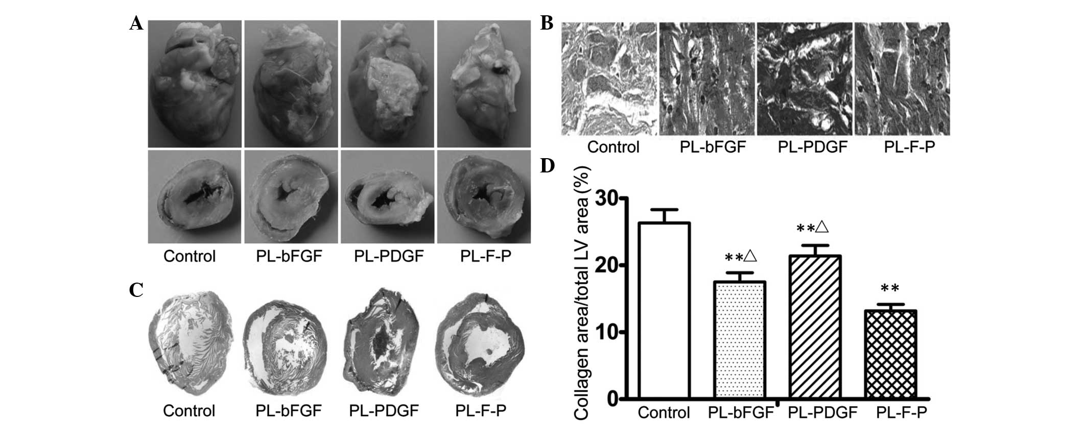
PL-F-P injection reduces fibrosis of the
infarcted myocardium
Histological examination revealed that the average
infarct size in all the therapy groups was smaller than that in the
PL-Null group at 28 days subsequent to infarction (Fig. 2). Compared with the infarct size in
the PL-Null group, the average infarct size in the PL-bFGF, PL-PDGF
and PL-F-P groups was reduced by 30.0, 18.0 and 46.7%,
respectively. PL-F-P showed the most marked effect on infarct size.
The average infarct size in the PL-F-P group was 23.7% smaller than
that in the PL-bFGF group and 33.4% smaller than that in the
PL-PDGF group.
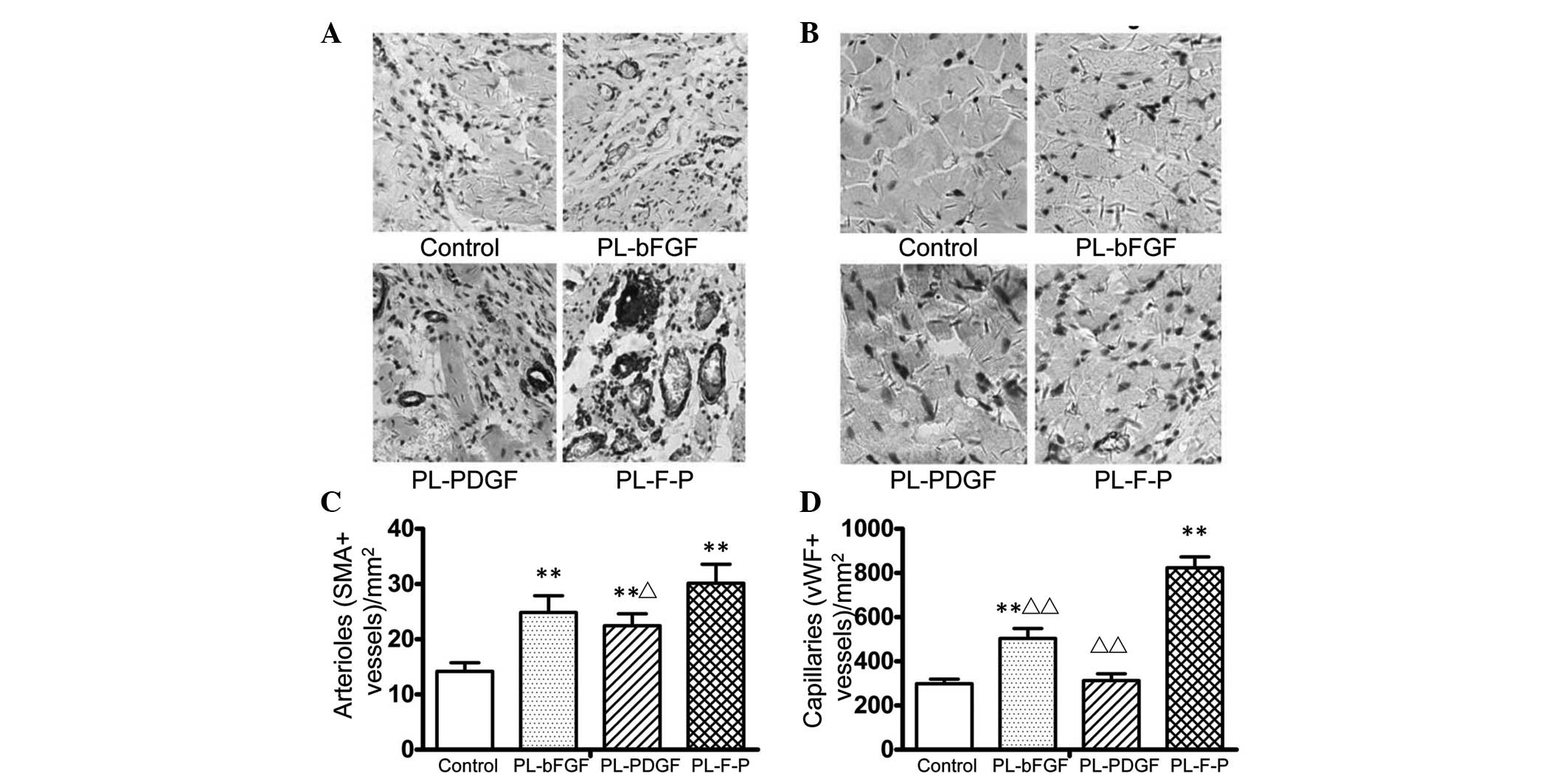
PL-F-P injection increases vascular
density
Immunostaining of the infarcted myocardium for the
expression of vWF and SMA demonstrated the augmentation of
neovascularization in all the therapy groups (Fig. 3). Among the three therapy groups,
PL-F-P showed the most marked vessel growth-stimulating effect. The
arteriole density (SMA-positive rate) in the PL-F-P group increased
by 23.9% (P<0.05) compared with that in the PL-bFGF group and
37.8% compared with that in the PL-PDGF group (P>0.05). However,
for the arterioles, the difference between the PL-F-P and PL-PDGF
groups was not statistically significant (Fig. 3C). The respective increases in
capillary density (vWF-positive rate) were 61.5 and 167% (both
P<0.01) compared with that of PL-bFGF and PL-PDGF groups
(Fig. 3D).
Discussion
Since therapeutic angiogenesis was suggested by
Höckel et al (14) in 1993,
studies on therapeutic angiogenesis have been performed globally. A
number of growth factors, including FGF-1, FGF-2, FGF-4, vascular
endothelial growth factor-121 (VEGF121), VEGF165 and VEGF-2, have
been shown to exert angiogenic effects in preclinical and clinical
studies (15–19). A small, double-blind, randomized
trial suggested the clinical efficacy of sustained-release FGF-2
implanted in the myocardium during surgery. In addition, a phase I
trial indicated that recombinant human VEGF improved myocardial
perfusion at rest, possibly via a dose-dependent effect (20). However, to date, all published
results from large clinical studies, including the FGF Initiating
RevaScularization Trial (FIRST) (21), the Vascular endothelial growth
factor in Ischemia for Vascular Angiogenesis (VIVA) trial (22) and the Angiogenic GENe Therapy-3 and
4 (AGENT-3 and -4) trials (15),
have revealed no or modest therapeutic effects, with no agent or
delivery strategy yet to demonstrate success in phase III testing.
The disappointing results of these large clinical trials are
reflective of the slow progress in the field of therapeutic
angiogenesis. One of the factors contributing to the failure of
large clinical trials may be that all the trials to date have been
based on a treatment strategy using a single growth factor. Given
the biological complexities of collateral blood vessel formation,
more sophisticated strategies using a combination of growth factors
may be required.
The data from the present study indicate that
compared with single gene therapy, plasmid-mediated dual gene
transfer of bFGF and PDGF resulted in a large increase in the
number of capillaries and arterioles in ischemic muscle 28 days
subsequent to infarction. In addition, compared with single
gene-treated groups, the dual gene treatment group showed much
larger arterioles, with increased pericyte coverage. These results
suggest a synergistic effect between bFGF and PDGF during the
process of neovascularization. A possible mechanism for this
synergistic effect may be that bFGF acts to stimulate a robust
angiogenic response and also upregulates PDGF receptor (R)-α and
PDGF R-β expression. In this manner, PDGF-BB (PDGF homodimer), an
active ligand for the PDGF-α and PDGF-β receptors, may subsequently
display increased and potent angiogenic activity (9).
Compared with viral methods, the delivery of plasmid
DNA is limited by lower gene transfer efficiency rates and a
shorter half-life. However, skeletal muscle has been indicated to
take up and express naked DNA more efficiently than other types of
tissues (23). The present study
showed that plasmid-mediated gene delivery is sufficient to achieve
meaningful therapeutic effects. In addition, the results of the
present study indicate that a short duration of exposure of
ischemic tissue to PDGF and bFGF is sufficient to establish stable
and functional blood vessels. This observation may lead to a change
in the current therapeutic strategy, based on sustained exposure to
angiogenic factors, to the ‘one-shot’ delivery of PDGF and bFGF.
The mechanism underlying these observations may be that PDGF
receptors aggregate on the cell surface, and although the initial
aggregates are triggered by PDGF, the remainder may become
autophosphorylated following the removal of PDGF. Such a mechanism
for PDGF receptor activation in the absence of PDGF has been
described previously (24). Thus,
constant activation of PDGF receptors on endothelial and mural
cells may lead to the increased stability of newly formed vessels,
even following the removal of genous PDGF and bFGF (9). The additional advantage of
plasmid-mediated gene transfer is that immune and inflammatory
responses induced following the delivery of viral vectors are not
stimulated following the transfer of naked plasmid DNA (25).
In the present study, the results of the
echocardiography and examination of infarct size were consistent
with those obtained from blood vessel measurements.
Echocardiography showed a significant improvement in cardiac
function in the dual gene-treated group. The examination of infarct
size revealed that the average infarct size was significantly
decreased in the dual gene-treated group. These results suggest
that neovascularization was important for recovery following
myocardial ischemia. The number of mature vessels determines the
number of surviving myocardial cells; these cells are important for
the improvement of cardiac function.
Recently, bFGF and PDGF have been shown to exert a
synergistic effect that promotes the proliferation and migration of
endothelial progenitor cells (EPCs) and increases VEGF release
(26). The results of the present
study were consistent with those of Cao et al (9), in which a synergistic effect was
identified between bFGF and PDGF in a mouse cornea model and a
rat/rabbit ischemic hind limb model (9). In addition, Hao et al
(25) showed that the combination
of bFGF and PDGF increased the number of capillaries and arterioles
in the rat myocardial infarction model (25). In the present study, instead of
using repeated injections of protein and multiple plasmids, the
growth factors were delivered using an intramyocardial injection of
single plasmid DNA with two transcription units, allowing the
combined expression of two genes of interest. A gene delivery
method was selected for a number of reasons, as follows: i) Using a
gene delivery method targets the desired cells or tissues and
minimizes signal propagation to non-target cells and tissues; ii)
the gene delivery method avoids the side-effects caused by the
systematic administration of recombinant proteins and viral
carriers (27); iii) the
expression of two genes of interest under a single promoter usually
leads to a lower expression efficiency of the second gene.
Furthermore, it is hard to control the expression efficiency and
quantity of two genes cloned in two separate vectors. However, a
multigenic cloning plasmid with two transcription units may be used
to achieve controllable and independent gene expression in
vivo.
Single plasmid-mediated dual gene therapy with bFGF
and PDGF was effective in stimulating the functional and
morphological maturation of the vasculature in cardiac muscle
following infarction. The results of the present study may be
relevant to future clinical trials involving angiogenic factors for
the treatment of myocardial ischemic disease.
Acknowledgements
This study was supported by the Technology Support
Program of the Science and Technology Department of Sichuan
Province, China (grant no. 2012SZ0038), the National Natural
Science Foundation of China (grant no. NSFC81202324), the National
Science and Technology Programs of Significant New Drugs to Create
(grant no. 2013ZX09301304-006) and the National 973 Program of
China (grant no. 2010CB529900).
References
|
1
|
Grines CL, Watkins MW, Helmer G, et al:
Angiogenic Gene Therapy (AGENT) trial in patients with stable
angina pectoris. Circulation. 105:1291–1297. 2002. View Article : Google Scholar : PubMed/NCBI
|
|
2
|
Li X, Tjwa M, Van Hove I, et al:
Reevaluation of the role of VEGF-B suggests a restricted role in
the revascularization of the ischemic myocardium. Arterioscler
Thromb Vasc Biol. 28:1614–1620. 2008. View Article : Google Scholar : PubMed/NCBI
|
|
3
|
Schweigerer L, Neufeld G, Friedman J,
Abraham JA, Fiddes JC and Gospodarowicz D: Capillary endothelial
cells express basic fibroblast growth factor, a mitogen that
promotes their own growth. Nature. 325:257–259. 1987. View Article : Google Scholar : PubMed/NCBI
|
|
4
|
Unger EF, Banai S, Shou M, et al: Basic
fibroblast growth factor enhances myocardial collateral flow in a
canine model. Am J Physiol. 266:H1588–H1595. 1994.PubMed/NCBI
|
|
5
|
Lopez JJ, Edelman ER, Stamler A, et al:
Basic fibroblast growth factor in a porcine model of chronic
myocardial ischemia: a comparison of angiographic,
echocardiographic and coronary flow parameters. J Pharmacol Exp
Ther. 282:385–390. 1997.PubMed/NCBI
|
|
6
|
Laham RJ, Chronos NA, Pike M, et al:
Intracoronary basic fibroblast growth factor (FGF-2) in patients
with severe ischemic heart disease: results of a phase I open-label
dose escalation study. J Am Coll Cardiol. 36:2132–2139. 2000.
View Article : Google Scholar
|
|
7
|
Laham RJ, Sellke FW, Edelman ER, et al:
Local perivascular delivery of basic fibroblast growth factor in
patients undergoing coronary bypass surgery: results of a phase I
randomized, double-blind, placebo-controlled trial. Circulation.
100:1865–1871. 1999. View Article : Google Scholar
|
|
8
|
Udelson JE, Dilsizian V, Laham RJ, et al:
Therapeutic angiogenesis with recombinant fibroblast growth
factor-2 improves stress and rest myocardial perfusion
abnormalities in patients with severe symptomatic chronic coronary
artery disease. Circulation. 102:1605–1610. 2000. View Article : Google Scholar
|
|
9
|
Cao R, Brakenhielm E, Pawliuk R, et al:
Angiogenic synergism, vascular stability and improvement of
hind-limb ischemia by a combination of PDGF-BB and FGF-2. Nat Med.
9:604–613. 2003. View
Article : Google Scholar : PubMed/NCBI
|
|
10
|
Hellström M, Kalén M, Lindahl P, Abramsson
A and Betsholtz C: Role of PDGF-B and PDGFR-beta in recruitment of
vascular smooth muscle cells and pericytes during embryonic blood
vessel formation in the mouse. Development. 126:3047–3055.
1999.PubMed/NCBI
|
|
11
|
Sun Q, Silva EA, Wang A, et al: Sustained
release of multiple growth factors from injectable polymeric system
as a novel therapeutic approach towards angiogenesis. Pharm Res.
27:264–271. 2010. View Article : Google Scholar : PubMed/NCBI
|
|
12
|
Levanon K, Varda-Bloom N, Greenberger S,
et al: Vascular wall maturation and prolonged angiogenic effect by
endothelial-specific platelet-derived growth factor expression.
Pathobiology. 73:149–158. 2006. View Article : Google Scholar : PubMed/NCBI
|
|
13
|
Li J, Wei Y, Liu K, et al: Synergistic
effects of FGF-2 and PDGF-BB on angiogenesis and muscle
regeneration in rabbit hindlimb ischemia model. Microvasc Res.
80:10–17. 2010. View Article : Google Scholar : PubMed/NCBI
|
|
14
|
Höckel M, Schlenger K, Doctrow S, Kissel T
and Vaupel P: Therapeutic angiogenesis. Arch Surg. 128:423–429.
1993.
|
|
15
|
Kapur NK and Rade JJ: Fibroblast growth
factor 4 gene therapy for chronic ischemic heart disease. Trends
Cardiovasc Med. 18:133–141. 2008. View Article : Google Scholar : PubMed/NCBI
|
|
16
|
Yue X and Tomanek RJ: Effects of VEGF(165)
and VEGF(121) on vasculogenesis and angiogenesis in cultured
embryonic quail hearts. Am J Physiol Heart Circ Physiol.
280:H2240–H2247. 2001.PubMed/NCBI
|
|
17
|
Kottakis F, Polytarchou C, Foltopoulou P,
Sanidas I, Kampranis SC and Tsichlis PN: FGF-2 regulates cell
proliferation, migration, and angiogenesis through an
NDY1/KDM2B-miR-101-EZH2 pathway. Mol Cell. 43:285–298. 2011.
View Article : Google Scholar : PubMed/NCBI
|
|
18
|
Carmeliet P: Fibroblast growth factor-1
stimulates branching and survival of myocardial arteries: a goal
for therapeutic angiogenesis? Circ Res. 87:176–178. 2000.
View Article : Google Scholar : PubMed/NCBI
|
|
19
|
Witzenbichler B, Asahara T, Murohara T, et
al: Vascular endothelial growth factor-C (VEGF-C/VEGF-2) promotes
angiogenesis in the setting of tissue ischemia. Am J Pathol.
153:381–394. 1998. View Article : Google Scholar : PubMed/NCBI
|
|
20
|
Hendel RC, Henry TD, Rocha-Singh K, et al:
Effect of intracoronary recombinant human vascular endothelial
growth factor on myocardial perfusion: evidence for a
dose-dependent effect. Circulation. 101:118–121. 2000. View Article : Google Scholar : PubMed/NCBI
|
|
21
|
Simons M, Annex BH, Laham RJ, et al:
Pharmacological treatment of coronary artery disease with
recombinant fibroblast growth factor-2: double-blind, randomized,
controlled clinical trial. Circulation. 105:788–793. 2002.
View Article : Google Scholar
|
|
22
|
Henry TD, Annex BH, McKendall GR, et al:
The VIVA trial: Vascular endothelial growth factor in Ischemia for
Vascular Angiogenesis. Circulation. 107:1359–1365. 2003. View Article : Google Scholar : PubMed/NCBI
|
|
23
|
Davis HL, Demeneix BA, Quantin B, Coulombe
J and Whalen RG: Plasmid DNA is superior to viral vectors for
direct gene transfer into adult mouse skeletal muscle. Hum Gene
Ther. 4:733–740. 1993. View Article : Google Scholar : PubMed/NCBI
|
|
24
|
Williams LT: Signal transduction by the
platelet-derived growth factor receptor. Science. 243:1564–1570.
1989. View Article : Google Scholar : PubMed/NCBI
|
|
25
|
Hao X, Mansson-Broberg A, Gustafsson T, et
al: Angiogenic effects of dual gene transfer of bFGF and PDGF-BB
after myocardial infarction. Biochem Biophys Res Commun.
315:1058–1063. 2004. View Article : Google Scholar : PubMed/NCBI
|
|
26
|
Sufen G, Xianghong Y, Yongxia C and Qian
P: bFGF and PDGF-BB have a synergistic effect on the proliferation,
migration and VEGF release of endothelial progenitor cells. Cell
Biol Int. 35:545–551. 2011. View Article : Google Scholar : PubMed/NCBI
|
|
27
|
Renault MA and Losordo DW: Therapeutic
myocardial angiogenesis. Microvasc Res. 74:159–171. 2007.
View Article : Google Scholar
|