Introduction
Moyamoya disease (MMD) is a progressive
cerebrovascular disease that is characterized by the formation of
excessive collateral vessels (moyamoya vessels) at the brain base
and occlusive or stenotic changes of the main cerebral arteries
(1). Moyamoya vessels are
hypothesized to be dilated and tortuous, perforating arteries and
compensating for regional hypoxia, with thickened or thinned
intima, sparse vascular smooth muscle cells (vSMCs) and fibrosis in
the media (2,3). Collateral formation plays a key role
in the pathogenesis of MMD (4).
Currently, the surgical treatments for MMD predominantly aim at
developing collateral vessels feeding from the external carotid
artery system (5).
Vascular endothelial growth factor (VEGF) is a
fundamental angiogenic factor in collateral vessel formation. In
cerebral ischemic diseases, cerebral angiogenesis is caused by the
release of VEGF (6). In addition,
VEGF contributes to the course of arteriogenesis, which is the
enlargement of preexisting arterioles, triggered by increased fluid
shear stress (7). VEGF has been
found to exhibit excessive expression in MMD patients (8–10),
however, the exact role of VEGF in the pathogenesis of MMD remains
unknown. Abundant VEGF does not appear to result in sufficient and
persistent collateral formation in MMD. A recent genetic study
revealed that among several VEGF gene polymorphisms, the CC
genotype of VEGF-634 may be specifically associated with better
collateral vessel formation in MMD following surgery (11). In addition to angiogenic factors,
such as VEGF, vessel formation is also regulated by antiangiogenic
cytokines and vascular stabilizing factors. Therefore, it is
hypothesized that VEGF-antagonizing antiangiogenic factors and
vascular stabilizing factors affected by VEGF may be involved in
the collateralization of MMD.
The aim of the present study was to investigate the
expression patterns of antagonists of VEGF and VEGF-affected vessel
stabilizing factors in MMD patients preoperatively and at day seven
following bypass surgery, to assess their involvement in the
pathogenesis of MMD. The association between these cytokines and
the six-month follow-up collateral vessel formation was also
analyzed.
Materials and methods
Study population
The study included 53 consecutive Chinese MMD
patients that had undergone indirect bypass surgery in the Stroke
Center of Beijing Tiantan Hospital (Beijing, China) between March
2012 and March 2013 (Table I). All
the patients were diagnosed by conventional angiography, according
to the Suzuki grading method (12). The mean age of the MMD patients was
35.22±11.47 years and 32 patients (60.38%) were male. The healthy
control group comprised 50 people, of which the mean age was
34.7±6.16 years and 30 individuals were male (60.00%). All the
operated hemispheres were demonstrated to have decreased cerebral
perfusion by perfusion computed tomography (CT) prior to surgery.
The National Institutes of Health Stroke Scale score was used to
assess the preoperative neurological status. Among the recruited
MMD patients, 46 patients received
encephalo-duro-arterio-synangiosis (EDAS) surgery, while EDAS plus
a multiple burr hole procedure was applied in seven patients.
Detailed surgical procedures were similar to previous descriptions
(13,14). Patients were excluded from the
study for the following reasons: i) Within 4 weeks of a
MMD-associated intracranial hemorrhage (ICH); ii) infection within
14 days prior to admission; iii) undergoing previous neurosurgical
treatment for MMD; and iv) unwilling to participate in the
study.
 | Table IClinical characteristics of the MMD
patients and healthy controls. |
Table I
Clinical characteristics of the MMD
patients and healthy controls.
| Characteristics | Healthy controls
(n=50) | MMD patients
(n=53) | P-value |
|---|
| Age (years; mean ±
SD) | 34.7±6.16 | 35.22±11.47 |
0.767a |
| Male gender (%) | 60 | 60.38 |
1.000b |
| Cerebral angiography
Suzuki’s grading (hemispheres, n) |
| II | - | 26 | |
| III | - | 31 | |
| IV | - | 37 | |
| V | - | 9 | |
| VI | - | 3 | |
| Location of ischemic
region (hemispheres, n) |
| ACA territory | - | 55 | |
| MCA territory | - | 87 | |
| PCA territory | - | 34 | |
| Categories by chief
symptom (n) |
| TIAs | - | 22 | |
| PSRs | - | 18 | |
| ICH | - | 9 | |
| Non-specific | - | 4 | |
| NIHSS on admission
(mean ± SD) | - | 1.11±2.19 | |
| Type of surgery
(n) |
| EDAS surgery | - | 46 | |
| EDAS surgery plus
multiple burr hole | - | 7 | |
The study was approved by the local Ethics Committee
(Institutional Review Board of Beijing Tiantan Hospital, Capital
Medical University) and all the procedures were conducted in
accordance with the Declaration of Helsinki. All the participants
and their close relatives were fully informed and provided written
consent.
Serum preparation and cytokine
detection
Peripheral venous blood samples were collected from
each patient on admission and at day seven following surgery
(healthy controls only once). The blood samples were collected in
plain serum tubes (BD Vacutainer, BD Vacutainer Systems, Plymouth,
UK) and were allowed to stand for 30 min for coagulation. Serum was
isolated using density gradient centrifugation for 10 min at 1,008
× g and stored at −80°C until assayed.
Quantitative measurements were performed using
various protein arrays to detect the serum levels of VEGF,
thrombospondin-2 (TSP-2), endostatin and angiopoietin-1 (Ang-1;
VEGF, TSP-2 and Ang-1, from Fluorokine MAP Multiplex Human
Angiogenesis Panel A base kit, R&D Systems, Minneapolis, MN,
USA), soluble VEGF receptor-1 (sVEGFR-1) and sVEGFR-2 (sVEGFR-1 and
sVEGFR-2, from Bio-Plex Pro™ Human Cancer Biomarker Panel, Bio-Rad
Laboratories, Hercules, CA, USA) and Ang-2 (Milliplex MAP Human
Angiogenesis/Growth Factor Magnetic Bead Panel, EMD Millipore,
Billerica, MA, USA). The level of TSP-1 was determined using a
commercial ELISA kit (Quantikine Human Thrombospondin-1
Immunoassay, R&D Systems, Minneapolis, MN, USA). All the
procedures were conducted by strictly following the manufacturer’s
instructions. Each sample was analyzed in duplicate and processed
at the first freeze-thaw cycle.
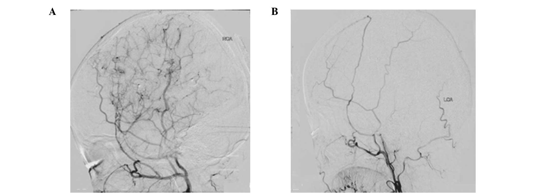
Follow-up collateral formation
assessment
Follow-up cerebral angiography was conducted
approximately six months following the bypass surgery. Newly
developed collateral vessels were evaluated according to the
grading method previously described by Matsushima et al
(15). In brief, grade A
represented a new collateral network covering more than one-third
of the middle cerebral artery (MCA) distribution; grade B
represented a network covering less than one-third of the MCA
distribution, but more than two cortical branches of the MCA were
supplied through the external carotid artery (ECA) system; grade C
indicated that only one cortical branch of MCA was fed by the ECA
system; and grade D indicated no collateral circulation. Due to the
limited sample size, the patients were further divided by
collateral vessel formation into good (collateral grade A) and poor
(collateral grade B, C and D) groups (Fig. 1).
Statistical analysis
Cytokine levels are presented as the mean ± standard
deviation. An independent t-test was used to compare the age and
cytokines levels between the MMD patients and the healthy controls.
An independent t-test was also used to compare the time interval
between surgery and follow-up angiography between the MMD patients
with good or poor collateral formation. A paired t-test was used to
compare the cytokines levels of MMD patients prior to and at day
seven following surgery. A two-sided χ2 test was used to
compare the male gender percentage between MMD patients and healthy
controls, and also between MMD patients had good or poor collateral
formation. Statistical analysis was conducted using Statistical
Package for the Social Sciences software (SPSS; SPSS, Inc.,
Chicago, IL, USA), where P<0.05 or P<0.005 was considered to
indicate a statistically significant difference, according to the
context.
Results
Clinical characteristics of the MMD
patients and healthy controls
Characteristics of the MMD patients and healthy
controls are described in Table I.
The MMD patients had no age or gender ratio discrepancy when
compared with the control group. Among the participants, 22 MMD
patients presented with transient ischemic attacks (TIAs), 18
patients presented with permanent stroke-related symptoms (PSRs),
nine individuals presented with ICH and four patients presented
with a non-specific headache. Preoperative cerebral angiography
indicated that the number of hemispheres graded as Suzuki’s grade
III or higher amounted to 79 (74.53%). In addition, the MCA
territory was the most often involved ischemic region according to
the perfusion CT scanning. The time interval between the initial
onset of symptoms to surgery was less than one year in 23 patients,
between one (included) and three years (not included) in 24
patients and more than three years (included) in six patients.
Comparison of baseline cytokines levels
between the MMD patients and healthy controls
Detailed immunoassay results are shown in Table II. Among the cytokines, the serum
level of VEGF (P<0.0001) was higher, while the levels of
sVEGFR-1 (P<0.0001) and sVEGFR-2 (P<0.0001) were
significantly lower in MMD patients when compared with the healthy
controls. Additional VEGF antagonists and vascular stabilizing
factors, including Ang-1 and Ang-2, were not statistically
different from the healthy controls.
| Table IIComparison of serum cytokines levels
between the healthy controls and preoperative MMD patients. |
Table II
Comparison of serum cytokines levels
between the healthy controls and preoperative MMD patients.
| Cytokines | Healthy controls
(pg/ml; n=50) | MMD prior to
surgery (pg/ml; n=53) | P-valuea |
|---|
| VEGF | 89.47±68.30 | 178.74±49.95 | <0.0001 |
| sVEGFR-1 | 108.80±33.47 | 71.15±18.00 | <0.0001 |
| sVEGFR-2 |
3009.10±1209.83 |
1401.59±1163.58 | <0.0001 |
| TSP-1 | 234.96±69.29 | 215.62±75.96 | 0.366 |
| TSP-2 |
12815.40±5209.16 |
8880.07±5414.17 | 0.014 |
| Endostatin |
42051.19±12964.90 |
48776.13±11708.16 | 0.063 |
| Ang-1 |
33612.53±11479.40 |
26821.96±7471.17 | 0.026 |
| Ang-2 | 1280.92±630.99 | 1235.68±610.21 | 0.801 |
Comparison of cytokine levels in MMD
patients at day seven and at the baseline
Detailed results are listed in Table III. At day seven following
surgery, sVEGFR-1 (P=0.019) and sVEGFR-2 (P=0.249) exhibited a
slight, but not significant, increase when compared with the level
at the baseline. VEGF (P<0.0001), TSP-2 (P<0.0001) and Ang-1
(P<0.0001) levels all significantly increased at day seven when
compared with the preoperative levels at the baseline.
| Table IIIComparison of cytokines levels
between MMD patients at the baseline and at day seven following
surgery. |
Table III
Comparison of cytokines levels
between MMD patients at the baseline and at day seven following
surgery.
| Cytokines | Baseline (pg/ml;
n=50) | Day 7 following
surgery (pg/ml; n=53) | P-valuea |
|---|
| VEGF | 178.74±49.95 | 361.00±199.34 | <0.0001 |
| sVEGFR-1 | 71.15±18.00 | 89.00±31.88 | 0.019 |
| sVEGFR-2 |
1401.59±1163.58 |
1792.70±1249.95 | 0.249 |
| Endostatin |
48776.13±11708.16 |
46332.85±14592.42 | 0.221 |
| TSP-1 | 215.62±75.96 | 187.29±66.83 | 0.043 |
| TSP-2 |
8880.07±5414.17 |
12605.01±7990.00 | <0.0001 |
| Ang-1 |
26821.96±7471.17 |
36194.26±14310.27 | <0.0001 |
| Ang-2 | 1235.68±610.21 |
1652.76±1274.39 | 0.030 |
Evaluation of collateral formation and
the association with sVEGFR-1 and -2 serum levels
Detailed results are listed in Table IV. Due to the limited number (n=7)
of patients that received EDAS plus multiple burr hole surgery,
only the patients that underwent EDAS were compared in this sector
(n=46). The patients were divided according to the follow-up
cerebral angiography and the good collateralization group comprised
21 cases (45.65%), while the poor group consisted of 25 cases
(54.35%). Age, male gender ratio and the time interval between
surgery and postoperative cerebral angiography had no discrepancy
between the two groups. The cases with good collateral formation
included 12 patients with TIAs, six patients with PSRs and one ICH
patient. The group with poor collateralization included 10 patients
with TIAs, 10 patients with PSRs and three ICH patients.
| Table IVClinical characteristics of the MMD
patients according to collateral formation grading. |
Table IV
Clinical characteristics of the MMD
patients according to collateral formation grading.
|
Characteristics | Good
collateralization group (n=21) | Poor
collateralization group (n=25) | P-value |
|---|
| Age (years; mean ±
SD) | 34.86±10.35 | 36.00±13.41 |
0.798a |
| Male gender
(%) | 57.14 | 62.50 |
1.000b |
| Interval between
surgery and postoperative cerebral angiography (months) | 5.40±0.54 | 6.35±0.47 |
0.166a |
| Categories by chief
symptom (n) | | | |
| TIAs | 12 | 10 | |
| PSRs | 6 | 10 | |
| ICH | 1 | 3 | |
| Non-specific | 2 | 2 | |
| Preoperative serum
level (pg/ml; mean ± SD) | | | |
| sVEGFR-1 | 63.61±12.75 | 77.74±19.66 |
0.029a |
| sVEGFR-2 | 951.42±504.27 |
1795.48±1429.18 |
0.035a |
| Postoperative serum
level (pg/ml; mean ± SD) | | | |
| sVEGFR-1 | 76.58±28.30 | 99.87±31.64 |
0.044a |
| sVEGFR-2 | 1328.57±731.30 |
2148.94±1355.09 |
0.047a |
On day seven following surgery, patients with good
collateralization exhibited no significant change in the levels of
sVEGFR-1 (P=0.142) or sVEGFR-2 (P=0.076) when compared with the
level at the baseline. Patients with poor collateralization had
increased sVEGFR-1 (P=0.006) levels, while sVEGFR-2 (P=0.472)
levels exhibited no significant change when compared with the
baseline levels. Patients with good collateralization presented
with lower levels of sVEGFR-1 and sVEGFR-2 preoperatively (P=0.029
and P=0.045, respectively) and at day seven following surgery
(P=0.044 and P=0.047, respectively) when compared with the group
with worse collateral formation.
Discussion
The aim of the present study was to investigate the
expression levels of VEGF-antagonizing cytokines and Ang-1 and
Ang-2 in MMD patients preoperatively and at day seven following
surgery. MMD patients were found to have significantly decreased
levels of sVEGFR-1 and sVEGFR-2. Lower levels of sVEGFR-1 and
sVEGFR-2 indicated better collateral formation at six months
following indirect bypass surgery.
Collateral formation plays a key role in the
pathogenesis and surgical treatment of MMD (4,5).
Collateral vessel enlargement (arteriogenesis) and capillary growth
(angiogenesis) are hypothesized to participate in the collateral
vessel formation induced by indirect bypass surgery in MMD
(16). VEGF is a specific and
critical molecule during vessel formation, promoting angiogenesis
and also participating in arteriogenesis (17). VEGF and VEGFR-2 binding is the
initial step of angiogenesis, which breaks the vascular quiescence
(18). Through VEGFR-2, VEGF
promotes endothelial cells to proliferate, migrate and inhibit
apoptosis (19). However, VEGFR-1
negatively regulates the angiogenic effects induced by VEGFR-2
(20). VEGFR-1 binds to VEGF and
reduces the availability of VEGF to combine with VEGFR-2 (21). In addition, VEGF contributes to the
course of arteriogenesis through a nitric oxide dependent pathway
(7).
Although increased VEGF expression has been
demonstrated in the dura matter (8), peripheral serum (9) and plasma (10) of MMD patients, the particular role
of VEGF in MMD remains unclear. Since VEGF is a pivotal angiogenic
factor, high concentrations in the serum of MMD patients should
induce the process of neovascularization. MMD tends to develop
collateral formation more easily than other cerebrovascular
occlusive diseases, but the moyamoya vessels disappear at the
terminal stage of MMD, depicted by the Suzuki’s grading system
(12,22). Previous studies have demonstrated
that VEGF deprivation impairs angiogenesis, and blocking of
VEGFR-1, VEGFR-2 not only inhibits arteriogenesis but also
partially decreases angiogenesis (23,24).
Therefore, it is reasonable to hypothesize that VEGF antagonists
(VEGF-associated antiangiogeneic factors) may play a role in the
pathological course of MMD.
In the present study, levels of sVEGFR-1 and
sVEGFR-2 were found to be lower in MMD patients than in healthy
controls, particularly sVEGFR-2 (less than half of the controls).
The expression pattern remained unchanged on day seven following
indirect bypass surgery. Thus, we hypothesize that these cytokines
may serve as biomarkers of MMD. sVEGFR-1 and sVEGFR-2 are soluble
forms of VEGFR-1 and VEGFR-2, respectively. sVEGFR-1 potently
inhibits angiogenesis by sequestering circulating VEGF, resulting
in less free VEGF available to bind to VEGFR-2 (25,26).
The affinity of VEGFR-1 to VEGF is >10-fold higher than VEGFR-2
(27). sVEGFR-1 is expressed and
deposited by adjacent endothelial cells and may serve a role in
maintaining vascular stability (28). To date, little is known about
sVEGFR-2, but based on previous studies, sVEGFR-2 may also have
antiangiogenic effects similar to sVEGFR-1 (26,29).
Thus, decreased sVEGFR-1 and sVEGFR-2 levels, in accordance with
the increased VEGF level, may facilitate collateral formation in
MMD, which is distinct from other cerebrovascular occlusive
diseases (22). During the course
of vessel maturation, sVEGFR-1 and sVEGFR-2 recruit mural cells via
a paracrine mechanism, which involves interplay in endothelial
cells between VEGF/VEGFR-2 and sphingosine-1-phosphate type-1
(S1P)/S1P1 pathways, resulting in the activation of endothelial
nitric oxide synthase (30). Thus,
lower sVEGFR-1 and sVEGFR-2 levels may also contribute to the
sparse vSMCs in the media of moyamoya vessels (2,3).
Inducing a well-developed collateral network is the
aim of surgical treatments for MMD. This network is not only able
to compensate for cerebral ischemia, but is also considered to
prevent the recurrence of ICH (31). In the present study, MMD patients
with better follow-up collateral formation had lower sVEGFR-1 and
sVEGFR-2 levels prior to and at day seven following surgery when
compared with the patients with worse collateralization. This
observation supports the hypothesis that sVEGFR-1 and sVEGFR-2
participate in collateral vessel formation in MMD. However, this
hypothesis is drawn with caution, as collateral formation is a
complex process that can be manipulated by a variety of cytokines.
The results of the present study are in accordance with the study
by Park et al, where the authors demonstrated that the CC
genotype of VEGF-634 contributes to better collateral formation in
MMD patients following bypass surgery (11).
Endostatin, TSP-1 and TSP-2 are all
VEGF-antagonizing cytokines. Endostatin inhibits VEGF-induced
angiogenesis partially through blocking VEGFR-2 signaling (32). TSP-1 and TSP-2 antagonize
VEGF-induced angiogenesis by CD36, CD47 and integrins, which
associate with VEGFR-2 to form a platform for the integration of
positive and negative signals for angiogenesis (33). Ang-1 and its receptor, Tie-2,
promote vascular stabilization by recruiting pericytes and
facilitate angiogenesis along with VEGF (34). Ang-2-mediated inhibition of Tie-2
signaling is required for vessel destabilization, leading to vessel
growth in the presence of VEGF or to vessel regression in the
absence of VEGF (35,36). However, the levels of these
cytokines failed to exhibit a significant difference when compared
between MMD patients and healthy controls. Considering the
VEGF-antagonizing mechanism of sVEGFR-1 and sVEGFR-2, future
studies on the role of VEGF in MMD should focus on the phase prior
to VEGF/VEGFR-2 binding.
In the present study, the increased level of VEGF on
day 7 following surgery marked an active vessel formation (19). Nakamura et al reported that
indirect bypass surgery-induced angiogenesis can be initiated
within seven days in a chronic cerebral ischemia model imitating
MMD (37). Besides VEGF,
angiogenesis-promoter Ang-1 and angiogenesis-blocker TSP-2 also
presented increased expression 7 days after bypass surgery in our
study. Thus, the study indicated that MMD patients preserve the
capacity to regulate the cytokine expression that is required by
vessel formation, at least partially.
There are certain limitations in the present study.
Firstly, the study did not include an age-matched cerebrovascular
occlusive disease control group, as usually these patients are
elderly. This impaired the strength of the observations. Secondly,
blood samples and cerebral angiographic assessment were not
analyzed at multiple time points during the postoperative
follow-up. Thus, how the levels of cytokines change and whether the
changes are consistent with the progression of Suzuki’s grading
remain unknown. Thirdly, the sample size was small, therefore, the
data should be interpreted with caution. In the future, the
observations require further validation by conducting studies with
larger cohorts.
In conclusion, the present study indicated that
antiangiogenic cytokines, including sVEGFR-1 and sVEGFR-2, may be
involved in collateral formation in MMD. In addition, the
preoperative and day seven postoperative serum levels indicated an
association with the six-month follow-up collateralization status
following indirect bypass surgery. However, the results indicated
that Ang-1 and Ang-2 may not be specifically involved in the course
of MMD.
Acknowledgements
The authors thank Dr Xiaojing Xu, Dr Yajie Wang and
Ms. Li Liu for their assistance with the immunoassay procedures.
The authors also thank Dr Xiaoxia Peng for the assistance in
statistical consulting.
References
|
1
|
Horev L, Lees MM, Anteby I, et al:
Oculoectodermal syndrome with coarctation of the aorta and moyamoya
disease: expanding the phenotype to include vascular anomalies. Am
J Med Genet A. 155:577–581. 2011. View Article : Google Scholar : PubMed/NCBI
|
|
2
|
Yamashita M, Oka K and Tanaka K:
Histopathology of the brain vascular network in moyamoya disease.
Stroke. 14:50–58. 1983. View Article : Google Scholar : PubMed/NCBI
|
|
3
|
Takebayashi S, Matsuo K and Kaneko M:
Ultrastructural studies of cerebral arteries and collateral vessels
in moyamoya disease. Stroke. 15:728–732. 1984. View Article : Google Scholar : PubMed/NCBI
|
|
4
|
Kuroda S and Houkin K: Moyamoya disease:
current concepts and future perspectives. Lancet Neurol.
7:1056–1066. 2008. View Article : Google Scholar : PubMed/NCBI
|
|
5
|
Vajkoczy P: Moyamoya disease:
collateralization is everything. Cerebrovasc Dis. 28:2582009.
View Article : Google Scholar : PubMed/NCBI
|
|
6
|
Sun Y, Jin K, Xie L, et al: VEGF-induced
neuroprotection, neurogenesis, and angiogenesis after focal
cerebral ischemia. J Clin Invest. 111:1843–1851. 2003. View Article : Google Scholar : PubMed/NCBI
|
|
7
|
Yang HT, Yan Z, Abraham JA and Terjung RL:
VEGF121- and bFGF-induced increase in collateral blood flow
requires normal nitric oxide production. Am J Physiol Heart Circ
Physiol. 280:H1097–H1104. 2001.PubMed/NCBI
|
|
8
|
Sakamoto S, Kiura Y, Yamasaki F, et al:
Expression of vascular endothelial growth factor in dura mater of
patients with moyamoya disease. Neurosurg Rev. 31:77–81. 2008.
View Article : Google Scholar : PubMed/NCBI
|
|
9
|
Rafat N, Beck GCh, Peña-Tapia PG,
Schmiedek P and Vajkoczy P: Increased levels of circulating
endothelial progenitor cells in patients with moyamoya disease.
Stroke. 40:432–438. 2009. View Article : Google Scholar : PubMed/NCBI
|
|
10
|
Kang HS, Kim JH, Phi JH, et al: Plasma
matrix metalloproteinases, cytokines and angiogenic factors in
moyamoya disease. J Neurol Neurosurg Psychiatry. 81:673–678. 2010.
View Article : Google Scholar : PubMed/NCBI
|
|
11
|
Park YS, Jeon YJ, Kim HS, et al: The role
of VEGF and KDR polymorphisms in moyamoya disease and collateral
revascularization. PLoS One. 7:e471582012. View Article : Google Scholar : PubMed/NCBI
|
|
12
|
Suzuki J and Kodama N: Moyamoya disease -
a review. Stroke. 14:104–109. 1983. View Article : Google Scholar
|
|
13
|
Smith ER and Scott RM: Surgical management
of moyamoya syndrome. Skull Base. 15:15–26. 2005. View Article : Google Scholar : PubMed/NCBI
|
|
14
|
Kuroda S and Houkin K: Bypass surgery for
moyamoya disease: concept and essence of sugical techniques. Neurol
Med Chir (Tokyo). 52:287–294. 2012.PubMed/NCBI
|
|
15
|
Matsushima T, Fukui M, Kitamura K, et al:
Encephalo-duro-arterio-synangiosis in children with moyamoya
disease. Acta Neurochir (Wien). 104:96–102. 1990. View Article : Google Scholar : PubMed/NCBI
|
|
16
|
Saito N and Imai H: Insights on the
revascularization mechanism for treatment of moyamoya disease based
on the histopathologic concept of angiogenesis and arteriogenesis.
World Neurosurg. 75:204–205. 2011. View Article : Google Scholar : PubMed/NCBI
|
|
17
|
Yancopoulos GD, Davis S, Gale NW, et al:
Vascular-specific growth factors and blood vessel formation.
Nature. 407:242–248. 2000. View
Article : Google Scholar : PubMed/NCBI
|
|
18
|
Carmeliet P: Mechanisms of angiogenesis
and arteriogenesis. Nat Med. 6:389–395. 2000. View Article : Google Scholar : PubMed/NCBI
|
|
19
|
Ferrara N, Gerber HP and LeCouter J: The
biology of VEGF and its receptors. Nat Med. 9:669–676. 2003.
View Article : Google Scholar : PubMed/NCBI
|
|
20
|
Zeng H, Dvorak HF and Mukhopadhyay D:
Vascular permeability factor (VPF)/vascular endothelial growth
factor (VEGF) receptor-1 downmodulates VPF/VEGF receptor-2-mediated
endothelial cell proliferation, but not migration, through
phosphatidylinositol 3-kinase-dependent pathways. J Biol Chem.
276:26969–26979. 2001. View Article : Google Scholar
|
|
21
|
Roberts DM, Kearney JB, Johnson JH, et al:
The vascular endothelial growth factor (VEGF) receptor Flt-1
(VEGFR-1) modulates Flk-1 (VEGFR-2) signaling during blood vessel
formation. Am J Pathol. 164:1531–1535. 2004. View Article : Google Scholar : PubMed/NCBI
|
|
22
|
Yoshimoto T, Houkin K, Takahashi A and Abe
H: Angiogenic factors in moyamoya disease. Stroke. 27:2160–2165.
1996. View Article : Google Scholar : PubMed/NCBI
|
|
23
|
Olfert IM, Howlett RA, Wagner PD and Breen
EC: Myocyte vascular endothelial growth factor is required for
exercise-induced skeletal muscle angiogenesis. Am J Physiol Regul
Integr Comp Physiol. 299:R1059–R1067. 2010. View Article : Google Scholar : PubMed/NCBI
|
|
24
|
Lloyd PG, Prior BM, Li H, Yang HT and
Terjung RL: VEGF receptor antagonism blocks arteriogenesis, but
only partially inhibits angiogenesis, in skeletal muscle of
exercise-trained rats. Am J Physiol Heart Circ Physiol.
288:H759–H768. 2005. View Article : Google Scholar : PubMed/NCBI
|
|
25
|
Kendall RL and Thomas KA: Inhibition of
vascular endothelial cell growth factor activity by an endogenously
encoded soluble receptor. Proc Natl Acad Sci USA. 90:10705–10709.
1993. View Article : Google Scholar : PubMed/NCBI
|
|
26
|
Jacobi J, Tam BY, Wu G, et al: Adenoviral
gene transfer with soluble vascular endothelial growth factor
receptors impairs angiogenesis and perfusion in a murine model of
hindlimb ischemia. Circulation. 110:2424–2429. 2004. View Article : Google Scholar
|
|
27
|
Ferrara N and Davis-Smyth T: The biology
of vascular endothelial growth factor. Endocr Rev. 18:4–25. 1997.
View Article : Google Scholar
|
|
28
|
Ito TK, Ishii G, Saito S, et al:
Degradation of soluble VEGF receptor-1 by MMP-7 allows VEGF access
to endothelial cells. Blood. 113:2363–2369. 2009. View Article : Google Scholar : PubMed/NCBI
|
|
29
|
Ebos JM, Bocci G, Man S, et al: A
naturally occurring soluble form of vascular endothelial growth
factor receptor 2 detected in mouse and human plasma. Mol Cancer
Res. 2:315–326. 2004.PubMed/NCBI
|
|
30
|
Lorquet S, Berndt S, Blacher S, et al:
Soluble forms of VEGF receptor-1 and -2 promote vascular maturation
via mural cell recruitment. FASEB J. 24:3782–3795. 2010. View Article : Google Scholar : PubMed/NCBI
|
|
31
|
Liu X, Zhang D, Shuo W, et al: Long term
outcome after conservative and surgical treatment of haemorrhagic
moyamoya disease. J Neurol Neurosurg Psychiatry. 84:258–265. 2013.
View Article : Google Scholar : PubMed/NCBI
|
|
32
|
Dixelius J, Cross MJ, Matsumoto T and
Claesson-Welsh L: Endostatin action and intracellular signaling:
beta-catenin as a potential target? Cancer Lett. 196:1–12. 2003.
View Article : Google Scholar : PubMed/NCBI
|
|
33
|
Lawler PR and Lawler J: Molecular basis
for the regulation of angiogenesis by thrombospondin-1 and -2. Cold
Spring Harb Perspect Med. 2:a0066272012. View Article : Google Scholar : PubMed/NCBI
|
|
34
|
Zacharek A, Chen J, Cui X, et al:
Angiopoietin1/Tie2 and VEGF/Flk1 induced by MSC treatment amplifies
angiogenesis and vascular stabilization after stroke. J Cereb Blood
Flow Met. 27:1684–1691. 2007. View Article : Google Scholar : PubMed/NCBI
|
|
35
|
Hanahan D: Signaling vascular
morphogenesis and maintenance. Science. 277:48–50. 1997. View Article : Google Scholar : PubMed/NCBI
|
|
36
|
Holash J, Wiegand SJ and Yancopoulos GD:
New model of tumor angiogenesis: dynamic balance between vessel
regression and growth mediated by angiopoietins and VEGF. Oncogene.
18:5356–5362. 1999. View Article : Google Scholar : PubMed/NCBI
|
|
37
|
Nakamura M, Imai H, Konno K, et al:
Experimental investigation of encephalomyosynangiosis using
gyrencephalic brain of the miniature pig: histopathological
evaluation of dynamic reconstruction of vessels for functional
anastomosis. J Neurosurg Pediatr. 3:488–495. 2009. View Article : Google Scholar
|