Introduction
The Hedgehog (Hh) signaling pathway is linked to
cell growth and differentiation. It is involved in embryonic
pattern formation and adult tissue homeostasis (1). Recently, the Hh signaling pathway has
been revealed to be dysregulated in several human malignancies and
to be critical for the maintenance and expansion of malignant stem
cells (2). Gli family zinc finger
(Gli) transcription factors constitute the final effectors of the
Hh signaling pathway. In a number of tumors, including those of the
pancreas, prostate, skin or lungs, the ectopic activation of Gli
proteins has been linked to tumorigenesis (3). There has also been evidence
suggesting additional, noncanonical mechanisms of Gli activation
(3). Certain studies have
suggested that in normal fibroblasts and keratinocytes, as well as
in various cancer cell lines, the Gli transcription factors are not
solely regulated by Hh/smoothened (Smo) signaling, but also by
other pathways, including transforming growth factor (TGF)-β
signaling. TGF-β has been shown to induce Gli2 expression in a
Smad3-dependent manner in various cell types and this effect was
demonstrated to be independent from the patched (Ptch)/Smo axis
(4).
In hematopoiesis, Hh family members play an
important role in the regulation of stem/progenitor cell expansion
in vitro and in vivo (5). Components of Hh signaling have been
detected in several leukemic cell lines including: Ptch and Smo,
which are expressed in Jurkat cells (6); sonic hedgehog (Shh) and Gli1, in
HL-60 and KG-1 cells (7); and Gli2
in U937 and HL60 cells (8).
Similarly to the Hh members, TGF-β plays an important role in
regulating the balance between proliferation and differentiation in
hematopoietic cells (9,10).
With the aim of identifying a new target for
leukemia treatment, in the present study it was hypothesized that
there is also cross-communication between the Hh signaling pathway
and TGF-β in leukemic cells. In the current study, the capacity of
TGF-β for modulating the expression of the Hh signaling molecule
Gli2 in the KG-1 human myeloid leukemia cell line was examined.
Targeting the interaction between Hh and TGF-β signaling may
provide novel therapeutic opportunities for leukemia treatment.
Materials and methods
Cell cultures and reagents
KG-1 human myeloid leukemia cells were donated for
use in the present study by the Institute of Hematology of the
Chinese Academy of Medical Sciences (Tianjin, China). They were
cultured in Iscove’s modified Dulbecco’s medium (IMDM, Solarbio,
Beijing, China) supplemented with 20% heat-inactivated fetal calf
serum (FCS), 0.1% penicillin (100 U/ml) and streptomycin (100
mg/ml), at 37°C in a humidified atmosphere of 5% CO2.
When the cells were in the logarithmic growth phase, the KG-1 cells
were seeded on starving medium (containing 2.5% FCS) at the same
cell density of 2×10⁵ cells/ml. They were treated with
human recombinant TGF-β1; 5 ng/ml; referred to as TGF-β), tumor
necrosis factor (TNF)-α; 5 ng/ml) and/or specific inhibitor of
smad3 (SIS3; 5 μM) for various time periods. TGF-β1 and TNF-α were
purchased from Peprotech Inc. (Rocky Hill, NJ, USA). SIS3 was
purchased from Merck KGaA (Darmstadt, Germany). The monoclonal
antibody anti-Gli2 was purchased from Santa Cruz Biotechnology Inc.
(Santa Cruz, CA, USA). The monoclonal antibody anti- TGF-βRI and
anti-TGF-βRII were purchased from Bioss Biotechnology Inc. (Bioss,
Beijing, China).
Reverse transcription (RT) and
quantitative (q) polymerase chain reaction (PCR)
The total RNA was extracted using an RNeasy mini kit
(Qiagen, Hilden, Germany). RNA (1 μg) was reverse-transcribed using
SuperScript™II (Invitrogen Life Technologies, Takara, Japan).
Reverse transcription was carried out on genomic DNA-free RNA using
random primers. The RT-PCR procedures were carried out on a GeneAmp
PCR System 9700 (Thermo Fisher Scientific, Waltham, MA, USA). The
qPCR was subsequently performed using SYBR Green PCR Core reagents
and a Rotor-Gene 6000 real-time rotary analyzer (Corbett Life
Science, Sydney, Australia) to quantify the steady-state levels of
Gli2 mRNA. The expression level of the housekeeping gene ABL was
used as a control. The process was repeated three times for each
sample. The final results were compared by the comparative ΔΔ Ct
method. The primer pairs for Gli2 (5′-TGGCCGCTTCAGATGACAGATGTTG-3′
and 5′-CGTTAGCCGAATGTCAGCCGTGAAG-3′) and ABL
(5′-CGAGAGCCTGGCCTACAACAA-3′ and 5′-CTAGCA GCTCATACACCTGGGACA-3′)
were designed and synthesized by Takara Bio, Inc. (Shiga, Japan).
PCR amplification was carried out using 45 cycles of 95°C for 60
sec, 95°C for 10 sec and 60°C for 30 sec.
Western blot analysis
At the end of the incubation period, cells were
washed twice in ice-cold phosphate-buffered saline (PBS), following
centrifugation at 200 g and 4°C. The cells were immediately
transferred to ice-cold lysis buffer (SunShineBio, Nanjing, China)
and phenylmethylsulfonyl fluoride (PMSF) was added. The solution
was shaken at 4°C for 30 min. Following this, the cellular extracts
were collected by centrifugation at 16,000 g for 5 min at 4°C and
the samples were immediately frozen at −80°C. The samples were
homogenized with a 4-(2-hydroxyethyl)-1-piperazineethanesulfonic
acid (HEPES) buffer solution (Gibco, Carslbad, CA, USA; 100
μg/lane), separated by 10% sodium dodecyl sulfate polyacrylamide
gel electrophoresis (SDS-PAGE) and blotted onto polyvinylidene
fluoride (PVDF) membranes. Subsequently, the membranes were blocked
with Tris-buffered saline and Tween 20 (TBST) containing 5% skimmed
milk. Gli2, TGF-βRI or TGF-βRII protein content was detected with
an anti-Gli2, anti-TGF-βRI or anti-TGF-βRII antibody. An antibody
directed against β-actin (Santa Cruz) was used to verify the equal
protein content in each sample. Following washing with TBST, the
membranes were incubated with goat anti-rabbit IgG (H+L)/TRITC
secondary antibodies (Santa Cruz). The membranes were detected with
an electrochemiluminescence (ECL) Western blotting system (Alpha
Diagnostic International, San Antonio, TX, USA).
Statistical analysis
The gene expression levels in the KG-1 cell line
were compared by one way analysis of variance (ANOVA). P<0.05
was considered to indicate a statistically significant difference.
Results obtained from multiple experiments are reported as the mean
± standard error of the mean.
Results
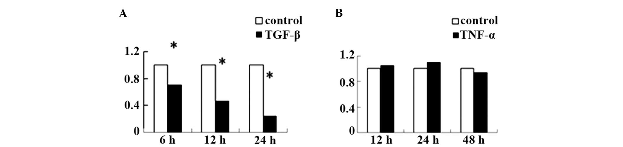
TGF-β affects the expression level of
Gli2 in the KG-1 cell line, while TNF-α does not
To determine whether TGF-β or TNF-α affected the
expression level of Gli2, KG-1 cells were incubated with TGF-β,
with TNF-α or without either (control). A significant reduction in
the expression level of Gli2 mRNA in response to TGF-β was observed
in the KG-1 cells (Fig. 1A). This
reduction was observed following 6 h of treatment and lasted for a
minimum of 24 h, with the TGF-β values significantly lower than
those in the control group during the experimental period. As shown
in Fig. 1B, TNF-α did not
demonstrate a significant effect on the expression level of Gli2
mRNA in the KG-1 cells.
TNF-α increases TGF-β type I receptor
(TGF-βRI) and TGF-β type II receptor (TGF-βRII) protein expression
levels in KG-1 cells
KG-1 cells were incubated with TNF-α in the TNF-α
(+) group, or were untreated in the control (−) group for 24 h.
Fig. 2 show that the TGF-βRI and
TGF-βRII protein expression levels in the KG-1 cells were
significantly higher in the TNF-α (+) group than in the control (−)
group.
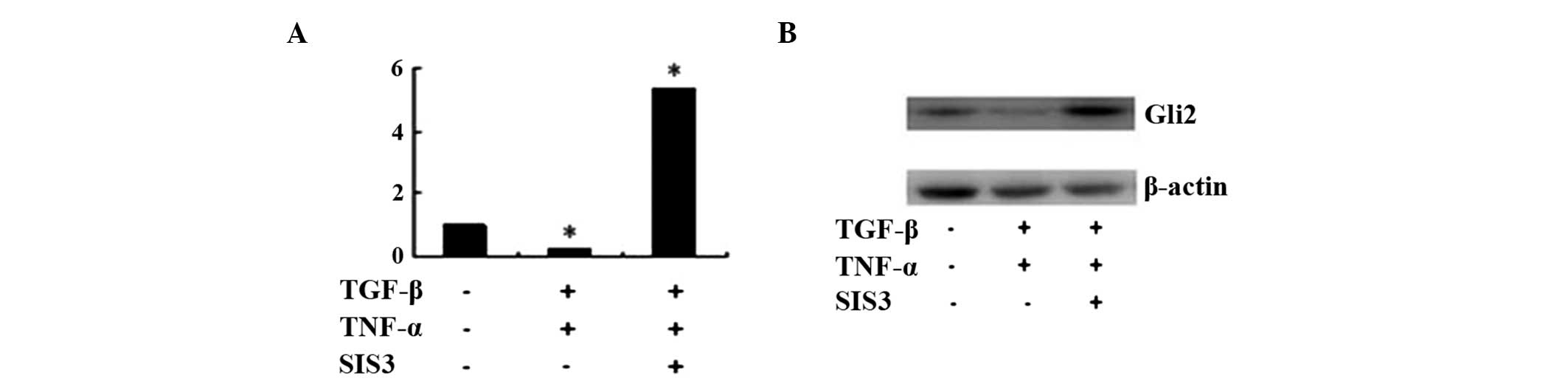
TGF-β strongly reduces the expression
level of Gli2 when KG-1 cells are incubated with TGF-β and TNF-α in
combination
To investigate whether a combination of TGF-β and
TNF-α is able to influence the expression level of Gli2 in KG-1
cells, the KG-1 cells were cultured with TGF-β, in the presence or
absence of TNF-α for 24 h. As shown in Fig. 3, the expression of Gli2 at the mRNA
and protein levels in the TGF-β + TNF-α group was much lower than
the expression of Gli2 in the TGF-β group and the control (−)
group. The expression level of Gli2 in the TGF-β group was lower
than that in the control (−) group.
Suppression of Gli2 expression by TGF-β
is Smad3-dependent
To determine whether the suppression of Gli2
expression by TGF-β was Smad3-dependent, SIS3 (11), a specific inhibitor of Smad3, was
used in combination with TGF-β and TNF-α to treat the KG-1 cells.
Fig. 4 shows that the expression
of Gli2 in the KG-1 cells at the mRNA and protein levels decreased
compared with the levels in untreated cells when they were treated
with TGF-β + TNF-α, and increased when they were treated with TGF-β
+ TNF-α + SIS3.
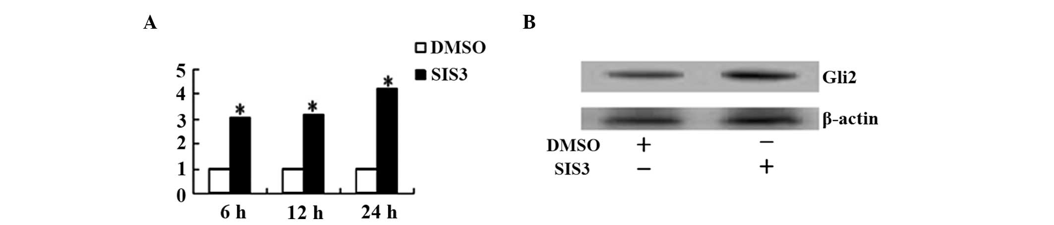
SIS3 increases the expression level of
Gli2 in KG-1 cells
To determine whether the TGF-β secreted by the KG-1
cells themselves is able to affect the expression level of Gli2,
the KG-1 cells were pretreated with SIS3 or a control (dimethyl
sulfoxide, DMSO). Fig. 5 shows
that the expression of Gli2 at the mRNA and protein levels in the
KG-1 cells treated with SIS3 was significantly higher than the
expression in those treated with DMSO.
Discussion
Hh signaling is critical in vertebrate development,
patterning, and cell fate induction (12). Deregulation of Hh signaling is
associated with different forms of human cancer (2). The Hh signaling pathway comprises Hh
ligands, Ptch and Smo receptors, and Gli zinc-finger protein
transcription factors (13). The
secreted protein Hh acts by binding to its receptor Ptch, which
then ceases to inhibit another transmembrane protein, Smo. This
activates downstream cytoplasmic transcription factors. In
Drosophila these are the Ci proteins and the mammalian
homologues of these are the Gli proteins (1). This downstream signaling initiates
the entry of cells into the cell cycle (14) in order to maintain the self-renewal
of stem cells in various tissues (15), inhibit apoptosis (16), modulate tissue polarity (17) and regulate the differentiation of
tissue stem cells (18). Evidence
has suggested that the Hh signaling pathway is prominent in cancer
stem cells (19). Recently,
potential methods of inhibiting the Hh signaling pathway have been
studied in order to develop a new class of possible therapeutics
for cancer treatment (20,21).
There are three related Gli proteins: Gli1, Gli2 and
Gli3 (22). Gli2 functions
upstream of Gli1 and is the primary mediator of Hh signaling
(23), inducing Gli1 expression
via direct binding to its promoter region (24). Gli3 genes inhibit the activating
functions of all coexpressed Gli genes (25).
Similarly to Hh members, as a family of growth
factors involved in various essential physiological processes,
TGF-β plays a complex role as a mediator in inflammation, tissue
repair, angiogenesis, and the regulation and differentiation of
cell growth (26). TGF-β binds to
a heteromeric cell-surface complex of type I (TGF-TβRI) and type II
(TGF-TβRII) serine/threonine kinase receptors (27). Following ligand binding, TβRII
recruits and activates TβRI, which phosphorylates Smad proteins.
The receptor-associated Smad proteins, Smad2 and Smad3,
heteromerize with Smad4 and later translocate into the nucleus and
act as transcription factors to regulate target gene expression
(28).
There is abundant data regarding the effect of TGF-β
on leukemic cells lines, ranging from inhibition of proliferation
and induction of differentiation, to altered expression of adhesion
molecules and cytokine receptors, and the induction of apoptosis
(29). Acting as an inhibitory
autocrine factor in the proliferation of leukemia cells (30), TGF-β1 is a primary negative
regulator of early hematopoiesis (31).
Hh receptor molecules are lost in several leukemia
cell lines, including KG-1 cells; however, the transcription factor
Gli2 is expressed (8). For these
cells, the blockade of the autocrine loop in Hh signaling would not
be available. Thus, Gli2 may be an alternative target for the
treatment of acute myeloid leukemia (AML). One possible strategy is
to use small interfering RNAs (siRNAs) that are specific to Gli2.
However, efficient transfer of siRNAs into the hematopoietic cells
is problematic (32). The present
study revealed that TGF-β, alone or combined with TNF-α, was able
to decrease the expression level of Gli2 in the KG-1 cell line,
thus suggesting that TGF-β may be one of the signaling molecules
that plays a key role in the regulation of the Gli2 gene in
leukemia cell lines.
Treatment with TGF-β significantly affected the
expression level of Gli2 in the KG-1 cells in the present study,
whereas no significant differences in the expression levels of Gli2
were observed between the control and TNF-α groups of KG-1 cells
(Fig. 1). A previous study has
revealed that TNF-α is able to affect Shh through the nuclear
factor κ-light-chain-enhancer of activated B cells (NF-κB) pathway
(33), and that Shh is able to
affect the expression level of Gli2. However, the Hh receptor
molecules (Ptch and Smo) are lost in KG-1 cells (8). This may explain why TNF-α did not
affect the expression level of Gli2 in the KG-1 cells. In the
present study, when the KG-1 cells were treated with TNF-α, it was
found that the TGF-βRI and TGF-βRII protein expression levels were
increased in the KG-1 cells (Fig.
2). This result is consistent with the results of a previous
study (34). Furthermore, the Gli2
levels in the KG-1 cells treated with TGF-β + TNF-α were different
from those in the cells only treated with TGF-β (Fig. 3). The expression level of Gli2 in
the KG-1 cells treated with TGF-β and TNF-α was lower than the
level in those treated with TGF-β alone (Fig. 3). It was possible that the
expression of TGF-β receptors in the KG-1 cells was increased by
TNF-α, when enabled TGF-β to strongly decrease the expression of
Gli2 expression.
The mechanism by which TGF-β decreases the
expression level of Gli2 in KG-1 cells involves Smad2 and Smad3. By
acting as a downstream signaling molecule for TGF-β, Smad3
contributes to the majority of Smad-dependent responses to TGF-β in
adults. Smad2, however, is critical during embryogenesis (35,36).
Specific inhibitor of Smad3 (SIS3) was used to inhibit the effects
of TGF-β. Fig. 4 shows that SIS3
inhibited the effects of TGF-β + TNF-α, which otherwise decreased
the expression levels of Gli2 in the KG-1 cells. Since Ptch and Smo
are not expressed in KG-1 cells (8), this effect was independent from the
Ptch/Smo axis. Thus, the suppression of Gli2 expression by TGF-β
occurs in a Smad3-dependent manner.
The present study also investigated the effect of
SIS3 on the expression level of Gli2 in KG-1 cells. As shown in
Fig. 5, SIS3 significantly
increased the expression level of Gli2 in the KG-1 cells. SIS3 is a
specific inhibitor of Smad3, which has been shown in the present
study to act via blocking the TGF-β signaling pathway. As this
effect on Gli2 was observed without adding TGF-β to the KG-1 cells,
this suggests that TGF-β secreted by the KG-1 cells is able to
decrease Gli2 expression.
TGF-β has been revealed to have growth-enhancing and
growth-inhibitory properties, the predominating effect being
dependent on the particular cell type and the other growth factors
present (37). The effect of TGF-β
on KG-1 cells in the current study differed from a previous study
which have reported that TGF-β increases the expression level of
Gli2 in various human cell types, including normal fibroblasts and
keratinocytes, as well as in various cancer cell lines (4). This variation may be due to the
characteristics of the different cell lines and the other growth
factors present.
In conclusion, the present study revealed that a
combination of TGF-β and TNF-α decreased the expression level of
Gli2 in KG-1 cell lines and that SIS3 inhibited the effect of
TGF-β. The suppression of Gli2 expression was Smad3-dependent.
These results demonstrate that TGF-β plays a role as a cytokine
that may be capable of decreasing Hh signaling in certain types of
leukemic cell lines. This may initiate the development of new
approaches for the efficient therapeutic treatment of certain types
of leukemia; however, further in vivo and in vitro
studies are required.
Acknowledgements
This study was supported by a project from the
Education Department of Liaoning Province (L2012302).
References
|
1
|
Lum L and Beachy PA: The Hedgehog response
network: sensors, switches, and routers. Science. 304:1755–1759.
2004. View Article : Google Scholar : PubMed/NCBI
|
|
2
|
Hao K, Tian XD, Qin CF, Xie XH and Yang
YM: Hedgehog signaling pathway regulates human pancreatic cancer
cell proliferation and metastasis. Oncol Rep. 29:1124–1132.
2013.PubMed/NCBI
|
|
3
|
Lauth M and Toftgård R: Non-canonical
activation of GLI transcription factors: implications for targeted
anti-cancer therapy. Cell Cycle. 6:2458–2463. 2007. View Article : Google Scholar : PubMed/NCBI
|
|
4
|
Dennler S, André J, Alexaki I, et al:
Induction of sonic hedgehog mediators by transforming growth
factor-β: Smad3-dependent activation of Gli2 and Gli1 expression
in vitro and in vivo. Cancer Res. 67:6981–6986.
2007.
|
|
5
|
Bhardwaj G, Murdoch B, Wu D, et al: Sonic
hedgehog induces the proliferation of primitive human hematopoietic
cells via BMP regulation. Nat Immunol. 2:172–180. 2001. View Article : Google Scholar : PubMed/NCBI
|
|
6
|
Detmer K, Walker AN, Jenkins TM, Steele TA
and Dannawi H: Erythroid differentiation in vitro is blocked
by cyclopamine, an inhibitor of hedgehog signaling. Blood Cells Mol
Dis. 26:360–372. 2000.
|
|
7
|
Bai LY, Chiu CF, Lin CW, Hsu NY, Lin CL,
Lo WJ and Kao MC: Differential expression of sonic hedgehog and
Gli1 in hematological malignancies. Leukemia. 22:226–228. 2008.
View Article : Google Scholar : PubMed/NCBI
|
|
8
|
Kobune M, Takimoto R, Murase K, et al:
Drug resistance is dramatically restored by hedgehog inhibitors in
CD34+ leukemic cells. Cancer Sci. 100:948–955. 2009.PubMed/NCBI
|
|
9
|
Fortunel N, Hatzfeld J, Kisselev S, et al:
Release from quiescence of primitive human hematopoietic
stem/progenitor cells by blocking their cell-surface TGF-β type II
receptor in a short-term in vitro assay. Stem Cells.
18:102–111. 2000.PubMed/NCBI
|
|
10
|
Fortunel NO, Hatzfeld A and Hatzfeld JA:
Transforming growth factor-β: pleiotropic role in the regulation of
hematopoiesis. Blood. 96:2022–2036. 2000.
|
|
11
|
Jinnin M, Ihn H and Tamaki K:
Characterization of SIS3, a novel specific inhibitor of Smad3, and
its effect on transforming growth factor-β1-induced extracellular
matrix expression. Mol Pharmacol. 69:597–607. 2006.PubMed/NCBI
|
|
12
|
Bale AE: Hedgehog signaling and human
disease. Annu Rev Genomics Hum Genet. 3:47–65. 2002. View Article : Google Scholar : PubMed/NCBI
|
|
13
|
Hui CC and Angers S: Gli proteins in
development and disease. Annu Rev Cell Dev Biol. 27:513–537. 2011.
View Article : Google Scholar : PubMed/NCBI
|
|
14
|
Duman-Scheel M, Weng L, Xin S and Du W:
Hedgehog regulates cell growth and proliferation by inducing Cyclin
D and Cyclin E. Nature. 417:299–304. 2002. View Article : Google Scholar : PubMed/NCBI
|
|
15
|
Lai K, Kaspar BK, Gage FH and Schaffer DV:
Sonic hedgehog regulates adult neural progenitor proliferation
in vitro and in vivo. Nat Neurosci. 6:21–27. 2003.
View Article : Google Scholar : PubMed/NCBI
|
|
16
|
Bigelow RL, Chari NS, Unden AB, et al:
Transcriptional regulation of bcl-2 mediated by the sonic hedgehog
signaling pathway through gli-1. J Biol Chem. 279:1197–1205. 2004.
View Article : Google Scholar : PubMed/NCBI
|
|
17
|
Watkins DN, Berman DM, Burkholder SG, Wang
B, Beachy PA and Baylin SB: Hedgehog signalling within airway
epithelial progenitors and in small-cell lung cancer. Nature.
422:313–317. 2003. View Article : Google Scholar : PubMed/NCBI
|
|
18
|
Spinella-Jaegle S, Rawadi G, Kawai S, et
al: Sonic hedgehog increases the commitment of pluripotent
mesenchymal cells into the osteoblastic lineage and abolishes
adipocytic differentiation. J Cell Sci. 114:2085–2094. 2001.
|
|
19
|
O’Flaherty JD, Barr M, Fennell D, Richard
D, Reynolds J, O’Leary J and O’Byrne K: The cancer stem-cell
hypothesis: its emerging role in lung cancer biology and its
relevance for future therapy. J Thorac Oncol. 7:1880–1890.
2012.PubMed/NCBI
|
|
20
|
Weiss GJ and Korn RL: Metastatic basal
cell carcinoma in the era of hedgehog signaling pathway inhibitors.
Cancer. 118:5310–5319. 2012. View Article : Google Scholar : PubMed/NCBI
|
|
21
|
Irvine DA and Copland M: Targeting
hedgehog in hematologic malignancy. Blood. 119:2196–2204. 2012.
View Article : Google Scholar : PubMed/NCBI
|
|
22
|
Kasper M, Regl G, Frischauf AM and Aberger
F: GLI transcription factors: mediators of oncogenic hedgehog
signalling. Eur J Cancer. 42:437–445. 2006. View Article : Google Scholar : PubMed/NCBI
|
|
23
|
Bai CB, Auerbach W, Lee JS, Stephen D and
Joyner AL: Gli2, but not Gli1, is required for initial Shh
signaling and ectopic activation of the Shh pathway. Development.
129:4753–4761. 2002.PubMed/NCBI
|
|
24
|
Ikram MS, Neill GW, Regl G, et al: GLI2 is
expressed in normal human epidermis and BCC and induces GLI1
expression by binding to its promoter. J Invest Dermatol.
122:1503–1509. 2004. View Article : Google Scholar : PubMed/NCBI
|
|
25
|
Ruiz I and Altaba A: Gli proteins encode
context-dependent positive and negative functions: implications for
development and disease. Development. 126:3205–3216.
1999.PubMed/NCBI
|
|
26
|
Roberts AB and Sporn MB: Transforming
growth factor-β. Adv Cancer Res. 51:107–145. 1988.
|
|
27
|
Massagué J: How cells read TGF-β signals.
Nat Rev Mol Cell Biol. 1:169–178. 2000.
|
|
28
|
Attisano L and Wrana JL: Signal
transduction by the TGF-β superfamily. Science. 296:1646–1647.
2002.
|
|
29
|
Piacibello W, Severino A, Stacchini A and
Aglietta M: Differential effects of transforming growth factor beta
1 on the proliferation of human lymphoid and myeloid leukemia
cells. Haematologica. 76:460–466. 1991.PubMed/NCBI
|
|
30
|
Chen Y, Lu L and Wang L: Study on gene
expression of TGF β1 and its receptor in leukemia cells and the
serum TGF β1 level in the patients with acute leukemia. Zhonghua
Xue Ye Xue Za Zhi. 19:576–580. 1998.(In Chinese).
|
|
31
|
Testa U, Masciulli R, Tritarelli E, et al:
Transforming growth factor-β potentiates vitamin D3-induced
terminal monocytic differentiation of human leukemic cell lines. J
Immunol. 150:2418–2430. 1993.
|
|
32
|
Gough PJ and Raines EW: Advances in
retroviral transduction of hematopoietic stem cells for the gene
therapy of atherosclerosis. Curr Opin Lipidol. 14:491–497. 2003.
View Article : Google Scholar : PubMed/NCBI
|
|
33
|
Kasperczyk H, Baumann B, Debatin KM and
Fulda S: Characterization of sonic hedgehog as a novel NF-κB target
gene that promotes NF-κB-mediated apoptosis resistance and tumor
growth in vivo. FASEB J. 23:21–33. 2009.PubMed/NCBI
|
|
34
|
De Benedetti F, Falk LA, Ellingsworth LR,
Ruscetti FW and Faltynek CR: Synergy between transforming growth
factor-β and tumor necrosis factor-α in the induction of monocytic
differentiation of human leukemic cell lines. Blood. 75:626–632.
1990.
|
|
35
|
ten Dijke P and Hill CS: New insights into
TGF-β-Smad signalling. Trends Biochem Sci. 29:265–273. 2004.
|
|
36
|
Massagué J, Seoane J and Wotton D: Smad
transcription factors. Genes Dev. 19:2783–2810. 2005.PubMed/NCBI
|
|
37
|
Roberts AB, Anzano MA, Wakefield LM, Roche
NS, Stern DF and Sporn MB: Type β transforming growth factor: a
bifunctional regulator of cellular growth. Proc Natl Acad Sci USA.
82:119–123. 1985.
|