Introduction
Laryngeal squamous cell carcinoma (LSCC) is one of
the most common malignancies in the head and neck region, which
leads to 350,000 mortalities worldwide each year (1,2).
Despite considerable advances in diagnosis and therapy, the
survival of patients with LSCC remains poor. At present, there are
no effective biomarkers for LSCC to assist early diagnosis or
monitor patient prognosis. Furthermore, traditional prognostic
markers, including tumor histological grade, clinical stages and
lymph node metastasis, may not fully evaluate the patient’s
survival (3). Therefore, improving
the understanding of the molecular mechanisms and gene alterations
involved in the development and progression of LSCC may be helpful
for the establishment of novel biomarkers to effectively monitor
patients with LSCC.
Artemin (ARTN) is a growth factor that belongs to
the glial cell line-derived neurotrophic factor (GDNF) family of
ligands (GFL), which consists of four members, including GDNF,
neurturin and persephin (4,5). GFL
family members (including ARTN) have been found to signal via
interaction with one or more of the GDNF receptor α family (GFRα),
which is comprised of four members, GFRα1–4 (5–7). In
addition to the role of neurotrophic factor (5–7),
ARTN has also been found to have an oncogenic role in promoting
tumor growth, migration, invasiveness and metastasis in a number of
types of human cancer (8–14). For example, it has been
demonstrated that the expression of ARTN is significantly increased
in breast cancer tissues compared with normal breast tissues, and
that high expression of ARTN is positively correlated with high
tumor stage and poor survival in breast cancer (9). In endometrial cancer, high expression
levels of ARTN have been observed be significantly associated with
high tumor grade and myometrial invasiveness in clinical tissue
specimens, and forced expression of ARTN has been demonstrated to
increase tumor cell growth and invasiveness in vivo and
in vitro (11). In
addition, the expression levels of ARTN and its receptors have been
observed to be upregulated in breast cancer and significantly
associated with disease progression (15). Furthermore, co-expression of ARTN
with its receptors has been found to produce synergistic increases
in the odds ratio for survival in patients with breast cancer
(15). These results suggest that
ARTN with its receptors may have an important role in human solid
tumors. However, to the best of our knowledge, the clinical impact
and prognostic significance of ARTN or its receptor expression in
human LSCC has not yet been investigated.
In the present study, the protein expression of ARTN
and one of its receptors, GFRα1, in LSCC was determined using
immunohistochemistry and the correlation between the expression
levels, clinicopathological features and patient survival outcome
was analyzed. The aim was to investigate whether ARTN and its
receptors may be potential biomarkers of disease progression and
prognosis in patients with LSCC.
Materials and methods
Patients and specimens
The patient population consisted of 76 consecutive
patients with LSCC and 26 consecutive patients with benign polyp,
who underwent surgery at the First Affiliated Hospital of Anhui
Medical University (Hefei, China) between 2007 and 2009. None of
the patients had undergone any chemotherapy or radiation therapy
prior to the surgery, or had a previous diagnosis of carcinoma or a
distant metastasis at the time of diagnosis. The pathohistological
diagnosis and tumor histological grade of the patients was based on
the World Health Organization (16). The pathological tumor staging
(pstage) was determined according to the TNM classification of
malignant tumors by the International Union Against Cancer (UICC,
2002) (17). The median time of
patient follow up was 60 months. This study was approved by the
institutional review board of the First Affiliated Hospital of
Anhui Medical University (Anhui, China) and written, informed
consent was obtained from all patients.
Immunohistochemistry
Formalin-fixed, paraffin-embedded tissues were
collected from each patient and cut into 4-μm-thick sections.
Immunohistochemical analysis of ARTN and GFRα1 protein expression
was performed using polyclonal antibodies against ARTN (1:100
dilution; R&D Systems, Minneapolis, MN, USA) and GFRα1 (1:100
dilution; Santa Cruz Biotechnologies, Santa Cruz, CA, USA) using
the peroxidase-conjugated streptavidin complex method
(Histostain-SP kit; Zymed, San Francisco, CA, USA), as previously
described (18).
Review and scoring
The results of the immunoreactivity of stained
sections were reviewed and scored for expression of ARTN and GFRα1
using a light microscope (Olympus American Inc., Melville, NY, USA)
by two pathologists in a blinded manner. The sections were scored
based on the staining intensity and the percentage of cells with
staining relative to the background (19). The evaluation of the extent of
staining was based on the percentage of positive-stained cells
among all the cells in the each case and scored from 0 to 4: 0, 0%,
1, 1–25%, 2, 26–50%, 3, 51–75% and 4, 76–100%. Similarly, the
intensity of staining was based on the color of the certain cells
in each case and scored from 0 to 3: 0, negative, 1, weak, 2,
medium and 3, strong. The sum score of the intensity and extent of
staining was used as the final score. Samples with a final score
>2 were considered positive.
Statistical analysis
All statistical analyses of results were performed
using SPSS software system for Windows (version 13.0; SPSS, Inc.,
Chicago, IL, USA). The chi-squared (χ2) test was used to
analyze the difference in the expression levels of ARTN and GFRα1
among different samples. Pearson’s correlation coefficient was
calculated to evaluate the association between the expression of
ARTN and GFRα1. Kaplan-Meier curves were produced to determine
patient relapse-free survival (RFS) and overall survival (OS)
rates. The statistical differences in survival among subgroups were
compared using the log-rank test. P<0.05 was considered to
indicate a statistically significant difference.
Results
Expression of ARTN and GFRα1 protein is
upregulated in LSCC tissue samples
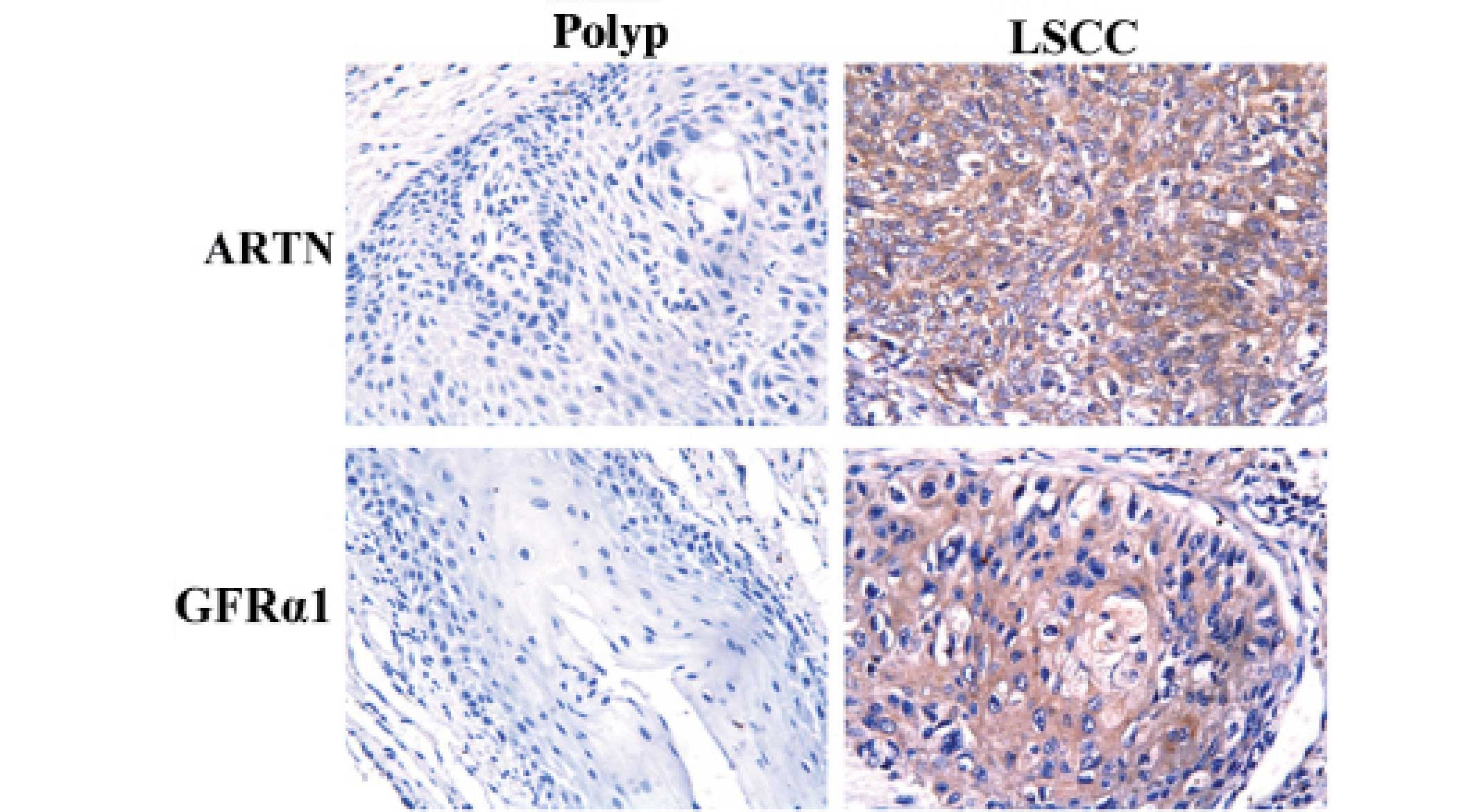
Immunohistochemistry was used to determine the
expression of immunoreactive protein for ARTN and GFRα1 in a cohort
of specimens. Positive signals were observed in the cytoplasm of
the squamous cell carcinoma cells or squamous epithelium of polyp
tissues (Fig. 1). As shown in
Table I, 53.9 and 51.3% of LSCC
samples were positive for ARTN and GFRα1, respectively, whilst only
26.9% of normal squamous epithelium from patients with polyp were
positive for ARTN and GFRα1 (P=0.015 and P=0.031,
respectively).
 | Table IExpression of ARTN and GFRα1 in LSCC
and polyp tissue specimens. |
Table I
Expression of ARTN and GFRα1 in LSCC
and polyp tissue specimens.
| | Expression, n
(%) |
|---|
| |
|
|---|
| Group | n | ARTN | GFRα1 |
|---|
| LSCC | 76 | 41 (53.9)a | 39 (51.3)b |
| Polyp | 26 | 7 (26.9) | 7 (26.9) |
Correlation between the expression of
ARTN and GFRα1 and clinicopathological features of LSCC
The association of tumor expression of ARTN and
GFRα1 with the clinicopathological features of LSCC was then
investigated. As observed in Table
II, the expression of ARTN and GFRα1 was significantly
associated with advanced pTNM stage (P=0.024 and P=0.006,
respectively). However, no significant association was observed
between the expression of ARTN and GFRα1 and any other
clinicopathological characteristics, including tumor site, tumor
differentiation and tumor lymph node metastasis (all
P>0.05).
| Table IIAssociation of ARTN and GFRα1
expression with clinicopathological parameters from patients with
LSCC. |
Table II
Association of ARTN and GFRα1
expression with clinicopathological parameters from patients with
LSCC.
| | ARTN | GFRα1 |
|---|
| |
|
|
|---|
| Parameter | n | Expression, n
(%) | P-value | Expression, n
(%) | P-value |
|---|
| Age (years) |
| ≤60 | 40 | 19 (47.5) | 0.235 | 17 (42.5) | 0.105 |
| >60 | 36 | 22 (61.1) | | 22 (61.1) | |
| Gender |
| Male | 70 | 39 (55.7) | 0.291 | 37 (52.9) | 0.358 |
| Female | 6 | 2 (33.3) | | 2 (33.3) | |
| Tumor site |
| Supraglottic | 22 | 14 (63.6) | 0.521 | 15 (68.2) | 0.131 |
| Glottic | 45 | 23 (51.1) | | 19 (42.2) | |
| Subglottic | 9 | 4 (44.4) | | 5 (51.3) | |
| Tumor
differentiation |
| Well | 28 | 17 (60.7) | 0.122 | 16 (57.1) | 0.681 |
| Moderate | 32 | 19 (59.4) | | 16 (50.0) | |
| Poor | 16 | 5 (31.3) | | 7 (43.8) | |
| pTNM stage |
| I–II | 35 | 14 (40.0) | 0.024a | 12 (34.3) | 0.006a |
| III–IV | 41 | 27 (65.9) | | 27 (65.9) | |
| Lymph node
metastasis |
| Yes | 26 | 13 (50.0) | 0.619 | 11 (42.3) | 0.257 |
| No | 50 | 28 (56.0) | | 28 (56.0) | |
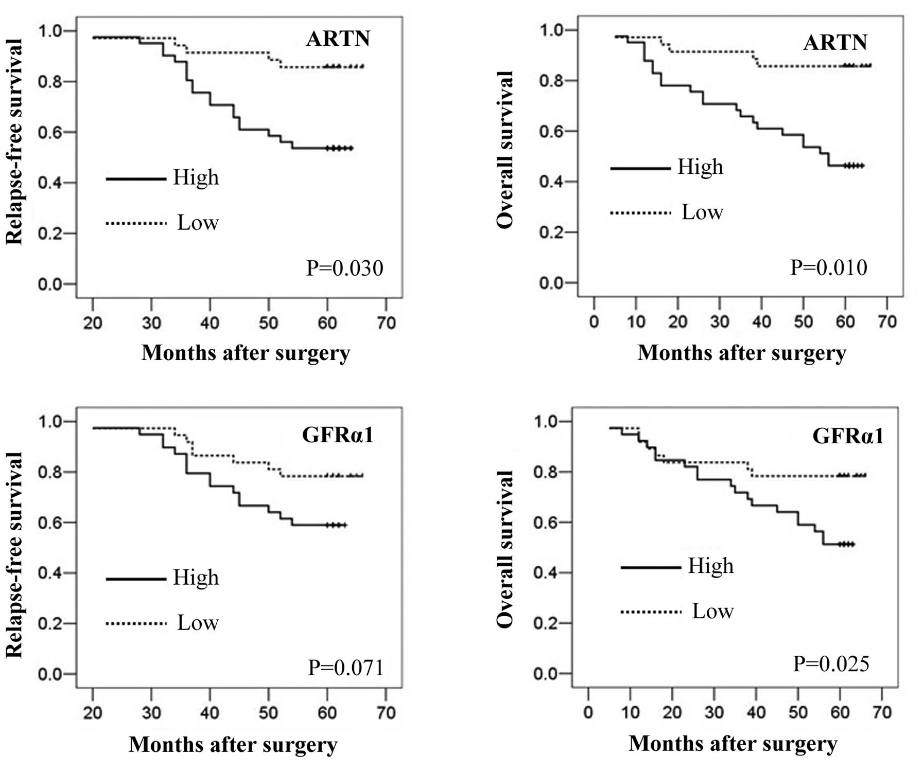
Correlation between ARTN and GFRα1
expression and patient survival
To determine the prognostic significance of ARTN and
GFRα1 expression in patients with LSCC, Kaplan-Meier analyses were
performed to correlate the expression of these proteins with the
RFS and OS of patients. As observed in Fig. 2, patients with LSCC whose tumors
were positive for expression of ARTN had a significantly lower
five-year RFS or OS compared with patients whose tumors were
negative for ARTN (P=0.030 and P=0.010, respectively). Similarly,
expression of GFRα1 protein also predicted a significantly lower
five-year OS compared with patients whose tumors were negative for
GFRα1 (P=0.025). In addition, patients whose tumors expressed GFRα1
protein exhibited a lower RFS compared with patients whose tumors
were negative for GFRα1 protein; however, this trend was not
significant (P=0.071).
Correlation between ARTN and GFRα1
expression
Correlation analysis was then conducted to determine
the correlation between ARTN protein expression and the expression
of GFRα1 protein in the same cohort of patients with LSCC. As was
expected, Pearson’s correlation analysis revealed that the
expression of ARTN was significantly correlated with the expression
of GFRα1 in these patients (rs=0.527, P=0.001).
Discussion
In this study, it was observed that a neurotrophic
factor, ARTN, was expressed at significantly higher levels in LSCC
compared with the levels in benign laryngeal polyp tissue samples.
Furthermore, the expression of ARTN was demonstrated to be
significantly associated with high tumor stage and poor survival.
In addition, previous studies have suggested that ARTN has a role
in the development and progression of diverse human carcinoma
(9,10,12,20–22).
In pancreatic ductal adenocarcinoma, ARTN has been reported to be
highly expressed compared with normal pancreases, and stimulates
the invasiveness of pancreatic cancer cells (13). Furthermore, the depletion of ARTN
expression inhibits survival, invasion and anchorage-independent
growth of both breast and endometrial cancer cells, while the
forced expression of ARTN promotes these cellular behaviors
(9,11). Molecularly, ARTN stimulates
survival and anchorage-independent growth of human non-small cell
lung cancer cells by upregulating BCL-2 expression (12). In addition, ARTN stimulates
estrogen receptor-negative breast cancer cell growth, migration and
metastasis by upregulating TWIST1 expression and activating the AKT
pathway (21). These studies are
in accordance with the results from the present study, suggesting
an oncogenic role of ARTN in the progression of human
malignancies.
ARTN has been reported to bind to and activate GFRα1
(5), a member of the GDNF receptor
α family. In order to determine whether GFRα1 mediates the effects
of ARTN in LSCC, the protein expression of GFRα1 in LSCC and
matched normal tissues was analyzed, and the correlation between
ARTN and clinicopathological features and patient survival outcome
was investigated. The results from the present study revealed that
the expression of GFRα1 was increased in cancerous tissues compared
with the expression level in normal tissues and was also
significantly associated with high tumor stage and poor survival of
patients, which indicates that GFRα1 has a similar role to that of
ARTN in LSCC. Furthermore, Pearson’s correlation analysis confirmed
that the expression of GFRα1 has a significantly high correlation
with ARTN expression. These results indicate that the functional
effects of ARTN in the progression of LSCC may be mediated by
GFRα1. Increased GFRα1 expression has been previously reported in
breast cancer, and its expression is associated with tumor lymph
node metastases and poor survival in patients (8,15).
In addition, the stimulation of GFRα1-positive breast cancer cells
with GDNF has been previously demonstrated to enhance cell
proliferation and survival in vivo (8). In human neuroblastoma, Yoong et
al demonstrated that GFRα1 promotes neurite outgrowth in tumor
cells via the activation of ERK1/2, Rac1 and Cdc42 (23).
In conclusion, to the best of our knowledge, this
study demonstrates for the first time the altered expression of
ARTN and GFRα1 in LSCC and the association of the expression levels
of these proteins with high stage disease and poor survival outcome
for patients. The expression levels of ARTN and GFRα1 protein may
therefore be useful as prognostic markers in LSCC. Whether other
receptors of ARTN may also mediate its effects on the progression
of LSCC remains to be determined.
Acknowledgements
This study was supported in part by a grant from the
First Affiliated Hospital of Anhui Medical University and by a key
program of the Educational Department in Anhui, China. (grant no.
KJ2012A162).
References
|
1
|
Jemal A, Bray F, Center MM, Ferlay J, Ward
E and Forman D: Global cancer statistics. CA Cancer J Clin.
61:69–90. 2011. View Article : Google Scholar
|
|
2
|
Chu EA and Kim YJ: Laryngeal cancer:
diagnosis and preoperative work-up. Otolaryngol Clin North Am.
41:673–695. 2008. View Article : Google Scholar : PubMed/NCBI
|
|
3
|
Tang XB, Shen XH, Li L, Zhang YF and Chen
GQ: SOX2 overexpression correlates with poor prognosis in laryngeal
squamous cell carcinoma. Auris Nasus Larynx. 40:481–486. 2013.
View Article : Google Scholar : PubMed/NCBI
|
|
4
|
Airaksinen MS, Holm L and Hätinen T:
Evolution of the GDNF family ligands and receptors. Brain Behav
Evol. 68:181–190. 2006. View Article : Google Scholar : PubMed/NCBI
|
|
5
|
Baloh RH, Tansey MG, Lampe PA, et al:
Artemin, a novel member of the GDNF ligand family, supports
peripheral and central neurons and signals through the
GFRalpha3-RET receptor complex. Neuron. 21:1291–1302. 1998.
View Article : Google Scholar : PubMed/NCBI
|
|
6
|
Airaksinen MS and Saarma M: The GDNF
family: signalling, biological functions and therapeutic value. Nat
Rev Neurosci. 3:383–394. 2002. View
Article : Google Scholar : PubMed/NCBI
|
|
7
|
Airaksinen MS, Titievsky A and Saarma M:
GDNF family neurotrophic factor signaling: four masters, one
servant? Mol Cell Neurosci. 13:313–325. 1999. View Article : Google Scholar : PubMed/NCBI
|
|
8
|
Esseghir S, Todd SK, Hunt T, et al: A role
for glial cell derived neurotrophic factor induced expression by
inflammatory cytokines and RET/GFR alpha 1 receptor up-regulation
in breast cancer. Cancer Res. 67:11732–11741. 2007. View Article : Google Scholar : PubMed/NCBI
|
|
9
|
Kang J, Perry JK, Pandey V, et al: Artemin
is oncogenic for human mammary carcinoma cells. Oncogene.
28:2034–2045. 2009. View Article : Google Scholar : PubMed/NCBI
|
|
10
|
Pandey V, Jung Y, Kang J, et al: Artemin
reduces sensitivity to doxorubicin and paclitaxel in endometrial
carcinoma cells through specific regulation of CD24. Transl Oncol.
3:218–229. 2010. View Article : Google Scholar : PubMed/NCBI
|
|
11
|
Pandey V, Qian PX, Kang J, et al: Artemin
stimulates oncogenicity and invasiveness of human endometrial
carcinoma cells. Endocrinology. 151:909–920. 2010. View Article : Google Scholar : PubMed/NCBI
|
|
12
|
Tang JZ, Kong XJ, Kang J, et al:
Artemin-stimulated progression of human non-small cell lung
carcinoma is mediated by BCL2. Mol Cancer Ther. 9:1697–1708. 2010.
View Article : Google Scholar : PubMed/NCBI
|
|
13
|
Ceyhan GO, Giese NA, Erkan M, et al: The
neurotrophic factor artemin promotes pancreatic cancer invasion.
Ann Surg. 244:274–281. 2006. View Article : Google Scholar : PubMed/NCBI
|
|
14
|
Li S, Li Z, Guo F, et al: miR-223
regulates migration and invasion by targeting Artemin in human
esophageal carcinoma. J Biomed Sci. 18:242011. View Article : Google Scholar : PubMed/NCBI
|
|
15
|
Wu ZS, Pandey V, Wu WY, Ye S, Zhu T and
Lobie PE: Prognostic significance of the expression of GFRα1, GFRα3
and syndecan-3, proteins binding ARTEMIN, in mammary carcinoma. BMC
Cancer. 13:342013.PubMed/NCBI
|
|
16
|
Thompson L: World Health Organization
classification of tumours: pathology and genetics of head and neck
tumours. Ear Nose Throat J. 85:742006.PubMed/NCBI
|
|
17
|
Sobin L and Wittekind C: TNM
classification of malignant tumours. 6th edition. Wiley-Liss; New
York, NY: pp. 392002
|
|
18
|
Yang Q, Liu Y, Huang Y, et al: Expression
of COX-2, CD44v6 and CD147 and relationship with invasion and lymph
node metastasis in hypopharyngeal squamous cell carcinoma. PLoS
One. 8:e710482013. View Article : Google Scholar : PubMed/NCBI
|
|
19
|
Masunaga R, Kohno H, Dhar DK, et al:
Cyclooxygenase-2 expression correlates with tumor
neovascularization and prognosis in human colorectal carcinoma
patients. Clin Cancer Res. 6:4064–4068. 2000.PubMed/NCBI
|
|
20
|
Banerjee A, Qian P, Wu ZS, et al: Artemin
stimulates radio- and chemo-resistance by promoting
TWIST1-BCL-2-dependent cancer stem cell-like behavior in mammary
carcinoma cells. J Biol Chem. 287:42502–42515. 2012. View Article : Google Scholar : PubMed/NCBI
|
|
21
|
Banerjee A, Wu ZS, Qian P, et al: ARTEMIN
synergizes with TWIST1 to promote metastasis and poor survival
outcome in patients with ER negative mammary carcinoma. Breast
Cancer Res. 13:R1122011. View
Article : Google Scholar : PubMed/NCBI
|
|
22
|
Banerjee A, Wu ZS, Qian PX, et al: ARTEMIN
promotes de novo angiogenesis in ER negative mammary carcinoma
through activation of TWIST1-VEGF-A signalling. PLoS One.
7:e500982012. View Article : Google Scholar : PubMed/NCBI
|
|
23
|
Yoong LF, Wan G and Too HP: GDNF-induced
cell signaling and neurite outgrowths are differentially mediated
by GFRalpha1 isoforms. Mol Cell Neurosci. 41:464–473. 2009.
View Article : Google Scholar : PubMed/NCBI
|