Introduction
The hardness of breast lesions is closely associated
with the degree of malignancy, and is the basis for the evaluation
of benign or malignant tumors by elastosonography. Elastosonography
reflects the hardness of lesions by detecting the differences in
hardness between various structures. Conventional elastography can
be used to estimate breast tissue elasticity (1) but there are certain disadvantages
with freehand compression: the elasticity map obtained is highly
dependent on the compressibility limits under stress of the organ
and on the extent of the tissue compression applied.
The diagnosis of breast lesions with
elastosonography using a 5-point scoring system is relatively
subjective (2), as the information
displayed relates to a local strain estimated at a given location
in the tissues, but this depends on the mechanical properties of
the surrounding tissues and it is not quantitative. It has been
shown that objective quantification of tissue elasticity could now
be possible using acoustic radiation force impulse (ARFI)
elastometry. With this system, instead of using external
compression, commercially available ultrasound scanners are used to
generate short-duration acoustic radiation forces that impart small
(1–10 mm) localized displacements in the tissue. The response of
the tissue to the radiation force is detected using conventional
B-mode imaging pulses in order to track the displacement of tissue
which correlates with the local stiffness of the tissue. As a
result, two-dimensional images of tissue displacement are generated
by the repetition of this process along multiple image lines
(3,4). The generated waves can provide a
qualitative response (gray-scale map) by virtual touch tissue
imaging or a quantitative response (shear wave velocity values,
measured in m/s) by virtual touch tissue quantification (VTQ),
dependent upon the interactions with the transducer (5). ARFI has been evaluated in various
tissues using both the grey-scale map and the shear wave speed
(6).
In this study, 86 patients with breast lesions
received routine ultrasound, elastosonography and VTQ examinations.
The aim of the present study is to investigate the diagnostic value
of elastosonography and VTQ technologies in the development of
benign or malignant breast tumors.
Materials and methods
General information
Between July 2011 and March 2012, 86 females who had
undergone surgical resections of breast lesions in Pudong New Area
People’s Hospital (Shanghai, China) were selected for the study.
The age of the patients was 18–72 years with a mean of 36.8±13.6
years. The lesion diameters ranged between 7 and 32 mm with an
average of 16±8 mm. All lesions were confirmed by histopathology.
This study was approved by the Ethics committee of Pudong New Area
People’s Hospital (Shanghai, China) and patient informed consent
was obtained prior to the study.
Instruments and methods
The Acuson S2000™ Ultrasound System with color
Doppler imaging (Siemens Healthcare, Erlangen, Germany) was used
for the elastosonography and VTQ technologies. A 9L4 linear array
probe was used with a frequency of 9 MHz. A routine ultrasound
examination was initially performed to observe the gray-scale
sonographic characteristics of the lesions, including shape, size,
edge, border, internal and posterior echo, and color Doppler flow
characteristics. The elastosonography function was then started to
determine the region of interest (ROI). The sampling frame used was
ideally greater than the scope of the lesion and the quality
control indicators were set between 60 and 80. The image with the
highest quality was selected for the elasticity score. The
elastosonography images were color-coded to represent the elastic
strain of the tissues. From soft to hard, the hardness was coded as
purple, blue, green, yellow and red. Green represented the average
hardness of tissue elasticity in the ultrasound sampling frame. Red
and yellow indicated that the hardness was greater than the average
hardness; purple and blue indicated that the hardness was less than
the average hardness. VTQ inspection was performed following
elastosonography. If the lesion was larger than the sampling frame,
the sampling frame was placed in the center of the lesion. If the
lesion was smaller than the sampling frame, the lesions were
located in the center of sampling frame. The VTQ function was
started, following which the machine automatically gave the
measured VTQ speed values by the ROI. Each lesion was measured five
times and the mean value was selected.
Diagnostic criteria
The conventional ultrasound diagnostic criteria for
malignant lesions comprised an irregular shape and burr-like edge,
internal microcalcifications, rear attenuation, internal rich blood
flow and a resistive index (RI) >0.75. The diagnostic criteria
for benign lesions included clear boundaries, insufficient blood
flow and an RI ≤0.75.
The flexible ultrasound diagnostic criteria were
based on the literature (7,8),
whereby the elasticity image was scored according to the different
colors in the lesions, as follows: 1 point, purple, green and red;
2 points, the lesion and surrounding tissue were green; 3 points,
red, green and yellow lesions, with >50% green; 4 points, red,
yellow and white lesions, with 50–90% red; 5 points, >90% of the
lesion area was red. Elastosonography scores of 1–3 points were
diagnosed as benign lesions and 4–5 points as malignant
lesions.
The pathological findings were utilized as the gold
standard. The sensitivity was taken as the vertical axis and one
minus the specificity as the horizontal axis. The receiver
operating characteristic curve was then created and the threshold
was analyzed. With regard to the diagnostic criteria for the three
techniques combined, lesions with at least two positive results
from the three techniques were defined as malignant, the rest as
benign.
Statistical analysis
Statistical Analysis System (SAS) 8.0 statistical
software (SAS Institute, Inc., Cary, NC, USA) was used for the
statistical analysis. The detections rates for the benign and
malignant elastosonography scores and the VTQ speeds in each group
were compared using a χ2 test. P<0.05 was considered
to indicate a statistically significant difference. Taking
pathological findings as the gold standard, the diagnostic
performance using a combination of the three methods of routine
ultrasound, elastosonography and VTQ technology was calculated.
Results
Pathology results
Benign breast lesions were histologically confirmed
in 47 out of 86 patients; out of these 47 patients, fibroadenoma
and adenosis were diagnosed in 38 and nine patients, respectively.
Malignant tumors were found in 39 patients; 35 patients had
invasive ductal carcinoma, one patient had invasive lobular
carcinoma, two patients had medullary carcinoma and one patient had
ductal carcinoma in situ.
Benign and malignant elastosonography
scores
The elastosonography scores are shown in Table I. The detection rate of benign
breast lesions with an elastosonography score of 1–3 points was
68.09%, which was significantly higher than that of malignant
tumors (P<0.05). The benign breast lesion elastosonography
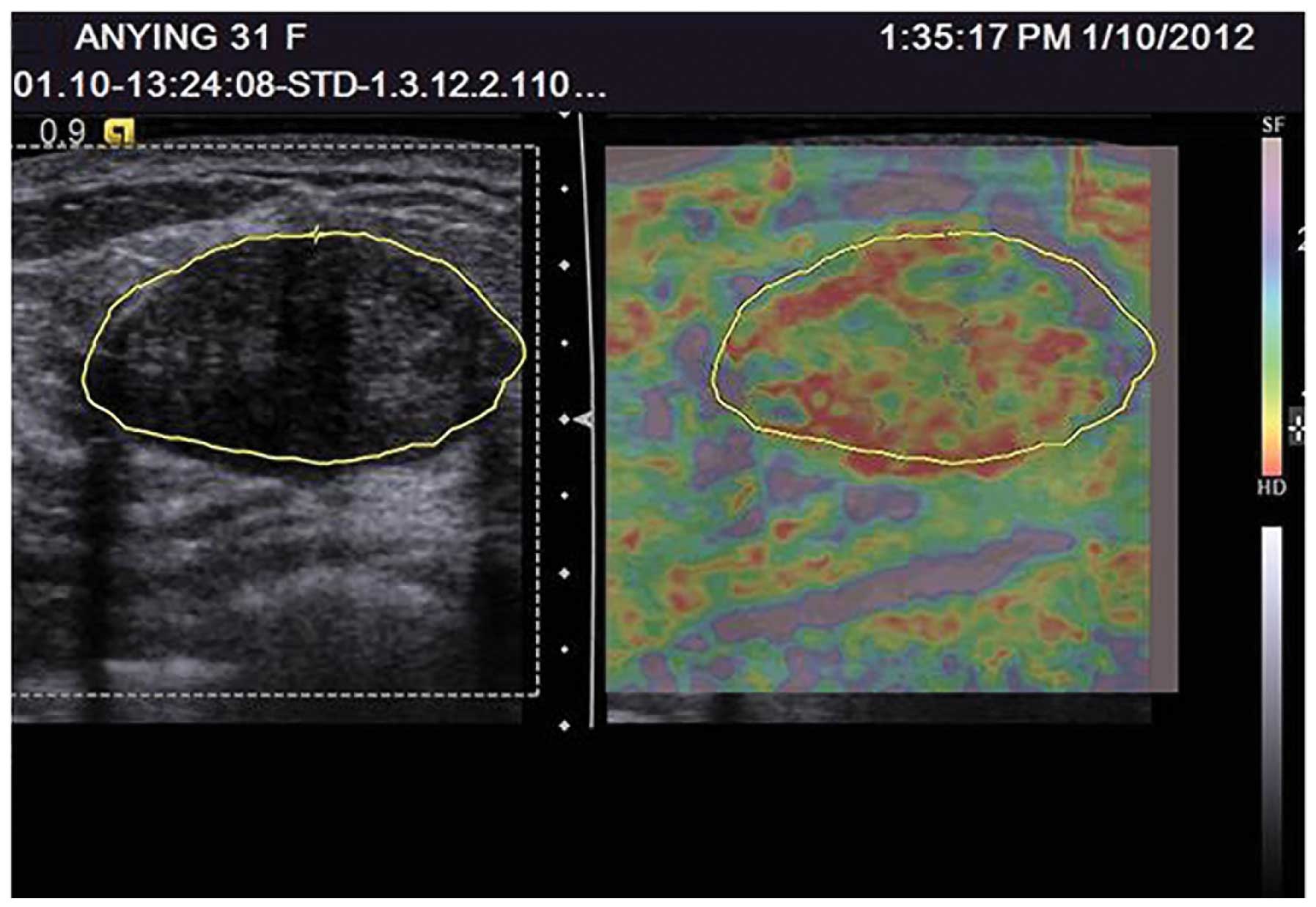
scores were mainly 1–3 (Fig. 1).
The detection rate of malignant tumors with an elastosonography
score of 4–5 points was 82.05%, which was significantly higher than
that of benign tumors (P<0.05), and the malignant breast lesion
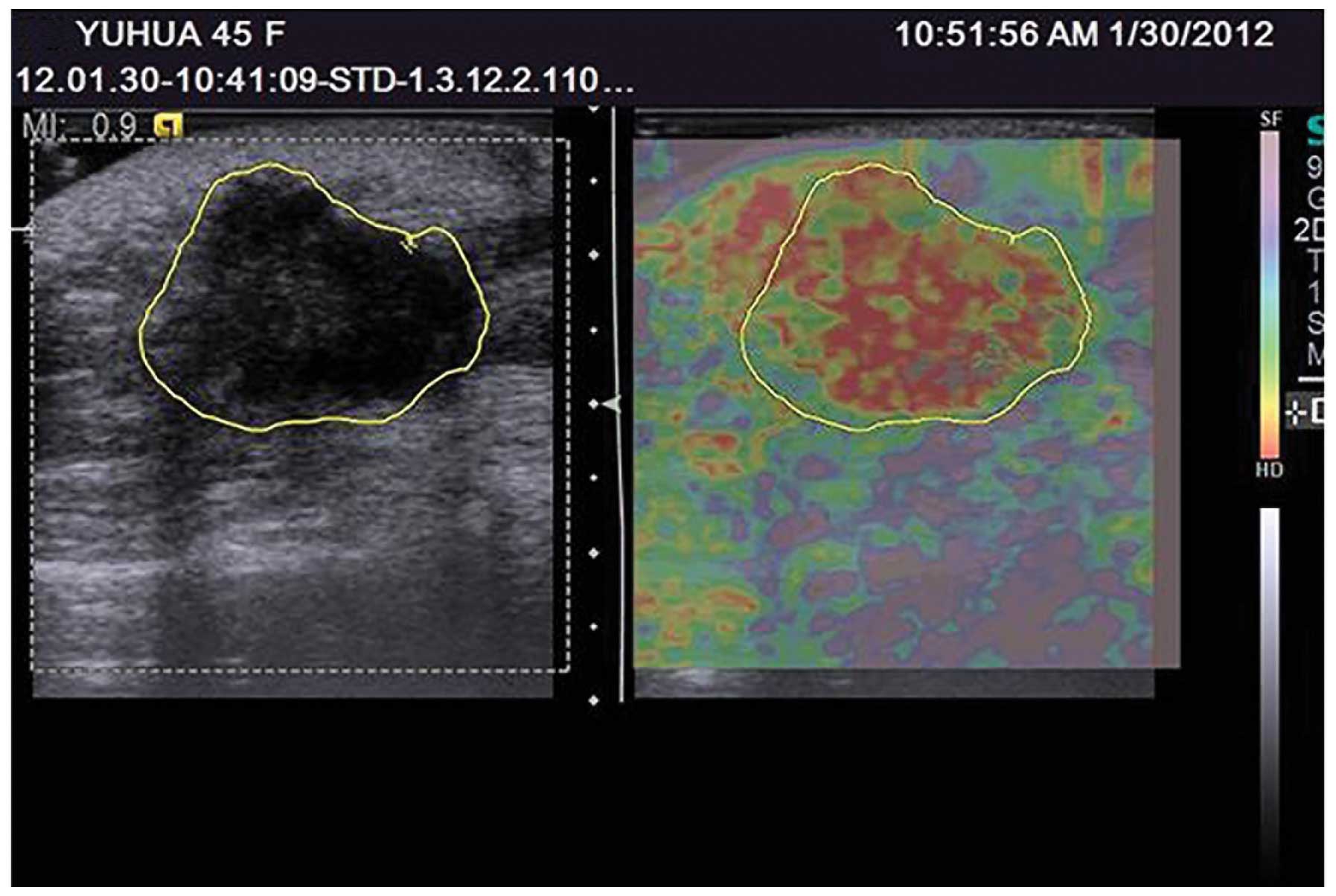
elastosonography scores were mainly 4–5 (Fig. 2). The detection rates of benign and
malignant tumors with an elastosonography score of 4 points were
21.28 and 20.51%, respectively; this difference was not
statistically significant (P>0.05).
 | Table IElastography scores of benign and
malignant breast lesions. |
Table I
Elastography scores of benign and
malignant breast lesions.
| | Ultrasound
elastography score |
|---|
| |
|
|---|
| | 1–3 | 4 | 4–5 |
|---|
| |
|
|
|
|---|
| Pathology | Number of cases
(n) | n | Detection rate
(%) | n | Detection rate
(%) | n | Detection rate
(%) |
|---|
| Benign | 47 | 32 | 68.09 | 10 | 21.28 | 15 | 31.91 |
| Malignant | 39 | 7 | 17.95 | 8 | 20.51 | 32 | 82.05 |
VTQ speed values in benign and malignant
tumors
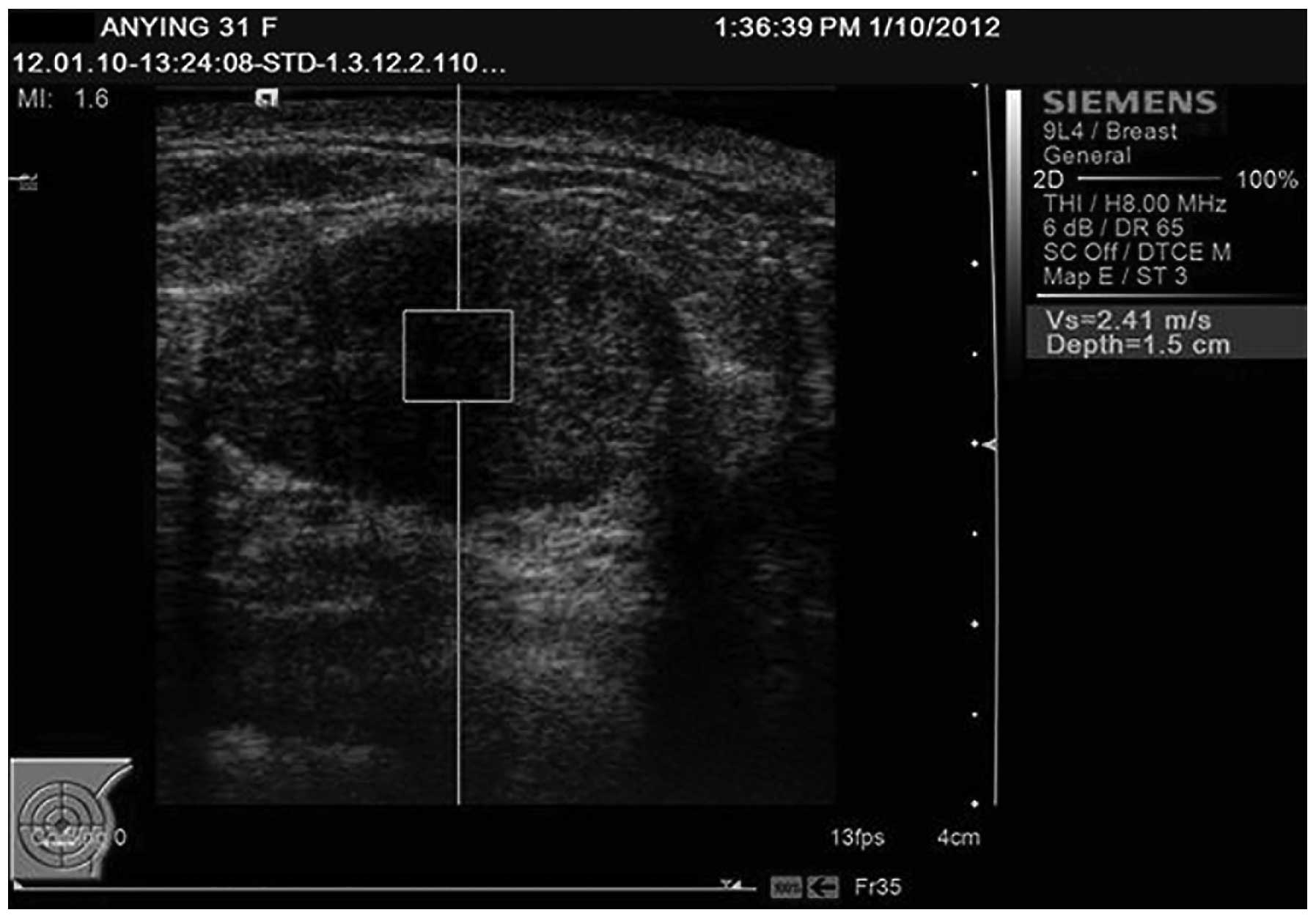
The VTQ speed results are presented in Table II. The detection rate of benign
breast tumors with a VTQ speed value of <2.98 m/sec was 74.47%,
which was significantly higher than that of the malignant tumors
(P<0.05); the VTQ speed of benign breast tumors was mainly
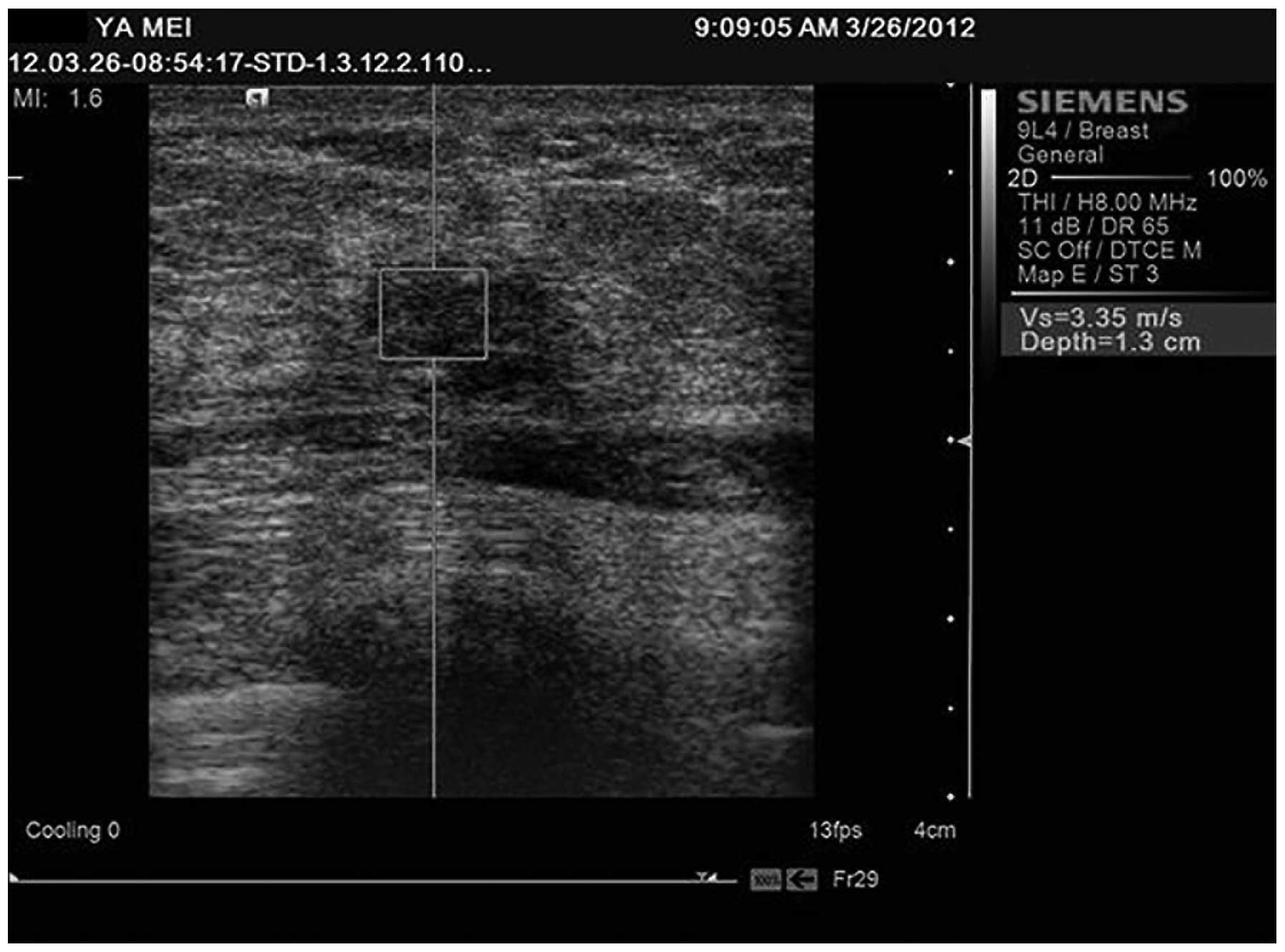
<2.98 m/sec (Fig. 3). The
detection rate of malignant breast tumors with a VTQ speed value of
≥2.98 m/sec was 79.49%, which was significantly higher than that of
the benign tumors (P<0.05); the VTQ speed of malignant breast
tumors was mainly ≥2.98 m/sec (Fig.
4).
| Table IIVTQ speed values of benign and
malignant breast lesions. |
Table II
VTQ speed values of benign and
malignant breast lesions.
| | VTQ speed value
(m/sec) |
|---|
| |
|
|---|
| | V<2.98 | V≥2.98 |
|---|
| |
|
|
|---|
| Pathology | Number of cases
(n) | n | Detection rate
(%) | n | Detection rate
(%) |
|---|
| Benign | 47 | 35 | 74.47 | 12 | 25.53 |
| Malignant | 39 | 8 | 20.51 | 31 | 79.49 |
Performance of the four types of
inspection methods in the diagnosis of benign and malignant
tumors
The diagnostic performance results are presented in
Table III. Statistically
significant differences were observed between the diagnostic
accuracy of conventional ultrasound, elastosonography, VTQ
technology and the three methods combined (P<0.05).
| Table IIIComparison of the diagnostic
performance of four types of inspection methods for benign and
malignant breast lesions. |
Table III
Comparison of the diagnostic
performance of four types of inspection methods for benign and
malignant breast lesions.
| Inspection
method | Preoperative
diagnosis | Postoperative
pathology | Sensitivity (%) | Specificity (%) | Accuracy (%) |
|---|
|
|---|
| Malignant (n=39) | Benign (n=47) |
|---|
|
|
|
|---|
| Malignant (n) | Benign (n) | Correct
diagnosis | Misdiagnosis | Correct
diagnosis | Misdiagnosis |
|---|
| Conventional
ultrasound | 41 | 45 | 31 | 8 | 37 | 10 | 79.49 | 78.72 | 79.07 |
| Elastosonography | 50 | 36 | 33 | 6 | 30 | 17 | 84.62 | 63.83 | 73.26 |
| VTQ | 41 | 45 | 30 | 9 | 36 | 11 | 76.92 | 76.60 | 76.74 |
| Combined
diagnosis | 40 | 46 | 36 | 3 | 43 | 4 | 92.31 | 91.49 | 91.86 |
Discussion
To date, there has been a constant pursuit of the
ultrasound characteristics of non-benign breast lesions, otherwise
referred to as malignant tumors. Two-dimensional gray-scale
ultrasound, color Doppler and pulsed Doppler techniques were
gradually developed to distinguish between benign and malignant
breast lesions. However, both in the two-dimensional gray-scale
images and in the images obtained using color Doppler flow imaging,
a large degree of overlap existed between benign and malignant
lesions, rendering it difficult for conventional ultrasound make an
accurate diagnosis. As a result, the emerging elastosonography and
VTQ technologies were applied to the study of breast disease
(9–11). Elastosonography took the biological
tissue elasticity (or stiffness) and the biological characteristics
of lesions as the theoretical basis, and provided a novel approach
for the differential diagnosis of breast lesions. The principle
underlying VTQ is that when the ROI tissues are affected by the
pulse wave, shear waves accompanied by lateral transfer movement
are produced; the probe pulse sequence then collects these subtle
changes and the system records and calculates its speed. This speed
is equivalent to or representative of tissue elasticity (12). VTQ is the absolute tissue
quantitative indicator, and can not only be contrasted with
neighboring tissues, but also with tissue images from different
patients. VTQ compensates for the defects of the qualitative or
semi-quantitative evaluation of previous elastosonography
methods.
The results of this study showed that the
elastosonography scores of the benign breast tumors were mainly 1–3
points, with a detection rate of 68.09%. The elastosonography
scores of the malignant breast tumors were mainly 4–5 points, with
a detection rate of 82.05%. The difference in the detection rate of
the elastosonography score between the benign and malignant tumors
was statistically significant (P<0.05). Elastosonography imaging
reflected the overall hardness level of the tumor tissue compared
with that of the surrounding normal breast tissue. The assessment
of VTQ technology showed that the benign breast tumor VTQ speeds
were mainly <2.98 m/sec, with a detection rate of 74.47%. The
VTQ values of the malignant breast tumors were mainly ≥2.98 m/sec,
with a detection rate of 79.49%. With regard to the VTQ speed
values, the detection rates of the benign and malignant breast
tumors showed a significant difference (P<0.05). The VTQ speed
values increased in parallel with increasing tissue hardness. These
findings may be explained by the fact that, in most cases, the
benign lesions had a soft texture whereas the malignant lesions had
a hard texture. The elastosonography scores were closely associated
with the pathological changes, but mainly reflected the elasticity
characteristics of the lesions. In 1998, Krouskop et al
(13) described the elasticity of
different breast tissues in decreasing order: Invasive ductal
carcinoma, non-invasive ductal carcinoma, breast fibrous tissue,
normal breast tissue and adipose tissue. It was speculated that the
larger the tissue elasticity coefficient, the greater the hardness.
The study by Konofagou (14)
showed that the malignant lesions had more complex and irregular
boundaries, therefore exhibiting poor mobility, reduced relative
strain and a large elasticity coefficient.
The results of this present study showed that the
benign and malignant tumors had a certain overlap in hardness.
Although in most cases, the malignant breast tumors had a harder
texture than the benign tumors, this was not absolute. In certain
histological types, including medullary carcinoma of the breast,
the pathology findings showed that the internal medullary carcinoma
tumor cells were in a follicular arrangement and contained a lower
fiber composition (15), therefore
leading to a relatively soft texture. In this study, two cases of
medullary carcinoma had elastosonography scores of 2 and 3 points,
and the VTQ velocity values were 2.06 and 2.23 m/sec, indicating
the soft texture of medullary carcinoma. Certain fibroadenomas with
high degrees of fibrosis exhibited a relatively high hardness. At
the same time, a variety of pathological changes within the breast
lesions may also have an impact on the hardness of the lesions. The
hardness may increase in benign lesions when fibrosis or
calcification occur and the hardness may decrease in malignant
lesions when necrosis or hemorrhage occur.
This study showed that the sensitivity, specificity
and accuracy of elastosonography in the diagnosis of breast lesions
were 84.62, 63.83 and 73.26%, respectively. The sensitivity,
specificity and accuracy of VTQ technology in breast lesions were
76.92, 76.60 and 76.74%, respectively. When elastosonography and
VTQ technology are used separately, they can only provide
information regarding lesions and tissue elasticity. The hardness
of benign and malignant tumors may overlap which would result in
misdiagnosis. In this study, the combined diagnosis using three
types of technology significantly improved the diagnostic accuracy
to ≤91.86%.
In conclusion, elastosonographies of breast lesions
and VTQ technology can indirectly and directly, respectively,
reflect the hardness of breast lesions. This can contribute to the
identification of benign and malignant tumors. However, due to the
overlap in the hardness of benign and malignant tumors, actual
clinical application would benefit from combining these methods
with conventional ultrasound to optimize the accuracy of breast
tumor diagnoses.
Acknowledgements
This study was supported by the Shanghai Pudong New
Area Leading Talents Training Plan (no. PWR12012-02), the Shanghai
Health Bureau Research Projects (no. 20114Y165), the Pudong New
Area Health Bureau of Health Science and Technology Development
(no. PW2011A-15) and the Pudong New Area Health System Training
Program for Outstanding Young Medical Personnel (no.
PWRq2010-09).
References
|
1
|
Itoh A, Ueno E, Tohno E, Kamma H,
Takahashi H, Shiina T, Yamakawa M and Matsumura T: Breast disease:
clinical application of US elastography for diagnosis. Radiology.
239:341–350. 2006. View Article : Google Scholar : PubMed/NCBI
|
|
2
|
Zhi H, Xiao XY, Yang HY, Ou B, Wen YL and
Luo BM: Ultrasonic elastography in breast cancer diagnosis: strain
ratio vs 5-point scale. Acad Radiol. 17:1227–1233. 2010. View Article : Google Scholar : PubMed/NCBI
|
|
3
|
Nightingale KR, Palmeri ML, Nightingale RW
and Trahey GE: On the feasibility of remote palpation using
acoustic radiation force. J Acoust Soc Am. 110:625–634. 2001.
View Article : Google Scholar : PubMed/NCBI
|
|
4
|
Nightingale K, Soo MS, Nightingale R and
Trahey G: Acoustic radiation force impulse imaging: in vivo
demonstration of clinical feasibility. Ultrasound Med Biol.
28:227–235. 2002. View Article : Google Scholar : PubMed/NCBI
|
|
5
|
Zhai L, Palmeri ML, Bouchard RR,
Nightingale RW and Nightingale KR: An integrated indenter-ARFI
imaging system for tissue stiffness quantification. Ultrason
Imaging. 30:95–111. 2008. View Article : Google Scholar : PubMed/NCBI
|
|
6
|
Cho SH, Lee JY, Han JK and Choi BI:
Acoustic radiation force impulse elastography for the evaluation of
focal solid hepatic lesions: preliminary findings. Ultrasound Med
Biol. 36:202–208. 2010.PubMed/NCBI
Song Y and Xia DZ: Ultrasound elastography
in observation of breast tissues for normal adult females. Zhong
Guo Yi Xue Ying Xiang Ji Shu. 28:1831–1834. 2012.(In Chinese).
|
|
7
|
Lin T, Lin HH, Wang JH and Xu YB: Analysis
of ultrasonic elastography in the diagnosis of benign and malignant
lesions of breast. Zhong Guo Lin Chuang Yi Xue Ying Xiang Za Zhi.
21:797–799. 2010.(In Chinese).
|
|
8
|
Luo BM, Yang HY, Xiao XY, Zhi H and Wen
YL: Prospective study of improved elasticity scores in the
differential diagnosis of malignant and benign breast lesions.
Zhong Hua Chao Sheng Ying Xiang Xue Za Zhi. 18:514–516. 2009.(In
Chinese).
|
|
9
|
Thomas A, Fischer T, Frey H, Ohlinger R,
Grunwald S, Blohmer JU, Winzer KJ, Weber S, Kristiansen G, Ebert B
and Kümmel S: Real-time elastography - an advanced method of
ultrasound: First results in 108 patients with breast lesions.
Ultrasound Obstet Gynecol. 28:335–340. 2006. View Article : Google Scholar : PubMed/NCBI
|
|
10
|
Wu XP, Song QH and Du DL: Two-dimensional
ultrasound, Doppler ultrasonography and ultrasonic elastography in
differential diagnosis of malignant and benign breast tumors. Zhong
Guo Yi Xue Ying Xiang Ji Shu. 27:971–974. 2011.(In Chinese).
|
|
11
|
Shen JH, Luo BM, Ou B and Yang HY:
Comparative study of ultrasonic elastography and conventional
ultrasonography in differential diagnosis of breast lesions. Zhong
Guo Yi Xue Ying Xiang Ji Shu. 23:540–542. 2007.(In Chinese).
|
|
12
|
Shen W, Ding H, Wang WP, Li C, Xu C and Xu
ZZ: Feasibility of virtual touch tissues quantification technique
in the assessment of liver fibrosis. Zhong Hua Chao Sheng Ying
Xiang Xue Za Zhi. 19:397–399. 2010.(In Chinese).
|
|
13
|
Krouskop TA, Wheeler TM, Kallel F, Garra
BS and Hall T: Elastic moduli of breast and prostate tissues under
compression. Ultrason Imaging. 20:260–274. 1998. View Article : Google Scholar : PubMed/NCBI
|
|
14
|
Konofagou EE: Quo vadis elasticity
imaging? Ultrasonics. 42:331–336. 2004. View Article : Google Scholar : PubMed/NCBI
|
|
15
|
Xu LZ: Breast Pathology. Shanghai Yi Ke Da
Xue Chu Ban She; Shanghai: pp. 1541999
|