Introduction
Bone defects caused by infection, trauma or tumors
remain intractable clinical issues. Study of implantable biomedical
materials is particularly important in the treatment of large bone
defects where self-repair of the human body is extremely difficult.
Currently, there are three main treatment methods for repairing
bone defects: autogenous bone grafts, allogeneic bone grafts and
implantation of biomedical materials (1). However, none of these methods are
perfect. For example, sources of autogenous bone grafts are limited
and additional trauma to the patient cannot be avoided during the
procedure. Although allogeneic bone grafts and biomedical materials
are easier to obtain, other problems exist, including the risk of
spreading disease, transplantation-associated immune reactions and
generally poor biodegradability and long-term outcomes.
The rapid development of bone tissue engineering has
generated new theories and technologies for repairing bone defects
in which seed cells, scaffolds and osteogenic factors are
considered to be the basic components for constructing
tissue-engineered bone (2,3). The scaffold material acts as an
artificial extracellular matrix (ECM) and provides support for cell
adhesion, growth, proliferation, metabolism and the formation of
new bone tissue. Currently, artificially synthesized inorganic
materials (including hydroxyapatite and calcium phosphate) and
polymer materials [including polylactic acid, polyglycolic acid
(PGA), polyphophazenes and chitosan] may be used as scaffolds for
bone tissue engineering (4–6).
However, all of these materials have certain limitations in
biocompatibility, biodegradability or mechanical matching (7–10).
Therefore, additional research is required to investigate other
types of composite materials.
In the present study, a new gel composite combining
strengthened β-tricalcium phosphate (β-TCP) with platelet-rich
plasma (PRP) was prepared. Its potential application as a scaffold
for repairing bone defects was evaluated by analyzing the
compressive strength, cell adhesion and cell cytotoxicity of the
material.
Materials and methods
Preparation of strengthened β-TCP
The β-TCP was prepared through chemical
precipitation as follows: i) 58.82 g
(NH4)2HPO4 and 141.69 g
Ca(NO3)2·4H2O were dissolved in
distilled water, respectively.
(NH4)2HPO4 solution was added to
the Ca(NO3)2 solution at a sustained rate of
1.5 ml/min, with the pH value adjusted to 7.0–7.5 by the addition
of NH4OH solution. The supernatant was removed 24 h
following the formation of a white precipitate, which was washed
with distilled water until the conductivity of the liquid was
<200 μS/cm; ii) The precipitate was transferred to a metal
container, compressively molded and calcined at 850°C for 10 h to
obtain the β-TCP sample; iii) the precipitate was dried at 120°C
and calcined at 850°C for 0.5 h to obtain the β-TCP powder. The
β-TCP powder was mixed with 4% hyaluronic acid (HA) nanoparticles
and added to distilled water together with 0.5% dispersant
polyethylene glycol to obtain a slurry (solid content, 60%). The
slurry was milled for 24 h and added to bioglass powder (total
solid content, 20%) for additional milling for 2 h. Surfactant
Tween 80 (Lantian, Xingtai, China) was added to the slurry, which
was subsequently stirred (rotation speed, 500 rpm) to generate foam
with a double-leaf stirring blade in a controlled N2
atmosphere. The foamed slurry was poured into a plaster cast for
molding. Once dried, the molding was sintered by first heating to
570°C at a rate of 2°C/min, maintained at this temperature for 1 h,
and subsequently heated to 1,000°C at a rate of 5°C/min, where it
was maintained for an additional 1 h. The strengthened β-TCP sample
was prepared using this method.
Preparation of PRP
To prepare the PRP, 10 ml venous blood was collected
from the hind limb of a beagle dog and placed in a 15-ml centrifuge
tube containing 1 mg sodium citrate. PRP was extracted using a
two-step centrifugation method. First, the blood sample was
centrifuged at 1,500 rpm for 10 min. The upper layer of plasma and
the erythrocytes located within 2 mm of the interface were
transferred to another centrifuge tube. Secondly, the sample was
centrifuged at 3,600 rpm for 10 min and the upper plasma layer,
containing a small number of suspended platelets, was removed. The
remaining plasma (~1 ml) was PRP. A total of 10 μl whole blood and
10 μl PRP were drawn from the centrifuge tube with a pipette and
diluted to 2 ml by platelet dilution. One drop of the mixture was
transferred to a clean counting chamber, incubated at room
temperature for 10 min and the cells were counted under a CX22
light microscope (Olympus, Tokyo, Japan) at high magnification
(x400). Additional PRP was prepared using this method and stored in
a refrigerator at −70°C. The current study, including the animal
ethics, was approved by the Ethics Committee of Xiangya Hospital
(Changsha, China).
Preparation of the composite combining
strengthened β-TCP and PRP
The strengthened β-TCP samples (cylindrical,
diameter, 1.0 cm; height, 4.0 cm) were cut into 0.5-cm slices using
a razor blade in an ultra-clean cabinet. The slices were repeatedly
washed with saline and sterilized at 180°C for 30 min after drying.
The slices were subsequently immersed in PRP until the tricalcium
phosphate material was completely enveloped by the plasma. The
activating agent (10% calcium chloride solution containing 100
μg/ml bovine thrombin) was added at a ratio of 1:1. The mixture was
incubated in a 37°C water bath to form the strengthened β-TCP/PRP
gel composite.
Isolation, cultivation, purification and
proliferation of beagle dog bone marrow stromal cells (BMSCs)
A 12-month-old adult beagle dog weighing 10 kg was
administered intravenous anesthesia with 3% pentobarbital (1.0
ml/kg) and 3 ml bone marrow was extracted using a sterile
technique. The marrow was washed and diluted with
phosphate-buffered saline (PBS) and slowly poured over Percoll
(1.079 g/ml; Pharmacia, Beijin, China) at a volume ratio of 1:1.
The mixture was centrifuged at 2,000 rpm for 15 min, during which
time the centrifuged mixture formed four layers in the tube. The
second white membrane-like layer was drawn with a syringe, washed
with PBS and centrifuged at 1,000 rpm for 5 min. Following removal
of the supernatant, Dulbecco’s modified Eagle’s medium-low glucose
(DMEM-LG) culture (containing 100 μg/ml penicillin and 100 μg/ml
streptomycin) supplemented with 10% fetal calf serum (FCS) was
added to resuspend the cells. The cells were seeded in a
25-cm2 culture flask at a density of 2×10⁵
cells/cm2 together with 4 ml of the culture medium and
placed in a CO2 cell culture incubator. The medium was
changed following the first 48 h of incubation and every three days
thereafter. Finally, 0.25% trypsin digestion was performed when the
adherent cell confluence reached 80–90%. Serial subculture was
performed at a 1:3 dilution. A control was prepared for the
adhesion rate assay by adding the BSMC suspension to the
composite.
BMSC adhesion rate and cell
cytotoxicity
Based on the method proposed by Kasten et al
(11), the strengthened β-TCP/PRP
gel composite was placed in 24-well culture plates. To the
composite was added dropwise 0.2 ml BMSC suspension (concentration,
1×10⁵ cells/ml) and cultured in a humidified cell
culture incubator (37°C, 5% CO2, saturated humidity).
The samples were collected at 4, 8, 12 or 24 h following incubation
(n=5 for each time point). The adherence rate (%) = (inoculation
cell number - number of cells lost - non-adherent cell
number)/(inoculation cell number - number of cells lost) ×100.
BMSCs were collected following the third passage and
the density of the cell suspension was adjusted to
1.0×10⁵ cells/l. The cell suspension was seeded onto the
strengthened β-TCP/PRP composite through a micropipettor with 50 μl
in each bracket to produce a cell-composite complex and incubated
in the cell culture incubator. BMSCs with the same density as the
cell-composite complex above were plated in wells as the control
group and five parallel samples were used for each group. The
proliferation rate was measured at 2, 4 and 8 days following
seeding using a
3-(4,5-dimethylthiazol-2-yl)-2,5-diphenyltetrazolium bromide (MTT)
assay. Briefly, the samples in each well were mixed with 100 μl 5%
MTT and cultured for an additional 4–6 h. The liquid in each well
was removed and 500 μl dimethyl sulfoxide (DMSO) was added. The
cells were incubated for 10 min and 200 μl supernatant from each
well was removed and plated in individual wells of a 96-well
culture plate. The absorbance was measured at a wavelength of 490
nm using a microplate reader (SpectraMax M5; Molecular Devices,
Sunnyvale, CA, USA). The mean absorbance was taken from six wells.
The relative growth rate (RGR) of each group was calculated using
the following formula: RGR (%) = [mean optical density (OD) value
of the experimental group/mean OD value of control group] × 100.
The RGR values for each group were converted into six-degree
cytotoxicities to assess the toxicity of the materials. These
cytotoxicities were on a scale of 0–5 as follows: 0, RGR >1; 1,
RGR 0.75–0.99; 2, RGR 0.5–0.74; 3, RGR 0.25–0.49; 4, RGR 0.01–0.24;
5, RGR 0.
Scanning electron microscopy (SEM)
examination of the seeded BMSCs on different scaffolds
The BMSCs were collected following the third passage
and the density of the cell suspension was adjusted to
4.0×10¹⁰ cells/l. The cell suspension was seeded onto
strengthened β-TCP and the β-TCP/PRP composite using a
micropipettor with 100 μl in each bracket and the cells were
incubated in the cell culture incubator for 4 h to allow for cell
adherence. Three experimental groups were assessed: i) BMSCs seeded
on strengthened β-TCP with low-glucose DMEM culture medium
containing 10% FCS; ii) BMSCs seeded on strengthened β-TCP/PRP with
low-glucose DMEM culture medium containing 10% FCS and; iii) BMSCs
seeded on strengthened β-TCP/PRP with an inducer (high-glucose DMEM
culture medium containing 10% FCS, 10−7 mol/l
dexamethasone, 10 mmol/l β-glycerophosphate and 500 mg/l ascorbic
acid).
Following culture of the cells for two weeks, the
composite was examined with a SEM (Jeol JSM-6360LV; Jeol, Inc.,
Tokyo, Japan) to observe cell growth and proliferation on the
materials as well as matrix secretion. The steps to prepare the
samples prior to SEM observation were as follows: i) cleaning, the
samples were immersed in saline for ultrasonic cleaning for 5–10
min; ii) fixing, the samples were fixed with 4% glutaraldehyde for
2 h and subsequently with 1% osmic acid for 2 h; iii) dehydration,
gradient dehydration was performed by incubating each of the
samples in 50, 70, 90 and 100% acetone for 10 min; iv)
substitution, performed by incubating the samples with 50, 70, 90
and 100% isoamyl acetate for 10 min each; v) drying, an HCP2
critical point dryer (Hitachi Ltd., Tokyo, Japan) was used for
drying; and vi) coating, Emitech K500X/K550X Sputter targets
(Quorum Technologies, Ltd., Laughton, UK) were used to plate gold
film onto the samples in a vacuum. Furthermore, the morphologies of
strengthened β-TCP and strengthened β-TCP/PRP composite were also
examined using SEM.
Biomechanical properties of β-TCP and
strengthened β-TCP/PRP composite
The β-TCP (five samples) and strengthened β-TCP/PRP
composite (five samples) were placed vertically in an Instron 8032
universal material tester (Instron, Norwood, MA, USA) for
compression analysis with a loading speed of 5 mm/min. The maximum
load (maximum external force that the sample was able to bear) and
the utmost intensity (calculated as maximum load/contact area) were
recorded during the experiment.
Statistical analysis
Results are presented as mean ± standard deviation
and statistical analyses were performed using SPSS software,
version 13.0 (SPSS, Inc., Chicago, IL, USA). A double sample
Student’s t-test was used to evaluate the differences between
groups and a difference was considered to be statistically
significant if two-tailed P<0.05.
Results
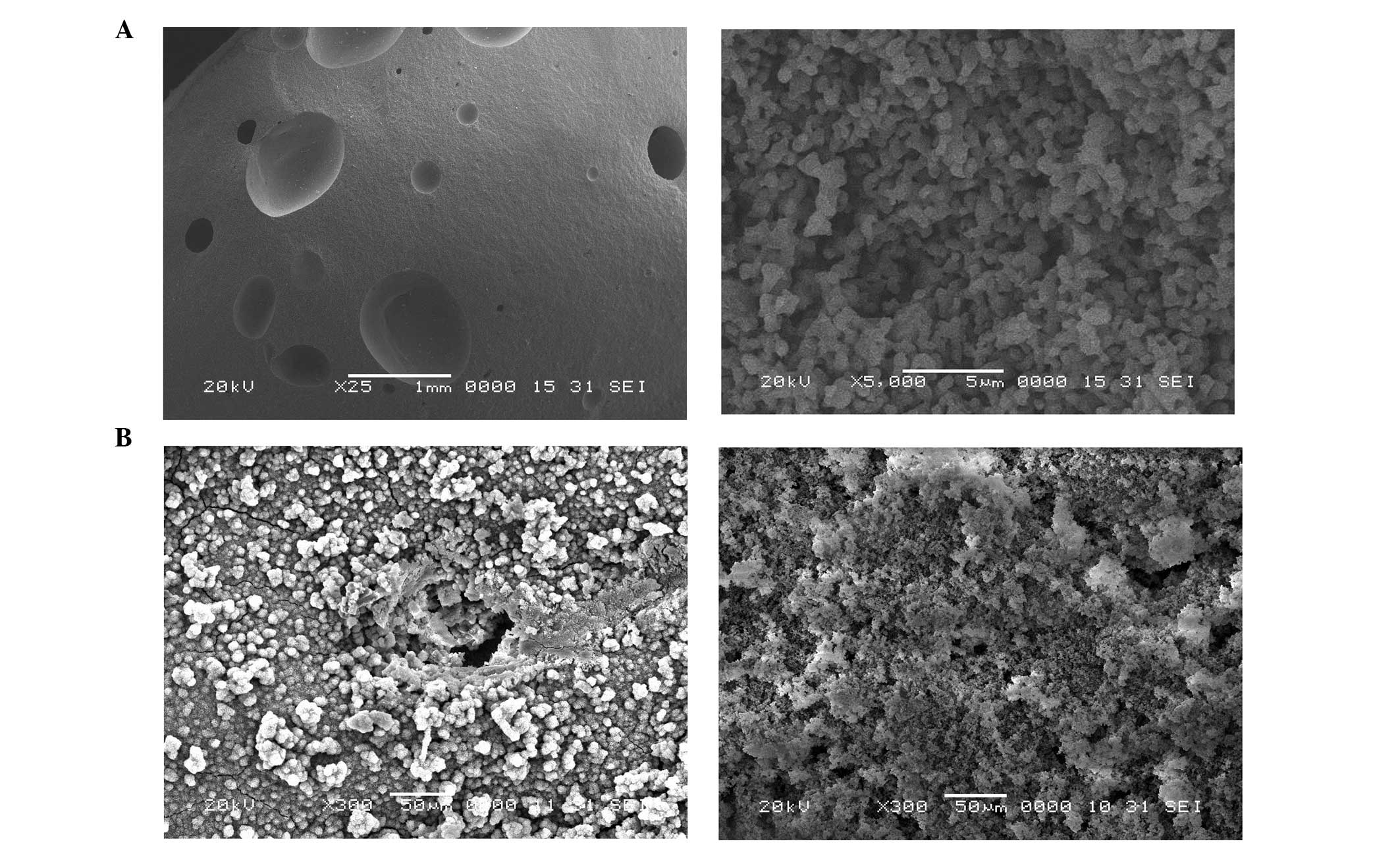
Morphological properties of the
strengthened β-TCP and strengthened β-TCP/PRP composite
The concentration of platelets present in the PRP
prepared using a two-step centrifugation procedure was calculated
to be 1,239.3±124.0×10⁹ cells/l, which was ~8.6-fold
higher compared with that of venous blood
(143.3±31.9×10⁹ cells/l). Strengthened β-TCP, a white
porous material with a variety of pore sizes (range, 100–400 μm),
was prepared in the form of a cylinder (diameter, 1 cm; height, 3–4
cm). SEM analysis revealed a granular appearance at high
magnification (x5,000; Fig. 1A).
The pores on the surface connected with each other and small
micropores were distributed with diameters of 1–10 μm. The PRP gel
appeared as particulate crystals and was well distributed on the
surface as well as the pores of the strengthened β-TCP/PRP
composite, giving the material an appearance like the fungus
Boletus kermesinus (referred to as kermesinus). The
micropores on the surface of the composite were observed to be
smaller due to the filling of the PRP particles when viewed by SEM
at high magnification (x300; Fig.
1B).
Adhesion rate and cell cytotoxicity of
BMSCs grown on strengthened β-TCP/PRP composite
The BMSC adhesion rate on the strengthened β-TCP/PRP
composite was 50±2, 80±1, 92±2 and 96±1% after 4, 8, 12 and 24 h,
respectively. There were no significant differences in the adhesion
rate at any of these time points compared with those in the control
(P>0.05). The OD and RGR values of the control and experimental
groups are shown in Table I.
According to the criteria used in the current study, the
cytotoxicity of the strengthened β-TCP/PRP composite was zero.
 | Table IOptical density (OD) and relative
growth rate (RGR) values of bone marrow stromal cells in the
control group and on the strengthened β-TCP/PRP composite. |
Table I
Optical density (OD) and relative
growth rate (RGR) values of bone marrow stromal cells in the
control group and on the strengthened β-TCP/PRP composite.
| OD | |
|---|
|
| |
|---|
| Day | Control group | Test group | RGR |
|---|
| 2 | 0.172 | 0.173 | 1.007 |
| 4 | 0.242 | 0.257 | 1.064 |
| 8 | 0.951 | 0.968 | 1.017 |
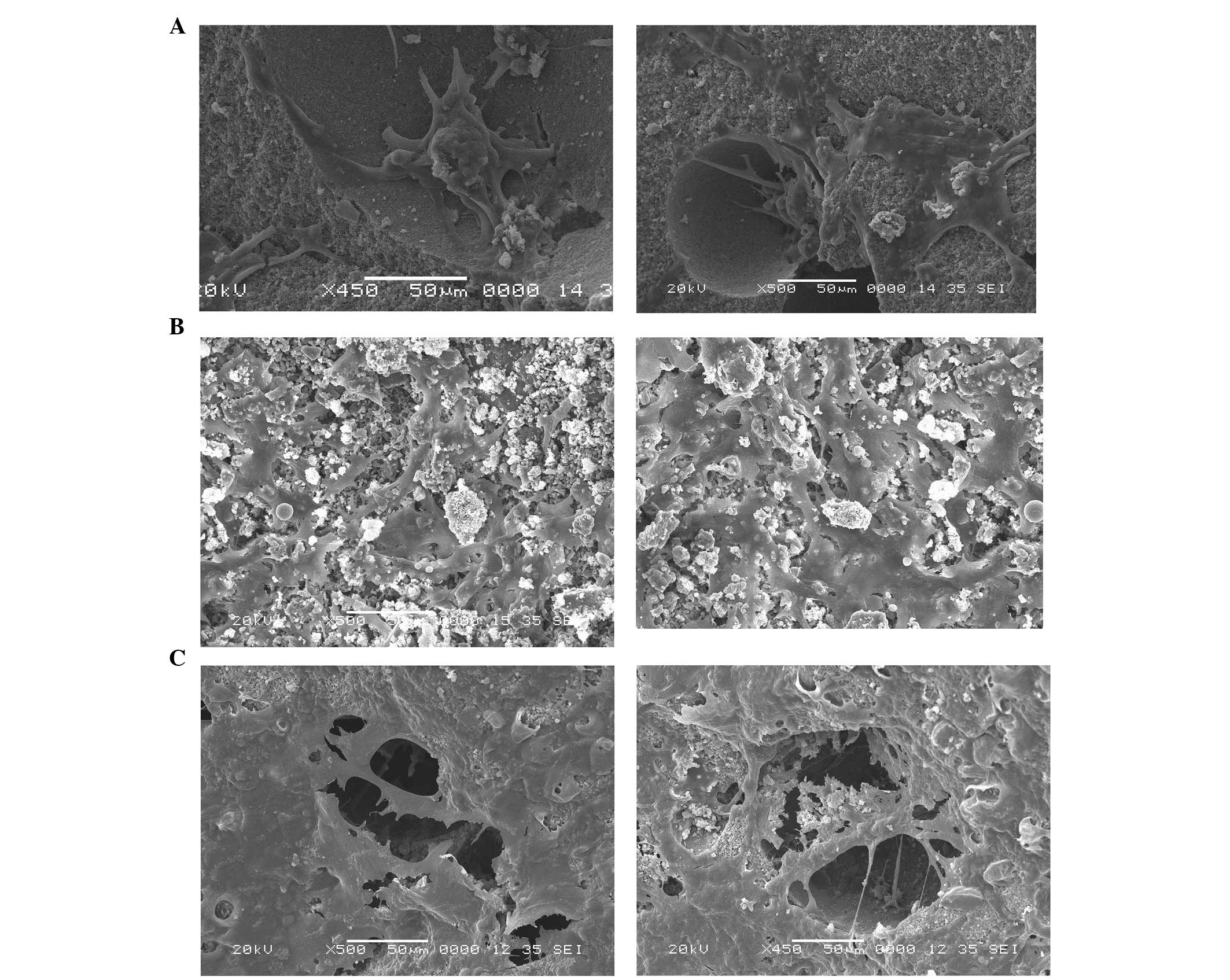
SEM examination of seeded BMSCs on
different scaffolds
There was a low number of BMSCs seeded on the
strengthened β-TCP grown in low-glucose DMEM culture medium and the
cells grew slowly with a fusiform morphology (Fig. 2A). The BMSCs seeded on strengthened
β-TCP/PRP in low-glucose DMEM culture medium grew faster and
exhibited fusiform, spherical and polygonal morphologies (Fig. 2B). For the strengthened β-TCP/PRP
in high-glucose DMEM culture medium, a large number of BMSCs were
attached to the surface and around the pores, and exhibited
fusiform, spherical and polygonal morphologies. Pseudopodia
extending from the cells were observed that connected with each
other and covered the pores; the secretion of ECM was also observed
(Fig. 2C).
Biomechanical properties of β-TCP and the
strengthened β-TCP/PRP composite
The maximum load and utmost intensity of the
strengthened β-TCP/PRP gel composite were 945.6±86.4 N and 13.1±0.5
MPa, respectively, which were significantly higher compared with
those of β-TCP (110.1±14.3 N and 1.6±0.2 MPa, respectively;
P<0.05). The utmost intensity of the strengthened β-TCP/PRP gel
composite was also slightly higher than that of human cancellous
bone (4–12 MPa).
Discussion
The ideal materials for bone tissue engineering
scaffolds should have the following characteristics: i) Effective
biocompatibility, non-toxic degradation products and no
inflammation; ii) suitable pore size for the in-growth of new bone
tissue, with an average diameter of 200–400 μm; iii) enough
mechanical strength to support new bone tissue with suitable
mechanical properties; iv) osteoconductive or osteoinductive
effects to promote bone deposition and growth and; v) adjustable
biodegradability. In the present study, β-tricalcium phosphate with
good physicochemical properties was used as the matrix, bioglass as
the binder and nano-HA was used for the dispersal phase to prepare
strengthened β-TCP using the physical foaming method. Strengthened
β-TCP had mutually-connected large pores and micropores, with a
relatively small pore size distribution. The diameters of the large
pores ranged between 750 and 850 μm, and a number of micropores
with a diameter of ~21 μm were also present on the walls of the
large pores. Furthermore, the composite material had a good
biocompatibility and mechanical properties; therefore, it has the
potential to be used as a scaffold in bone tissue engineering to
repair bone defects.
Artificial polymers and bioactive ceramics are
commonly used scaffold materials for bone tissue engineering. In a
study by Breitbart et al, PGA fiber scaffolds containing
rabbit periosteal osteoblasts completely repaired bone defects
(diameter, 1–5 mm) in a rabbit skull after 12 weeks (12). Sachlos et al increased the
hydrophilicity of scaffolds by introducing chemical functional
groups to their surface; local aseptic inflammation was observed in
certain patients following implantation of these scaffolds
(13). HA and tricalcium phosphate
(TCP) are common bioactive ceramics used in tissue engineering. In
a study by Morishita et al, BMSCs forced to differentiate
into osteoblasts on HA ceramics revealed a strong osteogenic
ability and repaired bone defects within six months (14). However, the brittleness and low
degradation rate of HA ceramics limits their application in bone
tissue engineering to a certain extent. TCP has effective
physicochemical properties, biocompatibility and biodegradability,
which makes it an efficient bone graft material that is able to
promote new bone formation and conduction, and repair bone defects.
Harris and Cooper loaded human mesenchymal stem cells onto cubes of
coral calcium carbonate-derived apatite, bovine bone-derived
apatite, synthetic HA/TCP (60/40%) or synthetic HA/TCP (20/80%) and
placed them into the dorsal regions of mice with severe combined
immunodeficiency (SCID) for five weeks (15). Histomorphometric analysis of bone
development within the cubes revealed an absence of bone formation
within the coral-derived and bovine bone-derived apatites. However,
bone formation within the synthetic HA/TCP scaffolds was revealed
to comprise 8.8 and 13.8% of the total tissue present for the 60:40
and 20:80% materials, respectively (15). TCP has certain disadvantages,
including an excessive degradation rate in vivo as well as
high rigidity and brittleness. For example, in a study by Wiltfang
et al, 3.5–4.7 ml cancellous bone defects were established
on the two sides of the tibial head in seven Goettingen minipigs
(16). After 16 and 28 weeks
post-filling of the defects with β-TCP, the degradation rate was 60
and 80%, respectively; after 68 weeks, only 5% of the β-TCP
remained (16). The degradation
rate of TCP was higher than the new bone formation rate, which is
likely to affect the repair of bone defects.
Growth factors are also important in bone tissue
engineering and play critical roles in the regulation of
osteogenesis, induction of osteogenic differentiation, cell
proliferation, collagen synthesis and vascularization. PRP contains
a variety of growth factors including platelet-derived growth
factor (PDGF), transforming growth factor-β (TGF-β), insulin-like
growth factor (IGF), vascular endothelial growth factor (VEGF) and
epidermal growth factor (EGF) (17–19).
As early as 1995, Slater et al revealed that PRP was able to
promote the proliferation of osteoblasts in vitro (20). PRP may also promote BMSC
differentiation and induction. It has been demonstrated that the
differentiation capability of human BMSCs reduces as the cell
passage number increases; however, the application of PRP
substantially enhances the proliferation activity (21–23).
In the present study, PRP was successfully isolated
from venous blood taken from the hind limb of a beagle dog using a
two-step centrifugation procedure. The platelet concentration was
~8.6 times greater than that of whole blood. Bovine thrombin was
added to the strengthened β-TCP/PRP gel composite in order to
activate platelets to release growth factors. The gel composite
extended the functional duration of the growth factors.
Furthermore, the adhesion rates of the BMSCs were >90 and
>95% at 12 and 24 h following implantation, respectively,
although there was no significant difference compared with the
control group. The MTT assay revealed that the toxic grade of the
composite was zero based on the criteria used in the current study.
The SEM micrographs demonstrated that the BMSCs adhered to the
surface of the scaffold, proliferated and secreted ECM. Cellular
proliferation and differentiation increased when an
osteogenesis-directed differentiation inducer was added to the
culture medium. The cell proliferation and secretion of ECM was
extremely strong to the extent that the scaffold was almost
completely covered and a three-dimensional structure was formed
where the cells had gradually grown into the scaffold. However, the
majority of the BMSCs were located on the surface of the scaffold,
which may be associated with the preparation of the composite, cell
concentration or the induction medium. Further study is required to
gain further insight into the mechanism. The in vivo
degradation behavior of the composite was not investigated in the
current study and further study with an animal model is required to
clarify the issue.
In the present study, a new composite combining
strengthened β-tricalcium phosphate with PRP was successfully
prepared with good biocompatibility and mechanical properties. The
new composite may potentially be used as a scaffold to treat bone
defects with bone tissue engineering technologies.
Acknowledgements
The present study was financially supported by the
Fundamental Research Funds for the Central Universities of China.
The authors would like to extend their thanks to the Central
Laboratory of National Hepatobiliary & Enteric Surgery Research
Center (Ministry of Health), The Key Laboratory of Tumor Proteomics
(Ministry of Health) and the Institute of Powder Metallurgy of
Central South University. The authors would like to thank Medjaden
Bioscience Limited (Hong Kong, China) for assisting in the
preparation of this study.
References
|
1
|
Park J, Kim JR and Yang KH: Treatment for
a femoral shaft bone defect using heterotopic bone formation as
autograft. Eur J Orthop Surg Traumatol. 22:135–138. 2012.
View Article : Google Scholar
|
|
2
|
Burg KJ, Porter S and Kellam JF:
Biomaterial developments for bone tissue engineering. Biomaterials.
21:2347–2359. 2000. View Article : Google Scholar : PubMed/NCBI
|
|
3
|
Cao H and Kuboyama N: A biodegradable
porous composite scaffold of PGA/beta-TCP for bone tissue
engineering. Bone. 46:386–395. 2010. View Article : Google Scholar : PubMed/NCBI
|
|
4
|
Ren T, Ren J, Jia X and Pan K: The bone
formation in vitro and mandibular defect repair using PLGA
porous scaffolds. J Biomed Mater Res A. 74:562–569. 2005.
|
|
5
|
Rezwan K, Chen QZ, Blaker JJ and
Boccaccini AR: Biodegradable and bioactive porous polymer/inorganic
composite scaffolds for bone tissue engineering. Biomaterials.
27:3413–3431. 2006. View Article : Google Scholar : PubMed/NCBI
|
|
6
|
Lohfeld S, Cahill S, Barron V, et al:
Fabrication, mechanical and in vivo performance of
polycaprolactone/tricalcium phosphate composite scaffolds. Acta
Biomater. 8:3446–3456. 2012.
|
|
7
|
Formigli L, Benvenuti S, Mercatelli R, et
al: Dermal matrix scaffold engineered with adult mesenchymal stem
cells and platelet-rich plasma as a potential tool for tissue
repair and regeneration. J Tissue Eng Regen Med. 6:125–134. 2012.
View Article : Google Scholar : PubMed/NCBI
|
|
8
|
Schuckert KH, Jopp S and Teoh SH:
Mandibular defect reconstruction using three-dimensional
polycaprolactone scaffold in combination with platelet-rich plasma
and recombinant human bone morphogenetic protein-2: de novo
synthesis of bone in a single case. Tissue Eng Part A. 15:493–499.
2009. View Article : Google Scholar
|
|
9
|
Kasten P, Vogel J, Luginbühl R, et al:
Influence of platelet-rich plasma on osteogenic differentiation of
mesenchymal stem cells and ectopic bone formation in calcium
phosphate ceramics. Cells Tissues Organs. 183:68–79. 2006.
View Article : Google Scholar : PubMed/NCBI
|
|
10
|
Kon E, Filardo G, Delcogliano M, et al:
Platelet autologous growth factors decrease the osteochondral
regeneration capability of a collagen-hydroxyapatite scaffold in a
sheep model. BMC Musculoskelet Disord. 11:2202010. View Article : Google Scholar
|
|
11
|
Kasten P, Luginbühl R, van Griensven M, et
al: Comparison of human bone marrow stromal cells seeded on
calcium-deficient hydroxyapatite, beta-tricalcium phosphate and
demineralized bone matrix. Biomaterials. 24:2593–2603. 2003.
View Article : Google Scholar : PubMed/NCBI
|
|
12
|
Breitbart AS, Grande DA, Kessler R, Ryaby
JT, Fitzsimmons RJ and Grant RT: Tissue engineered bone repair of
calvarial defects using cultured periosteal cells. Plast Reconstr
Surg. 101:567–574. 1998. View Article : Google Scholar : PubMed/NCBI
|
|
13
|
Sachlos E, Reis N, Ainsley C, Derby B and
Czernuszka J: Novel collagen scaffolds with predefined internal
morphology made by solid freeform fabrication. Biomaterials.
24:1487–1497. 2003. View Article : Google Scholar : PubMed/NCBI
|
|
14
|
Morishita T, Honoki K, Ohgushi H, Kotobuki
N, Matsushima A and Takakura Y: Tissue engineering approach to the
treatment of bone tumors: three cases of cultured bone grafts
derived from patients’ mesenchymal stem cells. Artif Organs.
30:115–118. 2006.PubMed/NCBI
|
|
15
|
Harris CT and Cooper LF: Comparison of
bone graft matrices for human mesenchymal stem cell-directed
osteogenesis. J Biomed Mater Res A. 68:747–755. 2004. View Article : Google Scholar : PubMed/NCBI
|
|
16
|
Wiltfang J, Merten HA, Schlegel KA, et al:
Degradation characteristics of alpha and beta tri-calcium-phosphate
(TCP) in minipigs. J Biomed Mater Res. 63:115–121. 2002. View Article : Google Scholar : PubMed/NCBI
|
|
17
|
Anitua E: Plasma rich in growth factors:
preliminary results of use in the preparation of future sites for
implants. Int J Oral Maxillofac Implants. 14:529–535.
1999.PubMed/NCBI
|
|
18
|
Carlson NE and Roach RB Jr: Platelet-rich
plasma: clinical applications in dentistry. J Am Dent Assoc.
133:1383–1386. 2002. View Article : Google Scholar : PubMed/NCBI
|
|
19
|
Huang S and Wang Z: Platelet-rich
plasma-derived growth factors promote osteogenic differentiation of
rat muscle satellite cells: in vitro and in vivo
studies. Cell Biol Int. 36:1195–1205. 2012. View Article : Google Scholar : PubMed/NCBI
|
|
20
|
Slater M, Patava J, Kingham K and Mason
RS: Involvement of platelets in stimulating osteogenic activity. J
Orthop Res. 13:655–663. 1995. View Article : Google Scholar : PubMed/NCBI
|
|
21
|
Marx RE, Carlson ER, Eichstaedt RM,
Schimmele SR, Strauss JE and Georgeff KR: Platelet-rich plasma:
growth factor enhancement for bone grafts. Oral Surg Oral Med Oral
Pathol Oral Radiol Endod. 85:638–646. 1998. View Article : Google Scholar : PubMed/NCBI
|
|
22
|
Fennis JP, Stoelinga PJ and Jansen JA:
Mandibular reconstruction: a histological and histomorphometric
study on the use of autogenous scaffolds, particulate
cortico-cancellous bone grafts and platelet rich plasma in goats.
Int J Oral Maxillofac Surg. 33:48–55. 2004. View Article : Google Scholar
|
|
23
|
Yoshimi R, Yamada Y, Ito K, et al:
Self-assembling peptide nanofiber scaffolds, platelet-rich plasma,
and mesenchymal stem cells for injectable bone regeneration with
tissue engineering. J Craniofac Surg. 20:1523–1530. 2009.
View Article : Google Scholar : PubMed/NCBI
|