Introduction
Hypertensive disorder complicating pregnancy (HDCP)
is a common clinical obstetric complication with an incidence rate
of 5% in China (1). Due to a lack
of effective means of prevention and treatment, HDCP threatens the
lives of pregnant females and their fetuses. The clinical
manifestations of HDCP are mainly hypertension, proteinuria and
edema, as well as convulsion, coma and heart or kidney failure when
aggravated (1). Studies on
molecular biomarkers for the early stages of HDCP development are
important for the prevention and treatment of HDCP. microRNA
(miRNA)-181b, a member of the miRNA-181 family, has been revealed
to be involved in the proliferation, invasion and apoptosis of
tumor cells that are associated with tumor invasiveness (2,3).
Plasminogen activator inhibitor-1 (PAI-1), a novel target for the
clinical treatment of cardiovascular disease (4), inhibits fibrinolysis, induces
barriers for extracellular matrix degradation, impacts the
invasiveness of cells (5) and
plays an important role in vascular structural and functional
changes (6).
Previous studies on the pathological mechanisms of
HDCP have demonstrated that the placenta plays an important role in
the occurrence and development of HDCP; the mechanism is associated
with the reduced invasiveness of trophoblastic cells and
pathological changes in small branches of the main uterine artery
(7,8). Furthermore, it is hypothesized that
the shallow invasion of trophoblastic cells into the uterus,
dysfunctional physiological recovery of the spiral arterioles and
placental ischemia and hypoxia are associated with HDCP (9). Studies have indicated that miRNA-181
has an effect on the proliferation of cells, the regulation of
angiogenesis, the conditioning of the immune system (10), tumor invasion and apoptosis
(11). Furthermore, miRNA-181 has
been shown to exhibit clinical value by regulating Bcl-2, OPN and
K-ras genes (12,13). Notably, miRNA-181b has been
demonstrated to promote the proliferation and invasiveness of
multiple tumor cells and is closely associated with tumor apoptosis
(14). In addition, a previous
study revealed that miRNA-181a promotes the invasiveness of
trophoblastic cells and is highly expressed in the placental tissue
of patients with HDCP (15).
However, the effect of miRNA-181b on the occurrence and development
of HDCP has not, to the best of our knowledge, been previously
studied.
PAI-1 is mainly expressed in vascular endothelial
cells and vascular smooth muscle cells (VSMCs), and plays an
important role in the hypertrophy, proliferation and remodeling of
the VSMCs (16). Bioinformatic
studies have indicated that the 3′-untranslated region of PAI-1
mRNA is complementary to miRNA-181b (17,18).
Therefore, we hypothesized that miRNA-181b may be capable of
regulating the expression of PAI-1, which affects vascular
structural remodeling and function, thereby promoting the
occurrence and development of HDCP. In the present study, the
mechanisms of action of miRNA-181b and PAI-1 in the occurrence and
development of HDCP were investigated.
Materials and methods
Subjects
Placental tissue was collected from 48 puerperae
with HDCP (HDCP group) and 40 puerperae with normal deliveries
(normal control group) between October 2011 and October 2013. In
the HDCP group, 28 females had gestational hypertension (high blood
pressure following 20 weeks of pregnancy; no proteinuria), 15
females had mild pre-eclampsia (systolic pressure ≥140 mmHg or
diastolic pressure ≥90 mmHg; 24 h proteinuria ≥300 mg) and five
females had severe pre-eclampsia (systolic pressure ≥160 mmHg or
diastolic pressure ≥100 mmHg; 24 h proteinuria ≥2 g). The ages of
the females in the HDCP group ranged between 24 and 34 years, with
an average age of 28.5 years and a median age of 27 years. The ages
of the females in the normal control group ranged between 25 and 30
years, with an average age of 26.5 years and a median age of 25
years. Written informed consent was obtained from all participants
and all experiments were approved by the Ethics Committee of
Shantou University (Shantou, China).
Reagents
VSMCs were purchased from the Cell Bank of Chinese
Academy of Sciences (Shanghai, China). Plasmids for
pGCMV/EGFP/miRNA-181b and the negative control were constructed by
Shanghai GenePharma Co., Ltd. (Shanghai, China). Rabbit anti-human
PAI-1 polyclonal antibody was purchased from Abcam (Cambridge, MA,
USA). An RNA extraction kit (spin column) was purchased from Sangon
Biotech Co., Ltd. (Shanghai, China). A reverse transcription kit
was purchased from Chengdu Boruike Biotech Co., Ltd. (Chengdu,
China). First-strand miRNA/cDNA synthesis kits were purchased from
Beijing CoWin Biotech Co., Ltd. (Beijing, China). SYBR Green
reverse transcription-quantitative polymerase chain reaction
(RT-qPCR) reagents were purchased from Kapa Biosystems (Boston, MA,
USA). Lipofectamine® 2000 was purchased from Invitrogen
Life Technologies (Carlsbad, CA, USA).
Total RNA extraction
Total RNA from the placental tissue and VSMCs was
extracted using the total RNA extraction kit in accordance with the
manufacturer’s instructions (Sangon Biotech Co., Ltd.). The quality
of the extracted RNA was confirmed by RNA molecular electrophoresis
and a NanoDrop ND-1000 UV-Vis spectrophotometer (NanoDrop
Technologies, Inc., Wilmington, DE, USA) at an optical density
ratio of 260/280 nm.
RT
Total RNA (1 μg) was used for RT. RNA template (5
μl), Escherichia coli poly(A) polymerase (0.4 μl), poly(A)
polymerase B buffer (2 μl), adenosine triphosphate solution (2 μl)
and RNase-free double distilled water (10.6 μl) were added into a
pre-cooled RNase-free Eppendorf tube and incubated at 37°C for 60
min prior to poly(A) modification. The mixture (4 μl) was
subsequently added to the RT reaction mixture (20 μl) containing RT
primer (2 μl), RT buffer (4 μl), deoxynucleotide triphosphate mix
(1 μl), TUREscriptH-RTase (0.9 μl) and RNase-free H2O
(8.9 μl). The RT reaction was maintained at 42°C for 50 min and
heated to 70°C for 15 min prior to termination. The cDNA was stored
at −20°C.
Western blot analysis
Placental tissue (100 mg) was obtained from
individuals in the HDCP and normal control groups and ground into a
powder in liquid nitrogen. Radioimmunoprecipitation assay lysis
buffer and protease inhibitors were added with thorough mixing
prior to incubation at 4°C overnight. The following day, the
samples were centrifuged at 15,680 × g at 4°C for 10 min and the
supernatants were stored in aliquots. Prior to electrophoresis,
sample loading buffer (2X) was mixed thoroughly with an aliquot of
the sample before denaturation for 5 min in a boiling water bath.
The mixture (10 μl) was loaded onto the gel for sodium dodecyl
sulfate polyacrylamide gel electrophoresis at a constant 80 V and
electrically transferred onto a polyvinylidene difluoride membrane
at constant 200 mA in an ice bath for 2 h. The membrane was blocked
by skimmed milk (50 g/l) for 1 h at room temperature. Primary
antibodies against PAI-1 (1:1,000; Abcam) and β-actin (1:5,000;
Abcam) were added prior to incubation with agitation overnight at
4°C. Following rinsing with phosphate-buffered saline (PBS) with
Tween 20 three times for 10 min, secondary antibodies labeled with
horseradish peroxidase (goat anti-mouse, 1:5,000; goat anti-rabbit,
1:2,000; Abcam) were added prior to incubation at room temperature
for 1 h. This was followed by rinsing with PBS with Tween 20 three
times for 10 min. The immunoreactive bands were visualized by
electrochemiluminescence.
RT-qPCR
U6 and GAPDH were used as internal controls for the
quantification of miRNA-181b and PAI-1, respectively. The
miRNA-181b reaction system was composed of the RT-qPCR mix (10 μl),
upstream and downstream primers (0.5 μl each; Invitrogen Life
Technologies), cDNA (5 μl) and double distilled H2O (13
μl). The amplification conditions were as follows: Initial
denaturation at 95°C for 10 min, denaturation at 95°C for 30 sec,
annealing at 65°C for 30 sec and extension at 72°C for 1 min,
repeated for 40 cycles. The samples were analyzed in triplicate.
The PAI-1 reaction system was composed of the RT-qPCR mix (10 μl),
upstream and downstream primers (0.5 μl each; Invitrogen Life
Technologies), cDNA (1 μl) and double distilled H2O (8
μl). The amplification conditions were as follows: Initial
denaturation at 95°C for 10 min, denaturation at 95°C for 1 min,
annealing at 58°C and 72°C for 30 sec, and extension at 72°C for 1
min, repeated for 40 cycles. The samples were analyzed in
triplicate.
Cell transfection
On the day prior to transfection, VSMCs
(1×10⁵) in the logarithmic phase were seeded onto
24-well plates and divided into the normal control, miRNA-181b-VSMC
and miRNA negative control groups. The cells were cultured in
antibiotic-free, high-glucose Dulbecco’s modified Eagle’s medium
(DMEM) and 10% fetal bovine serum (FBS). The cells were transfected
when 70% confluency was reached. The pGCMV/EGFP/miRNA-181b plasmid
(2 μg) and Lipofectamine 2000 (1 μl) were mixed individually with
Opti-MEM® I (50 μl; Invitrogen Life Technologies) in
separate Eppendorf tubes and left to stand for 5 min prior to being
mixed into one tube. After standing for 20 min at room temperature,
the mixture was added into the wells of the culture plates. After 6
h, the medium was replaced with fresh high-glucose DMEM and 10%
FBS. After 48 h, green fluorescence was observed under an inverted
fluorescence microscope (X51; Olympus Corporation, Tokyo, Japan)
for the preliminary analysis of transfection efficiency. After 48
and 72 h of transfection, the cells were analyzed to determine the
expression levels of the genes and proteins.
Statistical analysis
The results were analyzed using SPSS 16.0 software
(SPSS, Inc., Chicago, IL, USA). All results are expressed as the
mean ± standard deviation. Two groups of mean values were compared
using the Student’s t-test. P<0.05 was considered to indicate a
statistically significant difference.
Results
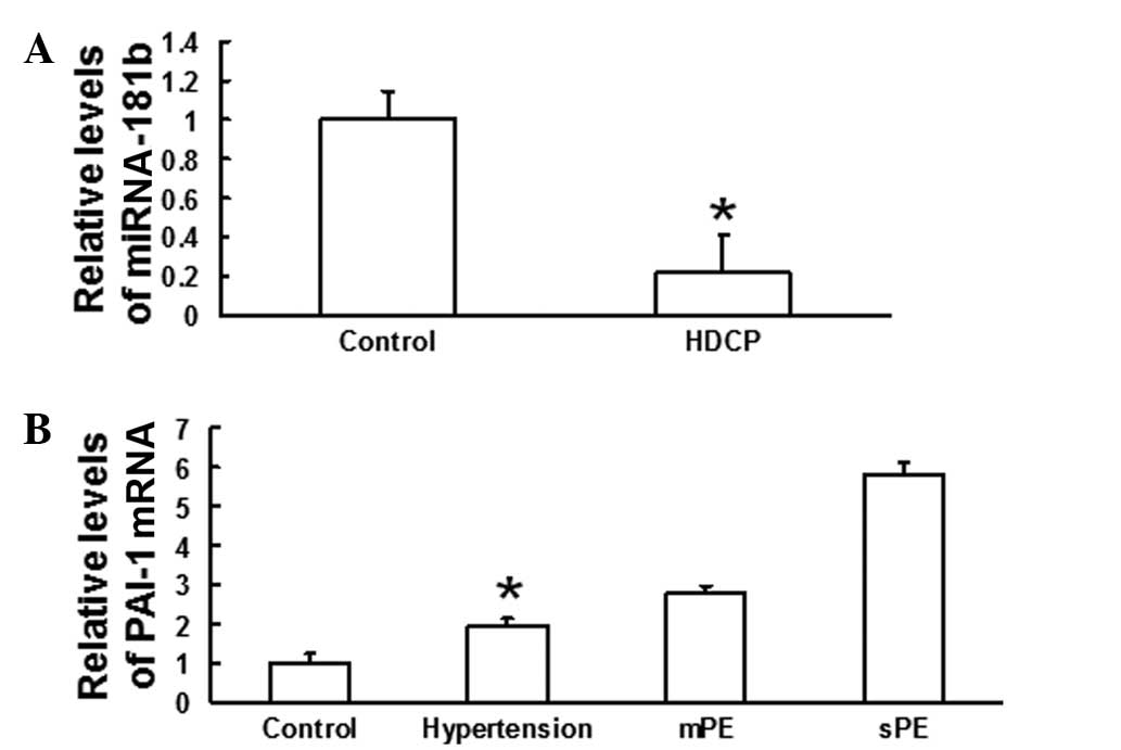
HDCP reduces the levels of miRNA-181b but
increases the levels of PAI-1 mRNA in placental tissue
To determine the levels of miRNA-181b and PAI-1 mRNA
in the placental tissue from pregnant females in the HDCP and
control groups, RT-qPCR was performed. The results revealed that
the level of miRNA-181b in the placental tissue from the HDCP group
was significantly lower than that in the placental tissue from the
control group (P<0.05; Fig.
1A). By contrast, the level of PAI-1 mRNA in the placental
tissue from the HDCP group was higher than that in the placental
tissue from the control group. The levels of PAI-1 mRNA in the
placental tissue from patients with gestational hypertension, mild
pre-eclampsia and severe pre-eclampsia increased 1.98-, 2.79- and
5.8-fold, respectively, compared with those from the control group
(Fig. 1B). These data indicated
that HDCP reduced the levels of miRNA-181b but increased the levels
of PAI-1 mRNA in placental tissue.
HDCP increases the protein expression of
PAI-1
To measure PAI-1 protein expression, western blot
analysis was performed. Western blotting revealed that the
expression of PAI-1 protein in placental tissue from the HDCP group
was significantly higher than that in placental tissue from the
normal control group (P<0.05; Fig.
2). This result suggested that HDCP increased the protein
expression of PAI-1.
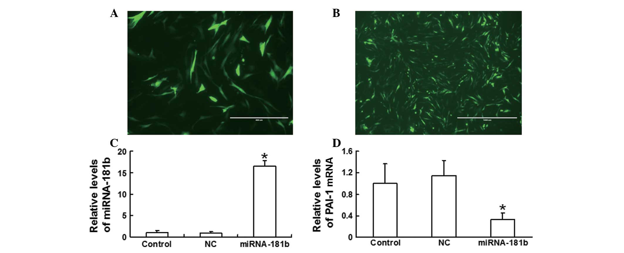
Transfection of VSMCs with the
pGCMV/EGFP/miRNA-181b plasmid enhances the levels of miRNA-181b and
reduces the levels of PAI-1 mRNA in VSMCs
To investigate the expression levels of miRNA-181b
and PAI-1 in the VSMCs, the pGCMV/EGFP/miRNA-181b plasmid was
transfected into the VSMCs and the expression of miRNA-181b was
detected using RT-qPCR. Microscopic observation following 48 h
(Fig. 3) of transfection revealed
that the fluorescence in the transfected VSMCs was distributed
evenly (Fig. 3B), with a wider
range and a higher density than that in the cells of the negative
control group (Fig. 3A). Analysis
by RT-qPCR demonstrated that the level of miRNA-181b in the VSMCs
following 48 h of transfection was significantly higher than that
in normal VSMCs (P<0.05; Fig.
3C). Furthermore, the levels of PAI-1 mRNA in the VSMCs
following 48 h of transfection were significantly lower than those
in normal VSMCs (P<0.05; Fig.
3D). These data demonstrated that transfection of VSMCs with
the pGCMV/EGFP/miRNA-181b plasmid enhanced the levels of miRNA-181b
in the VSMCs, in which the levels of PAI-1 mRNA were reduced.
Transfection of VSMCs with the
pGCMV/EGFP/miRNA-181b plasmid reduces PAI-1 protein expression
To determine PAI-1 protein expression in VSMCs
transfected with the pGCMV/EGFP/miRNA-181b plasmid, western
blotting was used to analyze the extracted proteins from VSMCs
following 72 h of transfection. The results revealed that the
protein expression of PAI-1 in VSMCs transfected with
pGCMV/EGFP/miRNA-181b was significantly lower than that in the
normal control and negative control groups (P<0.05; Fig. 4). These data indicated that
transfection of VSMCs with the pGCMV/EGFP/miRNA-181b plasmid
reduced PAI-1 protein expression.
Discussion
In the present study, the levels of miRNA-181b in
the placental tissue from the 48 patients with HDCP were lower than
those in the placental tissue of normal pregnant females. However,
the mRNA expression levels of PAI-1 in the placental tissue from
patients with HDCP were higher compared with those in the placental
tissue of normal pregnant females. In addition, the mRNA expression
levels of PAI-1 gradually increased with the development of HDCP at
different clinical stages (hypertension, mild pre-eclampsia and
severe pre-eclampsia). By contrast, the levels of miRNA-181b were
independent of the clinical stages of HDCP (data not shown).
Western blot analysis revealed that the protein
expression of PAI-1 in the HDCP group was significantly enhanced
compared with that in the control group, indicating that PAI-1
plays an important role in HDCP. By contrast, in hyperglycemia,
PAI-1 protein expression has been found to decrease (19). This inconsistency indicates that
PAI-1 protein expression may be regulated differently between
hyperglycemia and HDCP. The changes in the levels of miRNA-181b in
the placental tissue were contrary to the changes in the levels of
PAI-1, suggesting that miRNA-181b may be involved in the regulation
of PAI-1 expression.
Previous studies have demonstrated that miRNA-181
may regulate embryo implantation, placentation and decidualization
via the regulation of the focal adhesion signaling pathway
(20,21). These observations indicate that the
miR-181 family may play an important role in the development of the
embryo and placenta. Therefore, to further investigate the role of
miRNA-181b, an miRNA-181b eukaryotic expression model was
constructed by transfecting VSMCs with miRNA-181b. The RT-qPCR
results revealed that the levels of miRNA-181b increased by
16.5-fold, whereas the mRNA expression levels of PAI-1 were reduced
by 67% of the levels prior to transfection. Furthermore, the
protein expression level of PAI-1 decreased to 22% of the level
prior to transfection. These results demonstrated that PAI-1
expression was reduced when miRNA-181b expression was elevated,
indicating that miRNA-181b may be involved in the regulation of
PAI-1 expression. However, chromatin immunoprecipitation sequencing
is required prior to confirming that PAI-1 is the target gene for
miRNA-181b regulation.
In conclusion, miRNA-181b and PAI-1 were shown to be
pathologically expressed in the placental tissue of patients with
HDCP. Therefore, miRNA-181b plays an important role in the
occurrence and development of HDCP, possibly through participating
in the regulation of PAI-1, although the precise underling
mechanism requires further investigation. Thus, miRNA-181b and
PAI-1 have a potential clinical value in the early diagnosis and
prevention of HDCP.
Acknowledgements
This study was supported by The First Affiliated
Hospital of Shantou University Medical College. The authors thank
Dr Guohong Zhang for the invaluable instruction.
References
|
1
|
Zhou J, Xiao XM and Wu YH: Expression of
interferon-γ in decidual natural killer cells from women with
hypertensive disorder complicating pregnancy. J Obstet Gynaecol
Res. 40:670–676. 2014.
|
|
2
|
Yang L, Wang YL, Liu S, et al: miR-181b
promotes cell proliferation and reduces apoptosis by repressing the
expression of adenylyl cyclase 9 (AC9) in cervical cancer cells.
FEBS Lett. 588:124–130. 2014. View Article : Google Scholar : PubMed/NCBI
|
|
3
|
Shi L, Cheng Z, Zhang J, et al:
hsa-mir-181a and hsa-mir-181b function as tumor suppressors in
human glioma cells. Brain Res. 1236:185–193. 2008. View Article : Google Scholar : PubMed/NCBI
|
|
4
|
Simone TM, Archambeault J and Higgins PJ:
Small molecule targeting of PAI-1 function: a new therapeutic
approach for treatment of vascular stenosis. J Mol Genet Med.
7:10000592013. View Article : Google Scholar : PubMed/NCBI
|
|
5
|
Gomes-Giacoia E, Miyake M, Goodison S and
Rosser CJ: Targeting plasminogen activator inhibitor-1 inhibits
angiogenesis and tumor growth in a human cancer xenograft model.
Mol Cancer Ther. 12:2697–2708. 2013. View Article : Google Scholar
|
|
6
|
Boe AE, Eren M, Murphy SB, et al:
Plasminogen activator inhibitor-1 antagonist TM5441 attenuates
Nω-nitro-L-arginine methyl ester-induced hypertension and vascular
senescence. Circulation. 128:2318–2324. 2013.PubMed/NCBI
|
|
7
|
Li LX, Liu YL, Wen JG, et al: Expression
and significance of aquaporin 1 in placenta, placental membranes
and peritoneum of patients with hypertensive disorder complicating
pregnancy. Zhonghua Fu Chan Ke Za Zhi. 43:497–501. 2008.(In
Chinese).
|
|
8
|
Salmani D, Purushothaman S, Somashekara
SC, et al: Study of structural changes in placenta in
pregnancy-induced hypertension. J Nat Sci Biol Med. 5:352–355.
2014. View Article : Google Scholar : PubMed/NCBI
|
|
9
|
Okawara M, Seki H, Matsuoka K, et al:
Examination of the expression of cyclooxygenase-2 in placenta villi
from sufferers of pregnancy induced hypertension. Biol Pharm Bull.
32:2053–2056. 2009. View Article : Google Scholar : PubMed/NCBI
|
|
10
|
Henao-Mejia J, Williams A, Goff LA, et al:
The microRNA miR-181 is a critical cellular metabolic rheostat
essential for NKT cell ontogenesis and lymphocyte development and
homeostasis. Immunity. 38:984–997. 2013. View Article : Google Scholar : PubMed/NCBI
|
|
11
|
Zhu Y, Wu J, Li S, et al: The function
role of miR-181a in chemosensitivity to adriamycin by targeting
Bcl-2 in low-invasive breast cancer cells. Cell Physiol Biochem.
32:1225–1237. 2013. View Article : Google Scholar : PubMed/NCBI
|
|
12
|
Zhai XF, Fang FF, Liu Q, et al: MiR-181a
contributes to bufalin-induced apoptosis in PC-3 prostate cancer
cells. BMC Complement Altern Med. 13:3252013. View Article : Google Scholar : PubMed/NCBI
|
|
13
|
Wang XF, Shi ZM, Wang XR, et al: MiR-181d
acts as a tumor suppressor in glioma by targeting K-ras and Bcl-2.
J Cancer Res Clin Oncol. 138:573–584. 2012. View Article : Google Scholar : PubMed/NCBI
|
|
14
|
Kronski E, Fiori ME, Barbieri O, et al:
miR181b is induced by the chemopreventive polyphenol curcumin and
inhibits breast cancer metastasis via down-regulation of the
inflammatory cytokines CXCL1 and -2. Mol Oncol. 8:581–595. 2014.
View Article : Google Scholar : PubMed/NCBI
|
|
15
|
Yan JJ, Zhu XM, Yin GW, et al: Effect of
human miR-181a on function of trophoblast cell line HTR-8/SVneo.
Zhonghua Lin Chuang Yi Shi Za Zhi (Dian Zi Ban). 7:136–138.
2013.(In Chinese).
|
|
16
|
Simone TM and Higgins PJ: Low molecular
weight antagonists of plasminogen activator inhibitor-1:
therapeutic potential in cardiovascular disease. Mol Med Ther.
1:1012012. View Article : Google Scholar : PubMed/NCBI
|
|
17
|
Yang Z, Wan X, Gu Z, et al: Evolution of
the mir-181 microRNA family. Comput Biol Med. 52:82–87. 2014.
View Article : Google Scholar : PubMed/NCBI
|
|
18
|
Madonna R and De Caterina R: Cellular and
molecular mechanisms of vascular injury in diabetes - part I:
pathways of vascular disease in diabetes. Vascul Pharmacol.
54:68–74. 2011. View Article : Google Scholar : PubMed/NCBI
|
|
19
|
Cawyer RC, Horvat D, Leonard D, et al:
Hyperglycemia impairs cytotrophoblast function via stress
signaling. Am J Obstet Gynecol. May 1–2014.(Epub ahead of
print).
|
|
20
|
Su L, Liu R, Cheng W, et al: Expression
patterns of microRNAs in porcine endometrium and their potential
roles in embryo implantation and placentation. PLoS One.
9:e878672014. View Article : Google Scholar : PubMed/NCBI
|
|
21
|
Estella C, Herrer I, Moreno-Moya JM, et
al: miRNA signature and Dicer requirement during human endometrial
stromal decidualization in vitro. PLoS One. 7:e410802012.
View Article : Google Scholar : PubMed/NCBI
|