Introduction
Acute lung injury and acute respiratory distress
syndrome (ARDS) are life-threatening conditions involving acute
respiratory failure associated with extensive pulmonary infiltrates
(1–3). The infiltration of neutrophils and
activation of proinflammatory cytokines leads to the destruction of
the alveolar capillary barrier and subsequent lung fibrosis. Due to
the persistent respiratory failure, the morbidity and mortality
rates of affected patients remain high (1,3). The
reported overall mortality rate for individuals with acute lung
injury is ~30% (3), and the
currently available treatment for patients with acute lung injury
and ARDS is primarily palliative (2,4).
Erythropoietin (EPO) is the major regulator of
erythroid precursor cells. It elicits cytoprotective responses and
has been used in experiments to treat organ injury and
angiogenesis, inhibit apoptosis and fibrosis, and enhance tissue
regeneration (5–10). According to Kakavas et al
(11), advances in the
understanding of the biological and biochemical activities of EPO
may be useful in the management of patients with acute lung injury
and ARDS.
Yokomaku et al (12) revealed that the engineered EPO
derivative asialoerythropoietin (AEP), whose half-life is extremely
short, is non-hematopoietic but appears to retain
extra-hematopoietic effects and may, similarly to native EPO, be of
potential use in the treatment of patients with acute lung injury
and ARDS. In the present study a rabbit model of bleomycin-induced
acute lung injury was established to determine whether AEP
ameliorated the effects of pulmonary inflammation.
Materials and methods
Experimental procedures
The procedures in the present study were approved by
the Animal Experimentation Committee and performed according to the
Animal Care Guidelines of the Shiga University of Medical Science
(Otsu, Japan). The rabbits were anesthetized with intramuscular
injections of a mixture of ketamine hydrochloride (25 mg/kg
Ketalar® 50; Sankyo Yell Yakuhin Co., Ltd., Tokyo,
Japan) and medetomidine hydrochloride (0.1 mg/kg
Domitor®, Meiji Seika Co., Ltd., Tokyo, Japan) prior to
all experimental procedures. The rabbits were housed in a
temperature-controlled room (24±1°C) under a 12-h light/dark cycle.
Standard laboratory chow was available ad libitum.
Administration of bleomycin and AEP
Six adult female Japanese white rabbits (3.0 kg;
Japan SLC Inc., Tokyo, Japan) were randomly divided into two equal
groups (n=3). One group served as the control and was treated with
bleomycin only. At 30 min prior to the bleomycin injection, the
second group (AEP group) was pretreated with 80 ng/g AEP (Chugai
Pharmaceutical Co., Ltd., Tokyo, Japan) delivered by an intravenous
bolus injection via the auricular vein.
All rabbits were anesthetized with ketamine
hydrochloride and medetomidine hydrochloride. Bleomycin
hydrochloride (30 mg; Sigma-Aldrich, Tokyo, Japan) was dissolved in
2 ml physiological saline and 8 U/kg bleomycin was subsequently
injected into the trachea using a 22-gauge indwelling needle. To
obtain equal drug distribution throughout the lungs, four
injections of 2 U/kg each were delivered to each rabbit in the
prone and dorsal positions, on the right and left lateral
sites.
White blood cell (WBC) measurements
A total of 5 ml blood was collected from the
auricular vein of each rabbit. WBC counts were obtained prior to
any treatment and seven days post-treatment using a
Celltac-α™ analyzer (MEK-6358; Nihon Kohden, Tokyo,
Japan).
Computed tomography (CT) studies
Images were captured on a four-row multidetector CT
scanner (Toshiba Medical System Corporation, Otawara, Japan) prior
to, immediately after and seven days after the administration of
bleomycin. The scanning parameters were as follows: X-ray tube
voltage, 120 kV; X-ray tube current, 50 mA; collimation, 1 mm;
field of view, 100 mm; and helical pitch, 0.8. The CT images were
subsequently reconstructed; horizontal 1-mm cross-sections were
constructed at 5-mm intervals from the apex to the bottom of the
lungs in the lung field.
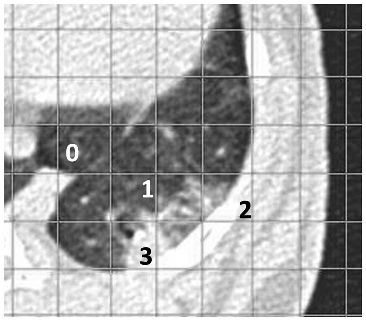
Scoring of CT images
Two of the authors analyzed the images with
Microsoft PowerPoint 2010 (Microsoft Corporation, Redmond, WA, USA)
using a 7×7 mm grid. Areas with abnormal density on the CT images,
reflective of consolidation, homogeneous ground-glass opacity and
reticulolinear shadows, were scored as a ratio of the grid as
follows: 0, normal; 1, abnormal area <1/4; 2, abnormal area ≥1/4
but <1/2; and 3, abnormal area ≥1/2 (Fig. 1).
Histopathological examination
Rabbits were sacrificed via an injection of
pentobarbital (Sumitomo Dainippon Pharma Co. Ltd., Tokyo, Japan)
into the heart on day 7 post-treatment. The lungs were resected,
fixed in formaldehyde and cut into 4-μm slices using a LEICA SM2000
R sliding microtome (Leica Microsystems, Tokyo, Japan). Consecutive
slices were mounted on glass slides and stained with hematoxylin
and eosin. One cross-section on each glass slide was selected from
the center of the craniocaudal axis in the bilateral anterior and
posterior lobes.
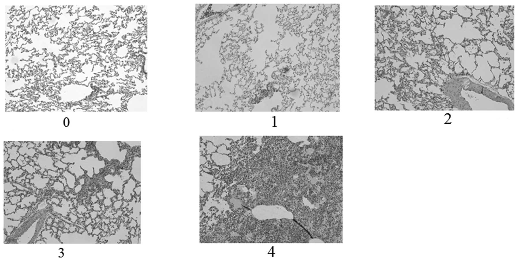
Scoring of pathological specimens
Using a Nikon ECLIPSE 90i (Nikon, Otawara, Japan),
images (magnification ×100) were evaluated by two blinded readers
who consensually scored the degree of inflammatory cell
infiltration in the alveolar wall and alveoli. A total of 10
sequential, non-overlapping fields from each lung specimen were
evaluated for inflammation as follows: 0, no inflammation; 1, focal
interstitial infiltrates; 2, diffuse interstitial infiltrates; 3,
focal alveolar infiltrates; and 4, confluent alveolar infiltrates
(Fig. 2).
Statistical analysis
SPSS 20.0 software (IBM Corp., Tokyo, Japan) was
used for data analysis. CT and pathology results of the two rabbit
groups were compared using the Student’s t-test. P<0.05 was
considered to indicate a statistically significant difference.
Results
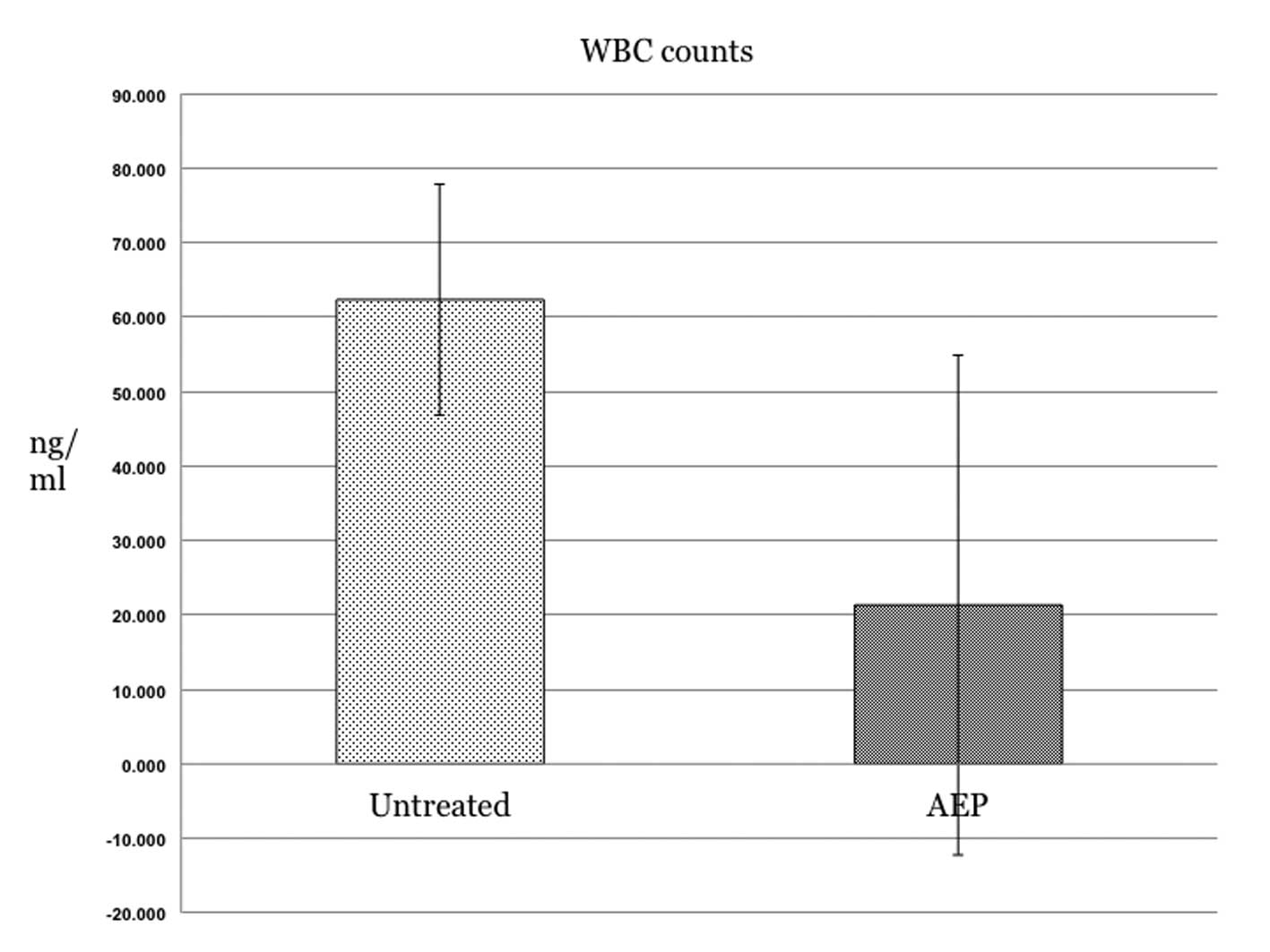
WBC count
The WBC count was lower in the AEP group than that
in the control group (21.3±33.6 vs. 62.3±15.5 ng/ml, respectively).
At P=0.127, the difference was not statistically significant
(Fig. 3).
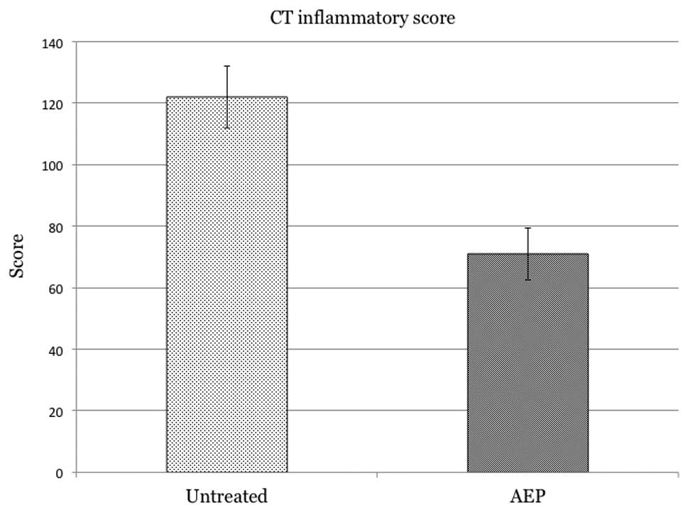
Inflammatory scores based on CT
images
The inflammation score was lower in the AEP-treated
group than that in the control group (71±8.5 vs. 122±10,
respectively; Fig. 4). The
difference between the two groups was significant (P=0.003).
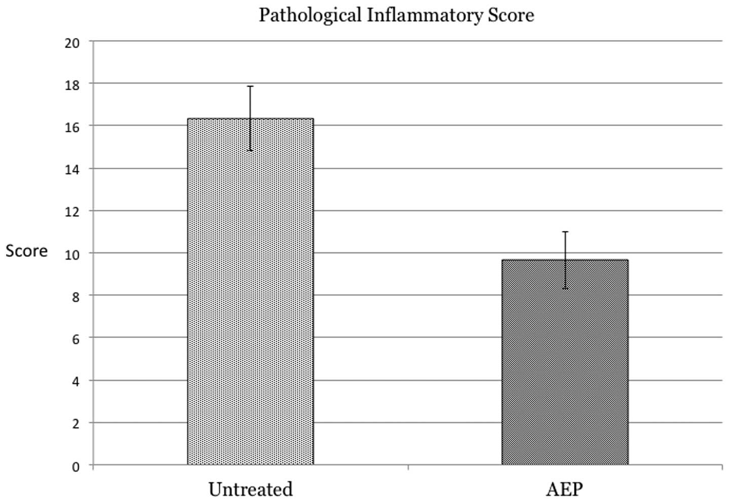
Pathological inflammatory scores
Macroscopically, the two groups did not differ in
inflammatory score. However, based on the microscopic results, the
average inflammatory score of the control rabbits was higher than
that of the AEP-treated rabbits (16.3±1.5 vs. 9.7±1.4; P=0.005;
Fig. 5).
Discussion
The present study demonstrated that AEP inhibited
the induction of inflammatory cells in the interstitial and
alveolar tissue of rabbits subjected to bleomycin-induced acute
lung injury. The direct injection of bleomycin into the airway
caused injury to the lung epithelium and endothelium and elicited
an inflammatory response (13).
Acute lung injury and ARDS have an early and late
phase. The early phase is characterized by an inflammatory response
and the late, fibroproliferative phase is characterized by collagen
deposition with tissue remodeling (14). The contribution of inflammatory
cells to acute lung injury has been previously demonstrated
(15). Activated neutrophils
release various cytotoxic mediators, including reactive oxygen
species.
In the present study, no significant difference was
observed between the WBC counts of the control and AEP-treated
rabbits. Even prior to the administration of bleomycin, two of the
six rabbits manifested high WBC counts (108 and 128 ng/ml,
respectively); however, CT studies confirmed that none had a
pre-existing inflammatory lung disease. As the rabbits were of a
mature age, the potential presence of inflammation in
extra-pulmonary organs cannot be ignored.
CT and microscopic studies revealed a significant
difference in the inflammatory scores of the control and
AEP-treated rabbits. On the CT images, the scores assigned to the
areas of abnormal density, reflective of severe alveolar and
interstitial edema and neutrophil infiltration, were significantly
lower for the AEP-treated rabbits than those for the control
animals. Microscopic inspection also revealed that the degree of
infiltration by inflammatory cells was lower in the AEP-treated
rabbits than that in the control group.
The results of the current study suggested that AEP
exerted anti-inflammatory effects on the rabbit model of
bleomycin-induced lung injury. Shang et al (8) reported that the pretreatment of rats
with EPO inhibited the production of tumor necrosis factor-α and
interleukin-1β, thereby decreasing the degree of pulmonary edema
and infiltration by neutrophils in the lung tissue. The
mechanism(s) of action of EPO and AEP may be similar. AEP may exert
ameliorative effects in the early phase of acute lung injury and
ARDS and may inhibit collagen deposition and tissue remodeling in
the late phase.
In the present study the ameliorative effects of AEP
were less pronounced than expected. AEP was administered 30 min
prior to the injection of 80 ng/g bleomycin following protocols
previously published (12,16), and the effect of this particular
time of delivery and dose of AEP requires further study.
To the best of our knowledge, no studies have been
published on the metabolism of AEP. Although Imai and Osawa
(17) observed that AEP binds to
its receptors faster than native EPO, the delivery of AEP at 30 min
prior to the administration of bleomycin may not have allowed
sufficient time for the manifestation of its ameliorative
effects.
The present preliminary study revealed that
pretreatment with AEP attenuated pulmonary inflammation in a rabbit
model of bleomycin-induced acute lung injury. Further studies are
underway to determine the role of the timing of delivery and dose
of AEP on its effects and to examine whether AEP may be a potential
therapeutic agent to treat acute lung injury. In practice, since
the mortality rate is high in elderly patients with ARDS, an
AEP-based treatment may be effective at improving the survival rate
of patients.
References
|
1
|
Saguil A and Fargo M: Acute respiratory
distress syndrome: diagnosis and management. Am Fam Physician.
85:352–358. 2012.
|
|
2
|
Cortés I, Peñuelas O and Esteban A: Acute
respiratory distress syndrome: evaluation and management. Minerva
Anestesiol. 78:343–357. 2012.
|
|
3
|
Brun-Buisson C, Minelli C, Bertolini G, et
al: Epidemiology and outcome of acute lung injury in European
intensive care units. Results from the ALIVE study. Intensive Care
Med. 30:51–61. 2004. View Article : Google Scholar : PubMed/NCBI
|
|
4
|
Ware LB: Clinical year in review IV: Acute
respiratory distress syndrome, radiology in the intensive care
unit, nonpulmonary critical care, and pulmonary infections in the
immunocompromised host. Proc Am Thorac Soc. 5:755–760.
2008.PubMed/NCBI
|
|
5
|
Mofidi A, Bader A and Pavlica S: The use
of erythropoietin and its derivatives to treat spinal cord injury.
Mini Rev Med Chem. 11:763–770. 2011. View Article : Google Scholar : PubMed/NCBI
|
|
6
|
Rjiba-Touati K, Ayed-Boussema I, Belarbia
A, Achour A and Bacha H: Recombinant human erythropoietin prevents
cisplatin-induced genotoxicity in rat liver and heart tissues via
an antioxidant process. Drug Chem Toxicol. 35:134–140. 2012.
View Article : Google Scholar
|
|
7
|
Watanabe D, Suzuma K, Matsui S, et al:
Erythropoietin as a retinal angiogenic factor in proliferative
diabetic retinopathy. N Engl J Med. 353:782–792. 2005. View Article : Google Scholar : PubMed/NCBI
|
|
8
|
Shang Y, Wu Y, Yao S, et al: Protective
effect of erythropoietin against ketamine-induced apoptosis in
cultured rat cortical neurons: involvement of PI3K/Akt and GSK-3
beta pathway. Apoptosis. 12:2187–2195. 2007. View Article : Google Scholar : PubMed/NCBI
|
|
9
|
Wang W, Horner DN, Chen WL, Zandstra PW
and Audet J: Synergy between erythropoietin and stem cell factor
during erythropoiesis can be quantitatively described without
co-signaling effects. Biotechnol Bioeng. 99:1261–1272. 2008.
View Article : Google Scholar : PubMed/NCBI
|
|
10
|
Srisawat N, Manotham K and Eiam-Ong S,
Katavetin P, Praditpornsilpa K and Eiam-Ong S: Erythropoietin and
its non-erythropoietic derivative: do they ameliorate renal
tubulointerstitial injury in ureteral obstruction? Int J Urol.
15:1011–1017. 2008. View Article : Google Scholar
|
|
11
|
Kakavas S, Demestiha T, Vasileiou P and
Xanthos T: Erythropoetin as a novel agent with pleiotropic effects
against acute lung injury. Eur J Clin Pharmacol. 67:1–9. 2011.
View Article : Google Scholar : PubMed/NCBI
|
|
12
|
Yokomaku Y, Sugimoto T, Kume S, et al:
Asialoerythropoietin prevents contrast-induced nephropathy. J Am
Soc Nephrol. 19:321–328. 2008. View Article : Google Scholar
|
|
13
|
Haslett C, Shen AS, Feldsien DC, et al:
111Indium-labeled neutrophil migration into the lungs of
bleomycin-treated rabbits assessed noninvasively by external
scintigraphy. Am Rev Respir Dis. 140:756–763. 1989. View Article : Google Scholar
|
|
14
|
Lamy ML, Fallat RJ, Koeniger EL, et al:
Pathophysiology of adult respiratory distress syndrome. Acta
Anaesthesiol Belg. 23(Suppl): 64–77. 1975.
|
|
15
|
Abraham E: Neutrophils and acute lung
injury. Crit Care Med. 31:S195–S199. 2003. View Article : Google Scholar : PubMed/NCBI
|
|
16
|
Nakazawa J, Isshiki K, Sugimoto T, et al:
Renoprotective effects of asialoerythropoietin in diabetic mice
against ischaemia-reperfusion-induced acute kidney injury.
Nephrology (Carlton). 15:93–101. 2010. View Article : Google Scholar : PubMed/NCBI
|
|
17
|
Imai Y and Osawa T: Elevation of the
activities of glycosyl transferases involved in
polylactosaminoglycan biosynthesis in autoimmune MRL lpr/lpr mouse
T cells. Mol Immunol. 27:335–342. 1990. View Article : Google Scholar : PubMed/NCBI
|