Introduction
Incontinentia pigmenti (IP), also known as
Bloch-Sulzberger syndrome or nuclear factor-κB essential modulator
(NEMO) syndrome, is an uncommon skin disorder characterized by an
X-linked dominant inheritance in the majority of cases (1). This syndrome is known as a
multi-systemic disease, which seriously affects skin, teeth, hair
and the central nervous system. Among these clinical signs, the
most notable symptom during the neonatal period is skin lesions,
which are characterized by a linear pattern of erythema with
vesicles and bullae appearing on the extremities and trunk
following birth.
The pathogenesis of IP syndrome has been defined as
a primary immunodeficiency with immune and non-immune
manifestations (2). Previous
clinical studies have reported the involvement of IP syndrome in
inflammatory and autoimmune diseases, including atypical
enterocolitis, Behçet’s syndrome and rheumatoid arthritis (3–5). The
immunodeficiency is particularly severe in surviving males, who are
typically affected by rare types of NEMO mutations that differ from
the more common deletion. Therefore, this mode of inheritance is
associated with male lethality in utero, and the majority of
live adult patients are female.
Since IP is a rare syndrome, multicenter or
large-scale clinical trials are impracticable. Several studies in
China have reported IP syndrome in neonates (6,7);
however, a number of these studies lacked a systematic summary and
detailed information of the different cases. Therefore, in the
present study, six cases from the Nanjing Children’s Hospital of
Nanjing Medical University (Nanjing, China) were described and the
available cases of neonatal IP in China were analyzed. The results
from this study may reflect the characteristics of the Chinese
population, in addition to providing useful information for
pediatricians.
Materials and methods
Data collection
A total of six patients diagnosed with IP in the
Department of Neonates were reviewed, and the data were compared
with those from previous studies in China. The Institutional Review
Board at Nanjing Children’s Hospital of Nanjing Medical University
approved this study. Parental informed consent was obtained using
an Institutional Review Board approved consent form. The diagnosis
of IP was confirmed in all cases using the diagnostic criteria
presented by Landy and Donnai (1),
including clinical manifestations, laboratory data and imaging
information. All previous cases were reported in Chinese journals
between January 1993 and December 2013. These reports were found
through a search using three Chinese medical journal search engines
[Cqvip (http://www.cqvip.com/), Wanfang Data
(http://www.wanfangdata.com/) and Science
China (http://www.scichina.com/)] using the
keywords ‘Incontinentia Pigmenti’ and ‘Infant’, and were then
retrospectively analyzed.
Results
Report of six cases
History
Between November 2003 and November 2013, six
patients with IP, including one male, were referred to the Nanjing
Children’s Hospital of Nanjing Medical University due to different
types of skin eruption (Tables I
and II). Age on admission ranged
between 4 h and 28 days. Gestational age and birth weight varied
between 37 and41 weeks, and 2,600 and 4,150 g, respectively. No
significant family history of IP was noted in any of the six
patients. The mothers of patients 2, 4 and 6 had abnormal amniotic
fluid, including oligohydramnios, polyhydramnios or turbidity. In
addition, the mothers of patients 2 and 6 suffered from influenza
and lupus separately during pregnancy. The mothers of patients 3
and 6 had a positive history of repeated abortion.
 | Table IAuxiliary examination of six
incontinentia pigmenti cases in the Department of Neonates (Nanjing
Children’s Hospital of Nanjing Medical University). |
Table I
Auxiliary examination of six
incontinentia pigmenti cases in the Department of Neonates (Nanjing
Children’s Hospital of Nanjing Medical University).
| Case no. | Eye | Brain ultrasound (ICH
stage) | Brain CT/MRI | EEG | X-raya | Skin biopsy | CSF | HIF, IgM/G/A
(g/l) | CIF, T/NK/B (%) | AAbs | TORCH | On admission | At discharge |
|---|
|
|
|---|
| WBC
(109/l) | EOS
(109/l) | CRP (mg/dl) | WBC
(109/l) | EOS
(109/l) | CRP (mg/dl) |
|---|
| 1 | N | II | N | N | N | T | N | 0.15/1.84/0.048 | 79.80/15.13/3.99 | NR | CMV (+) | 19.90 | 3.36 | N | 9.59 | 0.50 | N |
| 2 | N | II | N | N | N | T | N | NR | NR | (+) | (−) | 15.37 | 2.37 | N | 13.12 | 0.81 | N |
| 3 | N | II | N | N | N | T | N | NR | NR | (−) | CMV (+) | 14.01 | NR | N | NR | NR | NR |
| 4 | N | III | Swelling | Abnormal | N | T | N | NR | NR | (−) | (−) | 9.25 | 0.95 | N | 13.98 | 4.61 | N |
| 5 | Ret | II | N | N | N | T | N | 0.17/7.79/0.058 | 77.70/2.25/14.94 | (+) | (−) | 13.63 | 0.70 | N | 16.12 | 1.11 | 11 |
| 6 | N | I–II | N | N | N | T | N | 0.17/5.47/0.064 | 82.90/6.17/4.33 | (+) | (−) | 21.13 | 5.33 | N | 17.91 | 3.82 | 9 |
| Table IIClinical manifestations, outcomes and
prognosis of six incontinentia pigmenti cases in the Department of
Neonates (Nanjing Children’s Hospital of Nanjing Medical
University). |
Table II
Clinical manifestations, outcomes and
prognosis of six incontinentia pigmenti cases in the Department of
Neonates (Nanjing Children’s Hospital of Nanjing Medical
University).
| Case no. | Agea (days) | Gender | BW (g) | GA
(weeks+days) | Onset | Amniotic fluid | Maternal
health | Abortion
history | Familial
history | Alopecia | Skin lesion
(stage) | Seizure | Muscular
tension | Complication | Follow-up
(months) | Prognosis |
|---|
| 1 | 28 | M | 2700 | 37 | In
utero | Normal | Normal | - | - | (−) | II | (−) | Normal | MD; CHD | 5.0 | Alive |
| 2 | 4 | F | 2600 |
39+6 | In
utero | Oligohydramnios;
turbidity | Influenza;
lupus | | | (+) | I | (−) | Reduced | Lupus; CI | 6.0 | Alive |
| 3 | 21 | F | 3400 | 39 | In
utero | NR | Normal | Twice | Healthy (3
males) | (−) | II | (−) | Normal | Sepsis | 3.5 | Alive |
| 4 | 4 | F | 4150 | 40 | In
utero | Polyhydramnios;
turbidity | Normal | - | Healthy (1
male) | (−) | I | (+) | Reduced | CHD; sepsis | 2.0 | Alive;
recurrences |
| 5 | 17 | F | 2900 | 41 | In
utero | Normal | Normal | - | - | (−) | II | (−) | Normal | InI; LFD | 14.0 | Dead |
| 6 | 10 | F | 3600 |
38+4 | In
utero | Oligohydramnios;
turbidity | Lupus | Thrice | - | (−) | I | (−) | Normal | MD; CHD | 9.0 | Alive;
recurrences |
Clinical manifestations
Three patients showed typical vesicular-stage skin
changes on admission, with linear or ink-like vesicular eruption on
the extremities from birth. This skin lesion often contained
translucent or transparent material inside, and was surrounded by
redness and swelling on the surface of skin. A number of the
eruptions merged together or ulcerated with exudation. The main
manifestations in the other three infants were verrucous lesions on
admission. A few blisters were observed, as well as verrucous
hyperplasia with thickening of the texture and surface. Subsequent
skin biopsies all showed typical histological changes, including
aggregation of eosinophils (EOSs). In addition, the rash imprint
and smear exhibited infiltration of numerous EOSs (10.3–22.0%; data
not shown).
In addition to skin lesions, neurological changes,
ocular abnormalities and alopecia were the three other key symptoms
that were observed during the physical examination. Patient 4
suffered from several transient seizures following admission (at 4
days of age). The seizures were of the grand mal type, with mouth
twitching, staring eyes and stiffness of extremities. Decreased
muscle tone was found in patients 2 and 4. Alopecia was also
observed in patient 2, who showed sparse, thinning hair and
eyebrows following birth. Patient 5 was diagnosed with retinopathy
following a detailed fundus examination. All six patients had
varying degrees of complications. Patients 1 and 6 had myocardial
damage and congenital heart disease, while patients 2, 3, 4 and 5
suffered from infections of varying severity. Notably, patient 2
additionally suffered from an autoimmune disease, lupus.
Auxiliary examination
A series of auxiliary examinations associated with
IP were performed following admission. Routine chest and abdominal
X-rays showed normal results in these six infants. Although
cerebrospinal fluid tests were negative, brain ultrasounds revealed
intracranial hemorrhages (ICHs) in all six patients. Notably, as
well as ICH (stage III), high signal intensity lesions of the brain
stem and frontal and occipital lobes were observed in patient 4
using magnetic resonance imaging, indicating cerebral edema and
subcortical hemorrhage. The electroencephalogram revealed rhythmic
spikes and slow waves in this patient.
Compared with patients 5 and 6, patient 1 showed
lower immunoglobulin G levels and a higher natural killer cell
ratio, as indicated by humoral immune function and cellular immune
function tests. White blood cell (WBC) and EOS counts were recorded
on admission and at discharge. WBC counts on admission ranged
between 9.25×10⁹/l and 21.13×10⁹/l, and
between 9.59×10⁹/l and 17.91×10⁹/l at
discharge. EOS counts on admission ranged between
0.70×10⁹/l (5.1%) and 5.33×10⁹/l (25.2%), and
between 0.50×10⁹/l (5.2%) and 4.61×10⁹/l
(33.0%) at discharge. In patients 5 and 6, C-reactive protein
levels were normal on admission while moderately elevated at
discharge. In addition, autoantibody tests showed positive results
in patients 1, 4 and 6, and cytomegalovirus (CMV) infection was
found in patients 1 and 3 using the toxoplasmosis, rubella, CMV and
herpes simplex (TORCH) test.
Therapeutic procedure
Cod-liver oil was used for all patients to alleviate
the skin symptoms. Mupirocin ointment was also applied to prevent
cutaneous infection until discharge. In patients 1 and 2, a sharp
decrease in the EOS ratio was observed following symptomatic
treatments. For patients 3 and 4, antibiotics were administered to
treat bacterial infection according to the positive blood culture
results. In view of the myocardial damage and liver function damage
of patients 1, 5 and 6, creatine phosphate sodium and glutathione
were administered for organ protection. For patient 4,
phenobarbital and mannitol were applied together to control the
seizures and alleviate encephaledema.
Outcome and prognosis
The symptoms of the six patients were improved to
varying degrees at discharge. Patient follow-up ranged between 2
and 14 months with an average of 6.58 months. With the exception of
patient 5, the patients remained alive at the end of follow-up.
However, patients 4 and 6 presented with recurrence following
discharge.
Literature review
Distribution of neonatal IP in
China
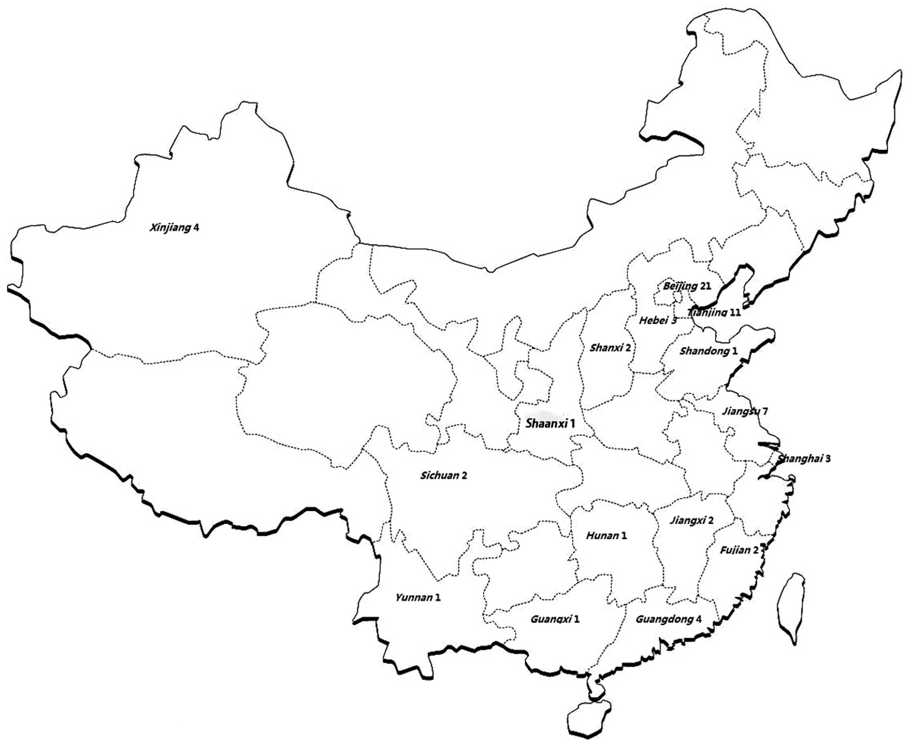
Among the 163 cases of IP reported previously in
Chinese journals, a total of 66 cases occurring during the neonatal
period were available for analysis (Tables III–V) (6–36).
Among them, 21 cases were from Beijing, 11 cases were from Tianjin,
seven cases were from Jiangsu province, four cases were from
Guangdong and Xinjiang provinces, respectively, three cases were
from Shanghai and Hebei province, respectively, two cases were from
Shanxi, Jiangxi, Fujian and Sichuan provinces, respectively, and
one case was from Yunnan, Guangxi, Hunan, Shaanxi and Shandong
provinces, respectively. The majority of the provinces and cities
were located near the eastern and southern coasts of China.
Fig. 1 shows the distribution of
the 66 cases.
| Table IIIGeneral information on 66
incontinentia pigmenti cases in China. |
Table III
General information on 66
incontinentia pigmenti cases in China.
| First author
(reference) | Location | Case no.
(year) | Gender | BW (g) | GA (weeks) | Onset (day) | Amniotic fluid | Maternal
health | Abortion
history | Familial
history |
|---|
| Cao (6) | Xinjiang | 7 (1999) | F | NR | 39 | 1 | NR | N | NR | + (Mother) |
| | 8 (1999) | F | NR | 38 | 2 | N | N | NR | − |
| Liu (7) | Tianjin | 9 (2000) | F | 2700 | 37 | 1 | N | N | − | + (Father) |
| | 10 (2000) | F | 3100 | 38 | 3 | N | N | NR | − |
| | 11 (2000) | F | 3350 | 38 | 1 | N | N | NR | − |
| | 12 (2000) | F | 2900 | 37 | 1 | N | N | − | − |
| | 13 (2000) | F | 3500 | 39 | 1 | N | N | NR | − |
| | 14 (2000) | M | 3000 | 38 | 1 | N | N | NR | − |
| Fan (8) | Shandong | 15 (2001) | F | 3000 | 39 | 1 | N | N | − | − |
| Liu (9) | Beijing | 16 (2002) | F | NR | NR | 1 | N | N | NR | NR |
| Chen (10) | Beijing | 17 (2004) | M | NR | 37 | 1 | NR | N | − | − |
| Xu (11) | Jiangxi | 18 (2004) | F | NR | 37 | 1 | NR | NR | − | NR |
| | 19 (2004) | F | NR | 37 | 1 | NR | NR | NR | NR |
| Han (12) | Beijing | 20 (2005) | M | NR | 38 | 1 | N | Diabetes | + | − |
| Song (13) | Beijing | 21 (2005) | F | NR | NR | 3 | NR | N | NR | − |
| Tang (14) | Beijing | 22 (2005) | F | 3300 | 39 | 1 | NR | NR | NR | NR |
| | 23 (2005) | F | 2800 | 38 | 1 | NR | NR | NR | NR |
| | 24 (2005) | F | 2500 | NR | 1 | NR | NR | NR | NR |
| | 25 (2005) | M | 3800 | 39 | 6 | NR | NR | NR | NR |
| | 26 (2005) | M | 3880 | 38 | 1 | NR | NR | NR | NR |
| | 27 (2005) | M | 4050 | 39 | 1 | NR | NR | NR | NR |
| Cui (15) | Yunnan | 28 (2006) | F | 2800 | 37 | 3 | NR | N | − | − |
| Liu (16) | Shanxi | 29 (2006) | F | NR | 34 | 1 | N | N | − | − |
| Zhang (17) | Beijing | 30 (2007) | F | 3310 | 41 | 1 | N | Influenza | − | − |
| Dong (18) | Hebei | 31 (2008) | F | NR | 37 | 3 | NR | N | − | − |
| He (19) | Guangdong | 32 (2008) | M | 2850 | 40 | 1 |
Oligohydramnios | N | + | − |
| | 33 (2008) | F | 3100 | 38 | 1 | Meconium
stained | Influenza | + | − |
| Zhou (20) | Xinjiang | 34 (2009) | F | NR | 37 | 4 | NR | N | − | + (Sister) |
| | 35 (2009) | F | 2500 | 37 | 1 | NR | N | − | + (Sister) |
| Tang (21) | Hunan | 36 (2009) | M | 2850 | 36 | 2 | N | N | + | − |
| Li (22) | Hebei | 37 (2009) | F | 3000 | 37 | 1 | NR | N | − | − |
| Wu (23) | Guangxi | 38 (2009) | F | 3400 | 37 | 3 | NR | N | − | − |
| Zhang (24) | Guangdong | 39 (2010) | F | 3140 | 39 | 1 | N | N | + | − |
| Luo (25) | Fujian | 40 (2010) | F | NR | NR | 1 | NR | NR | NR | NR |
| Hao (26) | Tianjin | 41 (2010) | F | 2700 | 38 | 1 | NR | N | NR | + (Mother) |
| | 42 (2010) | F | 2500 | 37 | 1 | NR | N | − | − |
| Hao (26) | Tianjin | 43 (2010) | F | 2900 | 39 | 1 | NR | N | NR | + (Sister) |
| | 44 (2010) | F | 4100 | 40 | 6 | NR | N | NR | − |
| | 45 (2010) | F | 3200 | 41 | 24 | NR | N | NR | − |
| Chen (27) | Sichuan | 46 (2010) | F | 2780 | 39 | 1 | Meconium
stained | N | + | − |
| Wang (28) | Shanghai | 47 (2010) | F | 4350 | 38 | 1 | N | N | + | − |
| | 48 (2010) | F | 2669 | 38 | 1 | N | N | − | + (Mother and
grandmother) |
| Li (29) | Shanghai | 49 (2010) | F | NR | 37 | 1 | NR | N | − | + (Mother) |
| Zhao (30) | Jiangsu | 50 (2010) | F | NR | NR | 1 | NR | NR | NR | NR |
| Yang (31) | Shanxi | 51 (2011) | F | 3310 | 41 | 2 | N | N | − | + (Sister) |
| | 52 (2011) | F | NR | 39 | 1 | N | N | − | + (Sister) |
| Huang (32) | Fujian | 53 (2012) | F | 3330 | 40 | 1 | NR | N | + | − |
| Zheng (33) | Beijing | 54 (2012) | F | 2750 | 37 | 1 | NR | NR | NR | − |
| | 55 (2012) | F | 2500 | 34 | 1 | NR | NR | NR | − |
| | 56 (2012) | F | 3310 | 41 | 3 | NR | NR | NR | − |
| | 57 (2012) | F | 3200 | 39 | 1 | NR | NR | NR | − |
| | 58 (2012) | F | 3000 | 39 | 1 | NR | NR | NR | − |
| | 59 (2012) | F | 3000 | 37 | 1 | NR | NR | NR | − |
| | 60 (2012) | F | 3150 | 37 | 1 | NR | NR | NR | + (Mother) |
| | 61 (2012) | F | 3300 | 38 | 3 | NR | NR | NR | − |
| | 62 (2012) | F | 3950 | 39 | 1 | NR | NR | NR | − |
| | 63 (2012) | F | 3100 | 37 | 1 | NR | NR | NR | − |
| Ma (34) | Hebei | 64 (2012 | F | 3000 | 38 | 1 | NR | N | − | − |
| Fang (35) | Guangdong | 65 (2013) | F | 3300 | 37 | 3 | N | N | − | +
(Grandmother) |
| Li (36) | Sichuan | 66 (2013) | F | 3400 | 38 | 1 | N | N | − | − |
| Table VSummary of the clinical
manifestations of the 66 incontinentia pigmenti cases. |
Table V
Summary of the clinical
manifestations of the 66 incontinentia pigmenti cases.
| Category | n (%) |
|---|
| Gender |
| Female | 57/66 (86.4) |
| Male | 9/66 (13.6) |
| Gestational
age |
| Term | 58/61 (95.1) |
| Pre-term | 3/61 (4.9) |
| History of
incontinentia pigmenti |
| Familial | 12/55 (21.8) |
| Sporadic | 43/55 (78.2) |
| Onset |
| <1 week | 65/66 (98.5) |
| >1 week | 1/66 (1.5) |
| Amniotic fluid |
| Normal | 22/28 (78.6) |
| Abnormality | 6/28 (21.4) |
| Abortion
history |
| Positive | 10/35 (28.6) |
| Negative | 25/35 (71.4) |
| Cutaneous
manifestations |
| Stage I | 59/90 (65.6) |
| Stage II | 28/90 (31.1) |
| Stage III | 3/90 (3.3) |
| Stage IV | 0 (0) |
| Extracutaneous
manifestations |
| Neurological | 18/66 (27.3) |
| Ocular | 9/50 (18.0) |
| Alopecia | 5/56 (8.9) |
| Histological
features |
| Typical | 64/66 (97.0) |
| Atypical | 2/66 (3.0) |
| TORCH |
| Positive | 3/29 (10.3) |
| Negative | 26/29 (89.7) |
| Autoantibodies |
| Positive | 3/10 (30.0) |
| Negative | 7/10 (70.0) |
| Blood
eosinophilia | 20/21 (95.2) |
Clinical manifestations of the 66
cases in China
Birth weight and gender. The mean gestational age at
delivery was 38.09±1.38 weeks (median, 38.09 weeks; range, 34–41
weeks). The average weight was 3,156.45±460.73 g (median, 3,156.45
g; range, 2,500–4,350 g). Among the 66 infants, three patients
(4.9%, 3/61) were <37 weeks. The patients included nine males
(13.6%) and 57 females (86.4%), and the female-to-male ratio was
6.33:1.
Family history. A total of 12 cases (21.8%, 12/55)
had a positive family history of IP. The mothers of 10 infants with
IP (28.6%, 10/35) had a history of recurrent spontaneous abortions.
The mothers of five patients (10.9%, 5/46) had infectious or
autoimmune diseases during pregnancy, and abnormal amniotic fluid
was observed in six cases (21.4%, 6/28).
Onset of symptoms. Clinical manifestations were
observed <1 week from birth in 65 cases (98.5%), and >1 week
in one case (1.5%). Cutaneous manifestations were observed at stage
I in 59 cases (65.6%), at stage II in 28 cases (31.1%) and at stage
III in three cases (3.3%) (multiple stages were recorded in certain
cases). No cutaneous manifestations were observed at stage IV.
Major manifestations of IP. In addition to the
typical skin lesions, neurological changes occurred in 18 cases
(27.3%), ocular changes were observed in 12 cases (30.0%, 12/40)
and alopecia was observed in five cases (8.9%, 5/56).
Auxiliary examination results of the 66
patients with IP
The results from the TORCH test were positive in
three cases (10.3%, 3/29). Tests for autoantibodies were positive
in three cases (30.0%, 3/10) and high blood EOS levels were
observed in 20 cases (95.2%, 20/21). Brain scans revealed positive
results in 16 cases (39.0%, 16/41), including edema and
hemorrhages.
Complications, outcomes and
follow-up
During the neonatal period, complications were
observed in 21 cases (32.3%, 21/65). A total of 34 cases were
followed for 1–6 months, six cases for 7–12 months and 17 cases for
13–84 months. Among them, 34 cases (59.6%) had no evidence of
recurrence (data not shown). Nine patients were not followed up
subsequent to discharge due to social and family factors. Five
patients, including one male, succumbed during the follow-up. IP
was found to persist in two cases after five years.
Discussion
IP is a rare hereditary disease, and the clinical
and epidemiological characteristics of IP in China remain almost
unknown compared with the fairly abundant historical data from
other countries. The incidence of IP is ~1/500,000 individuals per
year worldwide, and 50–96% of cases have a positive family history
(37). However, the specific
incidence of this disease is still not clear in China, even as a
rough estimate. Therefore, in the present study, the available data
on Chinese patients with IP were collected and analyzed.
In the present review, 60 cases were analyzed. The
majority of the IP cases were located near the eastern and southern
coasts of China, including Beijing, Tianjin and Jiangsu, Guangdong
and Hebei provinces; however, a small number of cases were also
reported in other regions. To the best of our knowledge, this is
the first report involving the distribution of IP in China. The
reason for this distribution disequilibrium is currently unknown;
however, economic and social factors, as well as the awareness and
misdiagnosis of this disease in different areas of China, may play
a role in the pathogenesis of this unusual disease in China.
Among the cases analyzed, females accounted for
86.4% (57/66), while males constituted 13.6% (9/66). Consistent
with an international study (41),
the majority of the patients were female. However, data from a
recent large-sample study in Serbia revealed a male-to-female ratio
of 91.83 to 5.85% (~15.70:1) (38), which is markedly higher than the
ratio found in the present study (6.33:1). Furthermore, in the
present study, the majority of the patients with IP were term
babies (58/61) and had a normal birth weight (51/51), suggesting
that IP syndrome is more common in term infants. The reason for
this is not clear; however, the mothers of IP patients appear to
have a higher chance of miscarriage. Therefore, the mortality of
fetuses caused by fetal death and stillbirth is higher in these
mothers, which contributes to a lower live birth rate in preterm
patients with IP. In the present study, the onset ages were almost
all <1 week (65/66), with the majority within 3 days (62/65),
which provides a good opportunity for early diagnosis and
treatment. However, international studies with accurate data
regarding onset age are lacking.
The physical condition of the mothers and the family
history were also investigated in this study. According to the
present results, abnormal maternal health was observed in five
cases (5/46). Notably, patient 2 and the mothers of patients 2 and
5 suffered from lupus erythematosus. In addition, patients 2, 5 and
6 all showed positive results for autoantibodies. As reported by
Piccoli et al (39),
chronic NEMO inhibition has an important role in the development of
complex immunological diseases, of which systemic lupus
erythematosus (SLE) is often considered the prototype. Therefore,
there may be a possible association between IP and certain
autoimmune diseases, including SLE. Similar to a previous study
(41), a history of abortion was
not uncommon in the mothers of the patients with IP (10/35).
However, as a result of the family planning policy, numerous
Chinese families volunteer to have only one child; therefore, the
birth rate is lower than that in foreign countries and the abortion
rate appeared lower than the real situation. Familial history is a
common characteristic in this syndrome, and, in the present study,
a positive rate of ≤21.8% (12/55) was observed. Cases 3 and 4 were
particularly noteworthy. These two female infants suffered from IP
while their brothers and mothers all appeared to be healthy. This
suggested that their fathers were symptomless carriers of the NEMO
gene. A similar situation was observed in the father of case 9, who
only presented with a mild cutaneous manifestation. Therefore, to a
certain degree, male carriers of the NEMO gene can have a nearly
normal quality of life. Although the specific mechanism is not yet
clear, the International IP Consortium proposed three mechanisms
for survival of these males: Hypomorphic alleles, the 47, XXY
karyotype (Klinefelter syndrome) and somatic mosaicism (40).
Among the clinical manifestations of IP, skin change
is a major diagnostic criteria. In the present study, the
constituent ratio was analyzed and skin lesions of stages I and II
were found in a large proportion of the cases. It was difficult to
distinctly separate the stages of the patients, since the skin
lesions of certain patients exhibited the characteristics of more
than one stage. Furthermore, several of the cases showed marked
melanin incontinence in the skin, which is a characteristic of
stage III and not common during the neonatal period (41). Therefore, in clinical practice it
is occasionally too difficult to separate the stages clearly based
on skin manifestation, and further discussion may be required. In
addition to skin lesions, neurological changes, ocular
abnormalities and alopecia were the other three key symptoms
observed. Hair anomalies were classified as a minor criterion for
IP (42). A total of 8.93% cases
of alopecia (5/56) were found in the present study, which was
significantly lower than the values in the literature (43,44).
This phenomenon could be explained due to hair anomalies typically
being more common in childhood. Ocular abnormalities, including
hyphema, retinal hemorrhage, retinal detachment, optic neuritis,
optic atrophy and retinopathy, were observed in 18.0% of the cases.
Although retinal anomalies are important components of the
diagnostic criteria of IP, it was found that non-retinal ocular
anomalies, such as optic neuritis and atrophy, were also not
uncommon in patients with IP. Therefore, when making the diagnosis
of atypical IP, it is possible that non-retinal ocular anomalies
should also be taken into account. Seizure was an important
clinical manifestation of neurological changes and was found in
27.27% of patients (18/66), which was near the value from previous
reports (38,45). However, it should be noted that
findings in the brain scan were quite inconsistent with the
clinical manifestations. For instance, certain patients with
apparent image abnormalities did not show neurological changes in
clinical practice (such as patients 23 and 31), and the reverse was
also common (such as patients 23 and 31). Although this
contradiction has lacked a reasonable explanation, it may be
associated with the time-point of scanning. Clinical manifestations
could be inconsistent with imaging results in the early and
recovery stages of certain diseases.
In conclusion, this study presents 66 cases of
neonatal IP in China. Among these, the majority of patients came
from the eastern and southern coasts of China. Approximately 98.5%
of IP cases occurred within <1 week of birth, particularly
within three days. The majority of the babies with IP were term
infants. The incidence of alopecia (8.93%) and the male-to-female
ratio (6.33:1) were lower than the values in the international
data. Although male patients exhibited a higher mortality rate,
they could almost have a normal quality of life. There appears to
be a potential association between IP and certain autoimmune
diseases, such as SLE and Sjögren’s syndrome. The skin lesions of
certain patients exhibited the features of two stages, and could
show marked characteristics of stage III, which is not common
during the neonatal period. Findings in the brain scan were also
relatively inconsistent with the clinical manifestations. Rare,
non-retinal ocular anomalies, such as optic neuritis and atrophy,
were also found in patients with IP. This indicated that more
considerations should be taken into account when making the
diagnosis of atypical IP. Clinicians such as dermatologists,
neonatologists and neurologists should thus be acquainted with IP,
and we suggest that the management of infants with IP requires a
skilled pediatric team with experience in the diagnosis and
management of IP in this young age group.
Acknowledgements
The authors would like to thank the parents of the
patients for their understanding, patience and endurance. The
authors are also grateful to Dr Jing-Jing Pan for her help in
preparing this manuscript.
References
|
1
|
Landy SJ and Donnai D: Incontinentia
pigmenti (Bloch-Sulzberger syndrome). J Med Genet. 30:53–59. 1993.
View Article : Google Scholar : PubMed/NCBI
|
|
2
|
Cheng LE, Kanwar B, Tcheurekdjian H, et
al: Persistent systemic inflammation and atypical enterocolitis in
patients with NEMO syndrome. Clin Immunol. 132:124–131. 2009.
View Article : Google Scholar : PubMed/NCBI
|
|
3
|
Sanz AB, Sanchez-Niño MD, Ramos AM, et al:
NF-kappaB in renal inflammation. J Am Soc Nephrol. 21:1254–1262.
2010. View Article : Google Scholar : PubMed/NCBI
|
|
4
|
Takada H, Nomura A, Ishimura M, Ichiyama
M, Ohga S and Hara T: NEMO mutation as a cause of familial
occurrence of Behçet’s disease in female patients. Clin Genet.
78:575–579. 2010.PubMed/NCBI
|
|
5
|
Brown KD, Claudio E and Siebenlist U: The
roles of the classical and alternative nuclear factor-kappaB
pathways: potential implications for autoimmunity and rheumatoid
arthritis. Arthritis Res Ther. 10:2122008. View Article : Google Scholar : PubMed/NCBI
|
|
6
|
Cao H and Liu SY: Two cases of
incontinentia pigmenti. Chinese Zhonghua Wei Chan Yi Xue Za Zhi.
3:972000.
|
|
7
|
Liu FL, Sun YE, Xu ZX and Zhang F: Six
cases of incontinentia pigmenti during neonatal period. Tianjin Yi
Xue Za Zhi. 28:563–564. 2000.
|
|
8
|
Fan CL and Liu CH: Incontinentia pigmenti.
Han Jian Shao Jian Bing Za Zhi. 4:60–61. 2001.
|
|
9
|
Liu F, Liu YH, Wang BZ, Ma DL and Fang K:
A case of incontinentia pigmenti during neonatal period. Zhonghua
Pi Fu Bing Xue Za Zhi. 6:4862002.
|
|
10
|
Chen J, Chen XX and Li RY: A case of male
incontinentia pigmenti. Lin Chuang Pi Fu Bing Za Zhi. 5:305–306.
2004.
|
|
11
|
Xu KG, Zhang LY and Li QP: Nursing care of
neonatal incontinentia pigmenti. Zhongguo You Sheng Yi Chuan Xue Za
Zhi. 12:1032004.
|
|
12
|
Han TY and Li ZL: A case of male
incontinentia pigmenti. Zhongguo You Sheng Yi Chuan Xue Za Zhi.
13:1092005.
|
|
13
|
Song Z and Li XX: A case of incontinentia
pigmenti with retinopathy. Zhongguo Yan Di Ji Bing Za Zhi.
6:4022005.
|
|
14
|
Tang ZZ, Hou XL, Zhou CL, Jiang Y and Li
JG: Clinical manifestations and follow-up of incontinentia pigmenti
during neonatal period. Shi Yong Er Ke Lin Chuang Za Zhi.
2:123–125. 2005.
|
|
15
|
Cui S, Li YF, Wu YQ, Gao J and Zhao M: A
case of incontinentia pigmenti during neonatal period. Zhongguo Xin
Sheng Er Ke Za Zhi. 21:1762006.
|
|
16
|
Liu Y and Li CX: A case of incontinentia
pigmenti with pust. Zhonghua Pi Fu Xing Bing Za Zhi. 1:47–48.
2006.
|
|
17
|
Zhang XF and Li Y: 1 case of incontinentia
pigmenti. Zhongguo Dang Dai Er Ke Za Zhi. 5:503–504. 2007.
|
|
18
|
Dong PY, Kang K, Wang J and Yao CH: A case
of incontinentia pigmenti. Zhongguo Ma Feng Bing Xue Za Zhi.
6:5012008.
|
|
19
|
He J, Zhou W, Huang XH and Lv H: Two cases
of incontinentia pigmenti during neonatal period. Zhongguo Ji Zhen
Er Ke Za Zhi. 5:495–496. 2008.
|
|
20
|
Zhou YP: Two cases of incontinentia
pigmenti in a family. Zhonghua Pi Fu Bing Xue Za Zhi.
11:7702009.
|
|
21
|
Tang JP, Shu Y and Wei Z: A case of
incontinentia pigmenti and follow-up. Zhonghua Pi Fu Bing Xue Za
Zhi. 10:7302009.
|
|
22
|
Li XL, Wang X, Sun M and Cheng YY: A case
of incontinentia pigmenti. Zhongguo Zhong Xi Yi Jie He Pi Fu Xing
Bing Xue Za Zhi. 8:2542009.
|
|
23
|
Wu CB and Zhou CL: 1 case of incontinentia
pigmenti during neonatal period. Zhongguo Yi Nan Bing Li Za Zhi.
8:5052009.
|
|
24
|
Zhang XL, Zhou XG, Song YZ and Zhao G: 1
case of incontinentia pigmenti. Guangdong Yi Xue Za Zhi.
5:6642010.
|
|
25
|
Luo MF, Wang YZ and Xie YC: Nursing care
of one newborn baby with incontinentia pigmenti. Zhongguo Hu Li Yan
Jiu Za Zhi. 7:17842010.
|
|
26
|
Hao LH, Rong LY, Ma QR and Xu Y: Clinical
characteristics of incontinentia pigmenti. Lin Chuang Jiao Dian Za
Zhi. 19:17112010.
|
|
27
|
Chen XR: Observation and one newborn
infant with incontinentia pigmenti. Zhongguo Hu Li Yan Jiu Za Zhi.
4:935–936. 2010.
|
|
28
|
Wang HB: Two cases of incontinentia
pigmenti during neonatal period. Zhonghua Wei Chan Yi Xue Za Zhi.
4:349–350. 2010.
|
|
29
|
Li XL, Shi YL, Li XJ and Liu ZY: A case of
incontinentia pigmenti during neonatal period. Zhonghua Pi Fu Xing
Bing Za Zhi. 24:6802010.
|
|
30
|
Zhao CL: Nursing care of incontinentia
pigmenti with pneumonia. Chinese General Nursing. 8:2812–2813.
2010.
|
|
31
|
Yang J: Incontinentia pigmenti in two
sisters. Zhong Yi Er Ke Za Zhi. 7:33–34. 2011.
|
|
32
|
Huang XL and Huang ZL: A case of
incontinentia pigmenti during neonatal period. Zhonghua She Qu Yi
Sheng Za Zhi. 20:2682012.
|
|
33
|
Zheng X and Liu H: 10 cases of
incontinentia pigmenti during neonatal period. Shi Yong Er Ke Lin
Chuang Za Zhi. 20:1576–1578. 2012.
|
|
34
|
Ma L: Nursing care of incontinentia
pigmenti during neonatal period. Zhongguo Hu Li Shi Jian Za Zhi.
10:575–576. 2012.
|
|
35
|
Fang SN, Li FJ, Yao YF, Du XH and Huang
XF: A case of incontinentia pigmenti during neonatal period.
Zhonghua Pi Fu Bing Xue Za Zhi. 4:2942013.
|
|
36
|
Li LL and Xiong Y: 1 case of incontinentia
pigmenti during neonatal period. Zhonghua Fu You Lin Chuang Yi Xue
Za Zhi. 9:4062013.
|
|
37
|
Orphanet Report Series. Prevalence of rare
diseases: Bibliographic data. 2013, http://www.orpha.net/uri.
Accessed Jan 24, 2014
|
|
38
|
Minić S, Trpinac D and Obradović M:
Systemic review of central nervous system anomalies in
incontinentia pigmenti. Orphanet J Rare Dis. 8:252013.
|
|
39
|
Piccoli GB, Attini R, Vigotti FN, et al:
NEMO syndrome (incontinentia pigmenti) and systemic lupus
erythematosus: a new disease association. Lupus. 21:675–681. 2012.
View Article : Google Scholar : PubMed/NCBI
|
|
40
|
Smahi A, Courtois G, Vabres P, et al:
Genomic rearrangement in NEMO impairs NF-kappaB activation and is a
cause of incontinentia pigmenti. The International Incontinentia
Pigmenti (IP) Consortium. Nature. 405:466–472. 2000. View Article : Google Scholar : PubMed/NCBI
|
|
41
|
Minić S, Trpinac D and Obradović M:
Incontinentia pigmenti diagnostic criteria update. Clin Genet.
85:536–542. 2014.PubMed/NCBI
|
|
42
|
Landy SJ and Donnai D: Incontinentia
pigmenti (Bloch-Sulzberger syndrome). J Med Genet. 30:53–59. 1993.
View Article : Google Scholar : PubMed/NCBI
|
|
43
|
Chen H: Incontinentia pigmenti. Atlas of
Genetic Diagnosis and Counseling. First edition. Humana Press Inc;
Totowa, NJ: pp. 539–544. 2006
|
|
44
|
Badgwell AL, Iglesias AD, Emmerich S and
Willner JP: The natural history of incontinentia pigmenti as
reported by 198 affected individuals. In: Abstract 38. American
College of Medical Genetics Annual Meeting; Nashville, TN. 2007
|
|
45
|
Kim BJ, Shin HS, Won CH, et al:
Incontinentia pigmenti: clinical observation of 40 Korean cases. J
Korean Med Sci. 21:474–477. 2006. View Article : Google Scholar : PubMed/NCBI
|