Introduction
Acupuncture has a long history in treating various
diseases and physiological malfunctions in Eastern medicine and is
gaining increasing popularity worldwide (1,2).
According to a review by the World Health Organization Consultation
on Acupuncture, stroke ranked highly among the ailments for which
acupuncture had proved to be effective (3). In addition, acupuncture is effective
in a broad spectrum of diseases and disorders, and is therefore
regarded as a nonspecific therapy in clinical practice (4). It is important to understand the
mechanism of acupuncture within the context of Traditional Chinese
Medicine (TCM). According to the principles of TCM, ‘qi’ is an
important constituent of the human body that is transported by the
meridian system (5). The meridian
system is a complex network of neurovascular bundles that are
characterized by distributed conductance, resistance, inductance
and capacitance as functions of time and frequency (6). In other words, qi interacts with time
and flows through the body. Based on this model, acupuncture works
by regulating the circulation of qi (7). Given the relationship among qi, time
and meridian, the method of Zi-Wu-Liu-Zhu is useful in calculating
the optimum time to administer acupuncture at a particular meridian
(8). For example, qi is strongest
on the stomach meridian of Foot-Yangming between 7:00 a.m. and 9:00
a.m., which indicates that the optimum time for acupuncture at the
Foot-Yangming is between 7:00 a.m. and 9:00 a.m. (9). Converging evidence from animal
studies (10,11) and clinical investigations (12–14)
indicates that the time to administer acupuncture determines the
effectiveness and success of the procedure. In the last five years,
time-related acupuncture has received increased focus and has been
developed as an interdiscipline of chronoacupuncture (8).
It has been suggested that the majority of
acupuncture effects are mediated via the brain (15). With the development of neuroimaging
techniques, functional magnetic resonance imaging (fMRI) is
becoming a useful method to investigate the neural mechanism of
acupuncture. An increasing number of fMRI studies have demonstrated
different brain networks for acupuncture performed at different
acupuncture points (16). Even for
a particular acupuncture point, differences were revealed between
healthy subjects and patients (17,18).
To the best of our knowledge, the neural mechanism underlying the
effect of timing on acupuncture has not been investigated. The
present study aimed to investigate brain activation patterns during
acupuncture at different time periods and to consider the timing
effects in stroke patients and healthy subjects. The acupuncture
point of Zusanli (ST36), which has been frequently used in the
treatment of stroke patients in clinical practice in China
(17,19,20),
was selected. In the literature, acupuncture stimulation at ST36 in
healthy individuals demonstrated effects in the somatosensory
(5,21) and motor (21–23)
areas, cerebellum (5,17,24),
limbic system (17,21,23–28)
and higher cognitive areas (5,17,21,23,28).
Compared with healthy controls, a weaker activation pattern of a
similar network was revealed for stroke patients (17). In the present study the timing
effect was tested by comparing acupuncture at the optimum time of
between 7:00 a.m. and 9:00 a.m. (the AM condition) and a
non-optimum time of between 3:00 p.m. and 5:00 p.m. (the PM
condition). It was hypothesized that acupuncture stimulation at
ST36 would differ during the time periods of 7:00–9:00 a.m. and
3:00–5:00 p.m., and the timing effect would differ between stroke
patients and healthy subjects.
Materials and methods
Subjects
The patient group was composed of 10 patients (five
males and five females; mean age, 58.10±9.34 years) with
subcortical ischemic stroke. The patients suffered from their
first-ever stroke >6 months prior to the enrollment. Descriptive
data for the patient group are summarized in Table I. The control group was composed of
10 healthy volunteer subjects (five males and five females; mean
age, 56.0±9.19 years). All subjects were right-handed and had no
acupuncture therapy experience. They did not have any history of
psychiatric or neurological disorders. The present study protocol
was approved by the Ethics Committee of Fujian University of
Traditional Chinese Medicine (Fuzhou, China). All subjects provided
written informed consent in accordance with the Medical Ethics
Committee of Fujian University of Traditional Chinese Medicine
(2013KY-004-02).
 | Table IDemographics of the stroke
subjects. |
Table I
Demographics of the stroke
subjects.
| | | | Ischemic
stroke |
|---|
| | | |
|
|---|
| Patient no. | FM-UL | Gender | Age (years) | Side | Area | Days after
stroke |
|---|
| 1 | 16 | M | 45 | R | Basal ganglia,
internal capsule | 221 |
| 2 | 12 | M | 66 | R | Basal ganglia,
internal capsule, corona radiate | 195 |
| 3 | 14 | F | 70 | R | Basal ganglia,
internal capsule | 243 |
| 4 | 13 | F | 57 | R | Basal ganglia,
internal capsule, corona radiate | 186 |
| 5 | 15 | F | 42 | R | Basal ganglia,
internal capsule | 210 |
| 6 | 11 | M | 69 | R | Basal ganglia,
internal capsule | 218 |
| 7 | 10 | F | 54 | R | Basal ganglia,
internal capsule, corona radiate | 237 |
| 8 | 12 | M | 59 | R | Basal ganglia,
internal capsule | 204 |
| 9 | 9 | M | 67 | R | Basal ganglia,
internal capsule, corona radiate | 188 |
| 10 | 15 | F | 52 | R | Basal ganglia,
internal capsule | 242 |
fMRI task design
The acupoint of ST36 is located on the tibialis
anterior muscle, four fingerbreadths below the lower margin of the
patella and one fingerbreadth across from the anterior crest of the
tibia (25). Manual acupuncture
manipulations at ST36 on the left leg were performed by the same
experienced and licensed acupuncturist. The stainless silver needle
was 0.3 mm in diameter and 40 mm in length. The needle was inserted
into the skin surface of ST36 at a depth of 1.0–2.0 cm with a
frequency of 2 Hz. ‘De qi’, a composite of unique sensations
(29), was to be achieved through
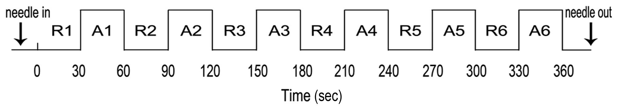
needle manipulation. The stimulation condition was alternated with
the rest condition in a block design, and each condition lasted 30
sec in duration (Fig. 1). To
ensure control was maintained for the duration of 30 sec, the
acupuncturist received instructions via audio signal through
headphones. In total, 12 blocks (six acupuncture blocks and six
rest blocks) were performed within a single session. All subjects
were scanned twice, once between 7:00 a.m. and 9:00 a.m. and again
between 3:00 p.m. and 5:00 p.m. The sequence of the two sessions
was randomized across the subjects. To avoid potential long-lasting
effects of acupuncture (30,31),
the two sessions were applied with an interval of seven days.
Following each scan, the feeling of ‘de qi’ was assessed and
checked for in each subject by interview.
fMRI acquisition
The fMRI series was acquired by a 3-T MRI machine
(GE HDXT; GE Healthcare Bio-Sciences, Pittsburgh, PA, USA). The
subjects were asked to adopt a supine position and instructed to
lie quietly and to keep their eyes closed. Foam cushions were used
to minimize head movement, and earplugs were used to reduce noise
interference. The functional T2* images were obtained
with an echo planar imaging sequence using the following
parameters: Repetition time (TR)/echo time (TE), 3,000 msec/40
msec; flip angle, 90°; field of view (FOV), 240×240 mm. Structural
T1-weighted images were obtained with a magnetization-prepared
rapid gradient echo sequence using the following parameters: TR/TE,
2,000 msec/24 msec; FOV, 240×240 mm.
fMRI data analysis
The fMRI data were analyzed using Statistical
Parametric Mapping (SPM) 8 (Wellcome Department of Imaging
Neuroscience, London, UK) and implemented in MATLAB®
(MathWorks, Natick, MA, USA). To avoid the nonequilibrium effects
of magnetization, the first eight scans were discarded.
Preprocessing included realignment of the functional time series to
correct for head movement, and subjects with head motion of >2.5
mm of translation or >2.5° of rotation in any direction were
excluded from this study. The resulting images were further
spatially normalized to the standard Montreal Neurological
Institute (MNI) space and were smoothed with a Gaussian kernel of 6
mm (full-width half-maximum).
At the individual level, the preprocessed data were
fitted to a general linear model in SPM 8 (32) using a box-car function. The
contrast of interest (acupuncture-rest) was obtained in each
subject on AM and PM occasions. In order to explore the timing
effect on acupuncture, the following contrast was also conducted at
the individual level to control for the effect of repeated
measurements (33,34):
(acupunctureAM-restAM) -
(acupuncturePM-restPM) and
(acupuncturePM-restPM) -
(acupunctureAM-restAM). At the group level,
whole brain analyses of one-sample t-tests with random effects were
performed (35). The statistical
threshold for the t-images was P<0.001 (uncorrected) at the
voxel level, with a cluster size of 20 voxels. Finally, all of the
activation maps were projected to MNI space for the identification
of regions involved in the contrasts of interest. The ‘Local Maxima
Labeling’ embedded in the automated anatomical labeling method was
used to label the peak coordinates of the blood oxygenation
level-dependent (BOLD) clusters (36).
Results
Brain activations of ST36
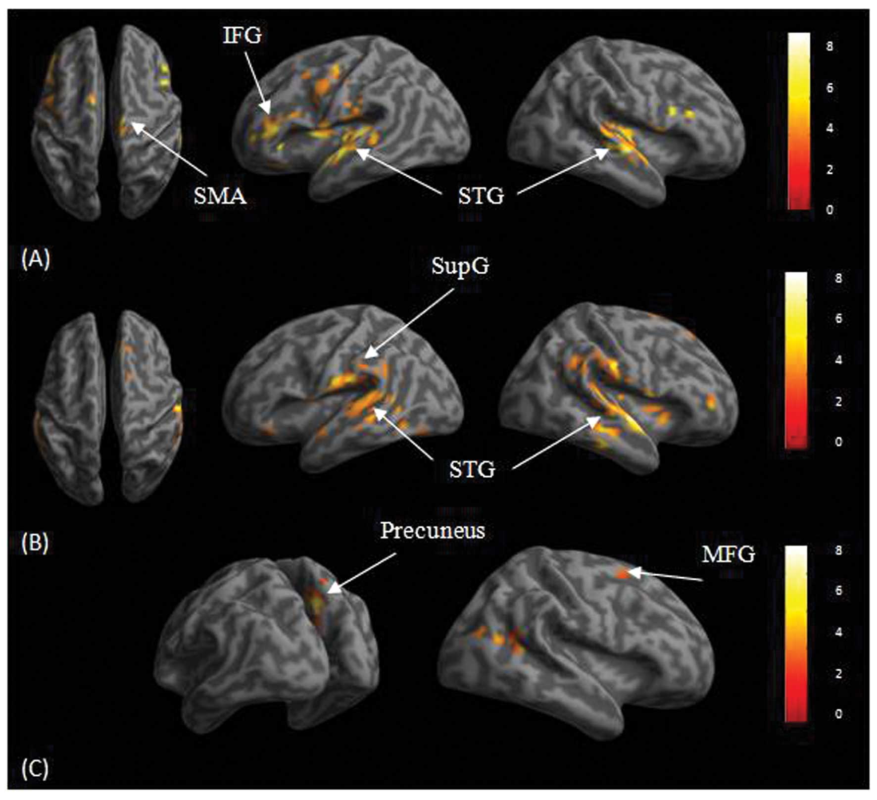
For the healthy subjects, group analyses on the AM
condition revealed activations in the bilateral superior temporal
gyrus, left hippocampus, bilateral inferior frontal gyrus, left
middle frontal gyrus and bilateral supplementary motor area
(Fig. 2). Activations were
revealed in the right superior temporal gyrus in the PM condition.
For the stroke subjects, results for the AM condition showed
activations in the bilateral superior temporal gyrus, right middle
frontal gyrus, right superior frontal gyrus, left supramarginal
gyrus, left putamen, left amygdala, right supplementary motor area
and occipital regions, including the cuneus and calcarine. Results
in the PM condition showed activations in the right precuneus,
right middle temporal gyrus and right middle frontal gyrus.
Timing effect
Group analysis for the healthy subjects revealed
stronger activations in the right middle frontal gyrus, right
superior frontal gyrus, left cingulum, right thalamus and right
cerebellum in the AM condition than those in the PM condition
(Table II). For the stroke
subjects, the results revealed stronger activations in the left
cuneus, right supplementary motor area and right inferior parietal
gyrus in the AM condition than those in the PM condition.
| Table IISummary of significant brain
activations for the longitudinal contrast of (AM-PM) and (PM-AM)
during acupuncture at the left ST36 in the healthy and patient
groups. |
Table II
Summary of significant brain
activations for the longitudinal contrast of (AM-PM) and (PM-AM)
during acupuncture at the left ST36 in the healthy and patient
groups.
| A, Healthy group
(n=10) |
|---|
|
|---|
| Coordinates x, y, z
(mm) | L/R | Label | Cluster size
(voxels) | T |
|---|
| AM>PM |
| 42, 36, 18 | R | Middle frontal
gyrus | 1076 | 8.93 |
| 6, 39, 45 | R | Superior frontal
gyrus | 777 | 8.03 |
| −12, −39, 18 | L | Posterior
cingulum | 91 | 7.68 |
| 9, −42, −15 | R | Cerebellum | 403 | 7.32 |
| 9, −30, 21 | R | Thalamus | 88 | 7.26 |
| 3, −21, −30 | R | Cerebellum | 67 | 6.35 |
| PM>AM |
| NA | | | | |
|
| B, Stroke group
(n=10) |
|
| Coordinates x, y, z
(mm) | L/R | Label | Cluster size
(voxels) | T |
|
| AM>PM |
| 3, −84, 39 | L | Cuneus | 84 | 9.40 |
| 9, 21, 45 | R | Supplementary motor
area | 95 | 6.42 |
| 54, −54, 42 | R | Inferior parietal
gyrus | 89 | 5.21 |
| PM>AM |
| NA | | | | |
Discussion
The present study investigated brain activation
patterns in healthy and stroke subjects receiving acupuncture at
the acupuncture point of ST36 during two time periods, one in the
morning (the AM condition) and one in the afternoon (the PM
condition). The main finding was a stronger activation in the AM
than in the PM condition in both subject groups. To the best of our
knowledge, this is the first neuroimaging report providing evidence
for the mechanism of chronoacupuncture.
Acupuncture of ST36 has consistently been shown to
elicit reduced neural activity in the limbic system (24,37).
Similar limbic reductions in neural activity have also been found
with other acupuncture points, such as LI4 (38), LV3 (39) and ST44 (23). These findings suggest a general
modulation effect of acupuncture on the limbic system. Given the
widespread anatomical and functional connections of the limbic
system (40), it is expected that
acupuncture affects the limbic system. The present results for the
AM condition revealed activations of the hippocampus in the healthy
subjects and putamen in the stroke subjects. The reversed pattern
of BOLD response could be attributed to different control
conditions. The studies mentioned previously used tactile
stimulation as the baseline condition (24,37),
whereas the present study used the resting state as the control
condition. In addition to limbic activations, the present results
showed activations for the AM condition in the superior temporal
gyrus, prefrontal cortex and supplementary motor area in both
healthy and stroke subjects. Prefrontal activations have been found
previously in healthy (37) and
stroke (41) subjects, suggesting
possible higher cognitive modulation effects. This is in agreement
with the theory that the cerebro-cerebellar system (42) could account for acupuncture
effects.
Notably, the present results revealed significantly
different activations following acupuncture of ST36 between the AM
and PM conditions. Stronger activations were found in the middle
and superior frontal gyrus, cingulum, thalamus and cerebellum in
the AM condition than those in the PM condition in healthy
subjects. Similar results of stronger activations in the AM
condition than those in the PM condition were revealed for the
stroke subjects: the regions included the left cuneus, right
supplementary motor area and right inferior parietal gyrus. The
results were in agreement with the theory of chronoacupuncture
stating that there is an optimum time for acupuncture to achieve
the most curative effect (8), and
this appears to apply to both healthy subjects and stroke patients.
The factor of timing in conducting acupuncture has been largely
ignored in previous neuroimaging studies, which may have
contributed to their heterogeneous results (16,43)
and low test-retest reliability (44). Although the factors of acupuncture
points, acupuncture manipulation and stimulation methods have been
investigated, the consideration of timing in conducting acupuncture
is also important.
The present results demonstrated different fMRI
activation patterns of acupuncture at ST36 at different time
periods in stroke patients and healthy subjects. Future studies
with larger sample sizes should further explore the timing effects
of acupuncture at different acupoints and in different
populations.
Acknowledgements
This study was supported by the Special Scientific
Research Fund of the Public Welfare Profession of China (no.
201307004) and the International S&T Cooperation Program of
China (no. 2011DFG33240).
Abbreviations:
|
BOLD
|
blood oxygenation level-dependent
|
|
fMRI
|
functional magnetic resonance
imaging
|
|
FM-UL
|
lower limb section of the Fugl-Meyer
Scale
|
|
IFG
|
inferior frontal gyrus
|
|
MFG
|
middle frontal gyrus
|
|
MNI
|
Montreal Neurological Institute
|
|
SMA
|
supplementary motor area
|
|
STG
|
superior temporal gyrus
|
|
SupG
|
supramarginal gyrus
|
|
TCM
|
Traditional Chinese Medicine
|
References
|
1
|
Diehl DL, Kaplan G, Coulter I, Glik D and
Hurwitz EL: Use of acupuncture by American physicians. J Altern
Complement Med. 3:119–126. 1997. View Article : Google Scholar : PubMed/NCBI
|
|
2
|
No authors listed. NIH Consensus
Conference. Acupuncture. JAMA. 280:1518–1524. 1998.PubMed/NCBI
|
|
3
|
World Health Organization. Acupuncture:
Review and Analysis of Reports on Controlled Clinical Trials. WHO
Western Pacific Region; Geneva, Switzerland: 2003
|
|
4
|
Mayer DJ: Acupuncture: an evidence-based
review of the clinical literature. Annu Rev Med. 51:49–63. 2000.
View Article : Google Scholar : PubMed/NCBI
|
|
5
|
Li L, Liu H, Li YZ, et al: The human brain
response to acupuncture on same-meridian acupoints: evidence from
an fMRI study. J Altern Complement Med. 14:673–678. 2008.
View Article : Google Scholar : PubMed/NCBI
|
|
6
|
Chang S: Physiological rhythms, dynamical
diseases and acupuncture. Chin J Physiol. 53:77–90. 2010.
View Article : Google Scholar : PubMed/NCBI
|
|
7
|
Lee MS, Lee YH, Shin BC, et al: Is there
any energy transfer during acupuncture? Am J Chin Med. 33:507–512.
2005. View Article : Google Scholar : PubMed/NCBI
|
|
8
|
Slopek A and Feng HT: Qi, time and
acupuncture. J Acupunct Tuina Sci. 7:75–79. 2009. View Article : Google Scholar
|
|
9
|
Li YM: Effect of warming-needle
acupuncture at different time of the day on superoxide dismutase
and T-cell subsets of old people. Zhong Yi Za Zhi. 22:1840–1842.
2011.(In Chinese).
|
|
10
|
Xie GG, Zhao CJ, Lu XQ, et al: Effects of
acupuncture at different Shichen (traditional twelve two-hour
periods) on serum SOD and MDA in guinea pigs. Zhongguo Zhen Jiu.
27:757–760. 2007.(In Chinese).
|
|
11
|
Wu F, Huang R and Xiong KR: Effect of
electroacupuncture intervention at different time-points in a day
on expression of c-fos and neuronal nitric oxide synthase in medial
prefrontal cortex in ketamine addiction rats. Zhen Ci Yan Jiu.
38:386–392. 2013.(In Chinese). PubMed/NCBI
|
|
12
|
Zhong XY, Su XX, Liu J and Zhu GQ:
Clinical effects of acupuncture combined with nimodipine for
treatment of vascular dementia in 30 cases. J Tradit Chin Med.
29:174–176. 2009. View Article : Google Scholar : PubMed/NCBI
|
|
13
|
Jiang H, Wang M, Guo J and Li Z: The
midnight-noon ebb-flow point selection for 30 cases of acute
ischemic cerebrovascular diseases. J Tradit Chin Med. 28:193–197.
2008. View Article : Google Scholar : PubMed/NCBI
|
|
14
|
Zhen JP, Liu C, He JZ, Wang GB, et al: The
research of brain fMRI in acupuncture of KI in different time.
Zhongguo Zhong Xi Yi Jie He Ying Xiang Xue Za Zhi. 6:325–331.
2008.(In Chinese).
|
|
15
|
Stux G and Hammerschlag R: Clinical
Acupuncture: Scientific Basis. Springer; New York, NY: 2001,
View Article : Google Scholar
|
|
16
|
Huang W, Pach D, Napadow V, et al:
Characterizing acupuncture stimuli using brain imaging with FMRI -
a systematic review and meta-analysis of the literature. PLoS One.
7:e329602012. View Article : Google Scholar :
|
|
17
|
Cho SY, Kim M, Sun JJ, et al: A comparison
of brain activity between healthy subjects and stroke patients on
fMRI by acupuncture stimulation. Chin J Integr Med. 19:269–276.
2013. View Article : Google Scholar : PubMed/NCBI
|
|
18
|
Li G, Jack CR Jr and Yang ES: An fMRI
study of somatosensory-implicated acupuncture points in stable
somatosensory stroke patients. J Magn Reson Imaging. 24:1018–1024.
2006. View Article : Google Scholar : PubMed/NCBI
|
|
19
|
Xue X, You Y, Tao J, et al:
Electro-acupuncture at points of Zusanli and Quchi exerts
anti-apoptotic effect through the modulation of PI3K/Akt signaling
pathway. Neurosci Lett. 558:14–19. 2014. View Article : Google Scholar
|
|
20
|
Tao J, Chen B, Gao Y, et al:
Electroacupuncture enhances hippocampal NSCs proliferation in
cerebral ischemia-reperfusion injured rats via activation of notch
signaling pathway. Int J Neurosci. 124:204–212. 2014. View Article : Google Scholar
|
|
21
|
Liu P, Zhou G, Zhang Y, et al: The hybrid
GLM–ICA investigation on the neural mechanism of acupoint ST36: an
fMRI study. Neurosci Lett. 479:267–271. 2010. View Article : Google Scholar : PubMed/NCBI
|
|
22
|
Cho SY, Jahng GH, Park SU, Jung WS, Moon
SK and Park JM: fMRI study of effect on brain activity according to
stimulation method at LI11, ST36: painful pressure and acupuncture
stimulation of same acupoints. J Altern Complement Med. 16:489–495.
2010. View Article : Google Scholar : PubMed/NCBI
|
|
23
|
Fang J, Jin Z, Wang Y, et al: The salient
characteristics of the central effects of acupuncture needling:
limbic-paralimbic-neocortical network modulation. Hum Brain Mapp.
30:1196–1206. 2009. View Article : Google Scholar
|
|
24
|
Hui KK, Liu J, Marina O, et al: The
integrated response of the human cerebro-cerebellar and limbic
systems to acupuncture stimulation at ST 36 as evidenced by fMRI.
Neuroimage. 27:479–496. 2005. View Article : Google Scholar : PubMed/NCBI
|
|
25
|
Bai L, Qin W, Tian J, et al: Time-varied
characteristics of acupuncture effects in fMRI studies. Hum Brain
Mapp. 30:3445–3460. 2009. View Article : Google Scholar : PubMed/NCBI
|
|
26
|
Claunch JD, Chan ST, Nixon EE, et al:
Commonality and specificity of acupuncture action at three
acupoints as evidenced by FMRI. Am J Chin Med. 40:695–712. 2012.
View Article : Google Scholar : PubMed/NCBI
|
|
27
|
Zhang WT, Jin Z, Luo F, Zhang L, Zeng YW
and Han JS: Evidence from brain imaging with fMRI supporting
functional specificity of acupoints in humans. Neurosci Lett.
354:50–53. 2004. View Article : Google Scholar
|
|
28
|
Bai L, Tian J, Zhong C, et al: Acupuncture
modulates temporal neural responses in wide brain networks:
evidence from fMRI study. Mol Pain. 6:732010. View Article : Google Scholar : PubMed/NCBI
|
|
29
|
Yang XY, Shi GX, Li QQ, Zhang ZH, Xu Q and
Liu CZ: Characterization of deqi sensation and acupuncture effect.
Evid Based Complement Alternat Med. 2013:3197342013. View Article : Google Scholar : PubMed/NCBI
|
|
30
|
Li K, Shan B, Xu J, et al: Changes in FMRI
in the human brain related to different durations of manual
acupuncture needling. J Altern Complement Med. 12:615–623. 2006.
View Article : Google Scholar : PubMed/NCBI
|
|
31
|
Yan B, Li K, Xu J, et al:
Acupoint-specific fMRI patterns in human brain. Neurosci Lett.
383:236–240. 2005. View Article : Google Scholar : PubMed/NCBI
|
|
32
|
Friston KJ, Holmes AP, Worsley KJ, et al:
Statistical parametric maps in functional imaging: a general linear
approach. Hum Brain Mapp. 2:189–210. 1994. View Article : Google Scholar
|
|
33
|
Poldrack RA: Imaging brain plasticity:
conceptual and methodological issues - a theoretical review.
Neuroimage. 12:1–13. 2000. View Article : Google Scholar : PubMed/NCBI
|
|
34
|
Poldrack RA and Gabrieli JD:
Characterizing the neural mechanisms of skill learning and
repetition priming: evidence from mirror reading. Brain. 124:67–82.
2001. View Article : Google Scholar : PubMed/NCBI
|
|
35
|
Holmes AP and Friston KJ:
Generalisability, random effects and population inference.
Neuroimage. 7:S7541998.
|
|
36
|
Tzourio-Mazoyer N, Landeau B,
Papathanassiou D, et al: Automated anatomical labeling of
activations in SPM using a macroscopic anatomical parcellation of
the MNI MRI single-subject brain. Neuroimage. 15:273–289. 2002.
View Article : Google Scholar : PubMed/NCBI
|
|
37
|
Napadow V, Makris N, Liu J, et al: Effects
of electroacupuncture versus manual acupuncture on the human brain
as measured by fMRI. Hum Brain Mapp. 24:193–205. 2005. View Article : Google Scholar
|
|
38
|
Hui KK, Liu J, Makris N, et al:
Acupuncture modulates the limbic system and subcortical gray
structures of the human brain: evidence from fMRI studies in normal
subjects. Hum Brain Mapp. 9:13–25. 2000. View Article : Google Scholar : PubMed/NCBI
|
|
39
|
Hui KK, Marina O, Claunch JD, et al:
Acupuncture mobilizes the brain’s default mode and its
anti-correlated network in healthy subjects. Brain Res.
1287:84–103. 2009. View Article : Google Scholar : PubMed/NCBI
|
|
40
|
Morgane PJ, Galler JR and Mokler DJ: A
review of systems and networks of the limbic forebrain/limbic
midbrain. Prog Neurobiol. 75:143–160. 2005. View Article : Google Scholar : PubMed/NCBI
|
|
41
|
Wang W, Qi JP, Xia YL, et al: The response
of human motor cortex to acupuncture of S36 and G34 as revealed by
functional MRI. Zhong Hua Wu li Yi Xue Yu Kang Fu Za Zhi.
26:472–475. 2004.(In Chinese).
|
|
42
|
Schmahmann JD: The cerebrocerebellar
system: anatomic substrates of the cerebellar contribution to
cognition and emotion. Int Rev Psychiatry. 13:247–260. 2001.
View Article : Google Scholar
|
|
43
|
Beissner F and Henke C: Methodological
problems in FMRI studies on acupuncture: a critical review with
special emphasis on visual and auditory cortex activations. Evid
Based Complement Alternat Med. 2011:6076372011. View Article : Google Scholar :
|
|
44
|
Kong J, Gollub RL, Webb JM, et al:
Test-retest study of fMRI signal change evoked by
electroacupuncture stimulation. Neuroimage. 34:1171–1181. 2007.
View Article : Google Scholar
|