Introduction
Chemotherapy is one of the primary treatments for
gastric cancer (1). Although many
new anticancer drugs and chemotherapies have been introduced, there
has been no significant progress in the treatment effect. The main
reason is that gastric cancer cells develop multidrug resistance to
chemotherapeutic drugs, which significantly limits the application
of chemotherapy drugs. 5-Fluorouracil (5-Fu) is an anti-metabolic
chemotherapeutic agent. It is the most frequently selected drug in
the clinical adjuvant chemotherapy and neoadjuvant chemotherapy of
tumors. It inhibits thymidylate synthase and thereby blocks the
transformation of deoxyuridylate into deoxythymidylate. It affects
DNA synthesis and leads to cell damage and death (2). However, the presence of drug
resistance in cancer patients reduces the efficacy of 5-Fu.
Celecoxib is a non-steroidal anti-inflammatory drug (NSAID), which
is a selective cyclooxygenase-2 (COX-2) inhibitor with
anti-inflammatory and analgesic effects (3). According to clinical and experimental
studies, celecoxib also has a role in tumor suppression; however,
the exact mechanism by which the NSAID acts as a specific antitumor
drug is unclear (4–6). Preliminary experiments of the present
study indicated that celecoxib can inhibit the proliferation of
SGC7901 human gastric cancer cells in vitro and may be
combined with 5-Fu to reduce the expression of cancer stem cell
markers such as hypoxia-inducible factor-2α (HIF-2α), ATP-binding
cassette transporter G2 (ABCG2) and octamer-binding transcription
factor 4 (Oct-4). On the basis of these previous experiments, the
current study used human gastric cancer cells transplanted in nude
mice to investigate the inhibitory effects of celecoxib on SGC7901
cell growth in vivo. Whether the combination of 5-Fu and
celecoxib is able to reduce the expression of stem cell markers
HIF-2α, ABCG2 and Oct-4 in human gastric carcinoma tumors
transplanted into nude mice and improve the resistance to 5-Fu
chemotherapy was also examined.
Materials and methods
Materials
The human gastric cancer cell line SGC7901 was given
as a gift from Professor Yuguang Feng of the Affiliated Hospital of
Weifang Medical College (Weifang, China). 5-Fu was acquired from
Jiangsu Zhenguo Pharmaceutical Co., Ltd. (Nantong, China).
Celecoxib was purchased from Pfizer (New York, NY, USA). Rabbit
anti-HIF-2α, Oct-4 and ABCG2 polyclonal antibodies were from Abcam
(HIF-2α, ab73895; Oct-4, ab18976; ABCG2, ab186770; Cambridge, MA,
USA). Immunohistochemistry kits (SP-9000) were purchased from
Zhongshan Golden Bridge Biotechnology Co., Ltd. (Beijing, China).
TRIzol reagent was from Invitrogen Life Technologies, and the
reverse transcription (RT) and polymerase chain reaction (PCR) kits
were purchased from Fermentas (Thermo Fisher Scientific). The
protein extraction kit was from Biyuntian Co. (Jiangsu, China). The
western blot enhanced chemiluminescence kit was from Thermo Fisher
Scientific. The 28 male nude mice (BALB/c nu/nu; age, 5–6 weeks)
were purchased from Beijing Weitong Lihua Laboratory Animal
Technology Co., Ltd. (Beijing, China). The mice weighed 18–22 g and
were raised in a specific pathogen-free environment.
Establishment of a tumor-bearing nude
mice model
A total of 28 male mice (BALB/c nu/nu) aged 5–6
weeks and weighing 18–22 g were used in the experiment. SGC7901
human gastric cancer cells in the logarithmic growth phase were
used to create a cell suspension with a concentration of
1×10⁷/ml. Under sterile conditions, 0.2 ml cell
suspension was inoculated subcutaneously into the nude mice, which
were continuously fed for two weeks to establish the nude mouse
model. The inoculated mice were randomly divided into four groups,
with seven mice in each group; the body weight difference between
groups was not significant. In the blank control group, an
intraperitoneal injection of saline (10 ml/kg) was performed every
other day. In experimental group 1 (the 5-Fu group), an
intraperitoneal injection of 5-Fu (60 mg/kg) was administered every
other day. In experimental group 2 (the celecoxib group), an
intraperitoneal injection of celecoxib (30 mg/kg) was given every
other day. In experimental group 3 (the combination group),
celecoxib (30 mg/kg) and 5-Fu (60 mg/kg) were administered by
injection every other day. All treatments were continued for 2
weeks. The diet, activity, urine and tumor growth of the nude mice
were observed every day. At the end of the experiment, the mice
were weighed and the tumor size was measured. Mice were sacrificed
by cervical dislocation, the tumor was stripped with scissors and
the tumor weight was documented. The procedures carried out in the
present study were approved by the Medical Ethics Committee of the
Affiliated Hospital of Weifang Medical University (Weifang,
China).
Body weight change and calculation of
tumor inhibition rate
On the day after the final administration, the
animals were sacrificed by cervical dislocation, the tumor tissue
was excised and the tumor mass was weighed using electronic scales.
The inhibition rate in each group was calculated according to the
following formula: Inhibition rate (%) = (average tumor weight in
the control group - average tumor weight in experimental
group)/average tumor weight in control group × 100. The short and
long diameters of the tumor were measured and the tumor volume was
calculated using the following formula: Tumor volume (V) = (L ×
S2)/2, where L is the long diameter and S is the short
diameter of the tumor.
Expression of HIF-2α, ABCG2 and Oct-4
mRNA and protein
Immunohistochemistry
Mice xenograft specimens from each group were cut (5
μm) and fixed with 10% formalin. The sections were
paraffin-embedded, stained with hematoxylin and eosin and observed
under a light microscope. The immunohistochemical staining of tumor
tissue from each group was conducted using an SP-9000 kit according
to the instructions of the manufacturer. HIF-2α, ABCG2 and Oct-4
antibody staining was carried out (dilution 1:50) following which
the tissues were placed in a 4°C refrigerator overnight.
3,3′-Diaminobenzidine color rendering, dehydration, transparency
were performed and the tissues were mounted with neutral gum.
Phosphate-buffered saline replaced the primary antibody to act as
the negative control. Staining for HIF-2α, Oct-4 and ABCG2 was
considered to be positive when brown particles were observed within
the cytoplasm.
RT-PCR
The TRIzol method was used for extraction of total
RNA from the tumor tissue. The RNA was dissolved in 30 μl
diethylpyrocarbonate-treated water. First-strand cDNA was reversely
synthesized, using an RT reaction system (20 μl) as follows: 9 μl
deionized water with no RNA enzyme, 2 μl template RNA, 181 μl Oligo
(dT), 4 μl 5× reaction buffer, 1 μl RNase inhibitor (20 U/μl), 2 μl
dNTP mix (10 mmol/l) and 1 μl M-MuLV RT. The reaction conditions
were as follows: 70°C for 5 min and then placed on ice; 37°C for 5
min; 37°C for 60 min; 70°C for 10 min and then placed on ice for
subsequent testing or preservation at −150°C.
The primers used were HIF-2α, forward: 5′-CTTGGA
GGGTTTCATTGCTGTGGT-3′ and reverse: 5′-GTGAAG TCAAAGATGCTGTGTCCT-3′,
with a product length of 123 bp; ABCG2, forward: 5′-CCCTTATGATGG
TGGCTTATTC-3′ and reverse: 5′-GTGAGATTGACC AACAGA CCAT-3′, with a
product length of 132 bp; Oct-4, forward:
5′-CCCGAAAGAGAAAGCGAACC-3′ and reverse: 5′-CAGAACCACACTCGGACCAC-3′,
with a product length of 151 bp; and GAPDH, forward: 5′-GCACCACCA
ACTGCTTAGCAC-3′ and reverse: 5′-GCAGCGCCA GTAGAGGCAGG-3′, with a
product length of 143 bp. In the 50-μl PCR reaction system, 1 μl
template cDNA, 1 μl each of upstream and downstream primers, 1 μl
Taq DNA polymerase, 5 μl dNTPs (2 mmol/l), 2 μl MgCl2
(25 mmol/l), 5 μl 10× PCR buffer and 34 μl ddH2O were
maintained at 94°C for 5 min; 94°C for 30 sec; 50°C for 30 sec and
72°C for 60 sec for 40 cycles. Extension was carried out at 72°C
for 10 min and 4°C for +∞. Then, 1.5% agarose gel electrophoresis
was used for identification of the product. A digital gel imaging
system was used to capture images and the optical density of the
amplification products was analyzed. Through analysis of the
HIF-2α, ABCG2 and Oct-4 mRNA and GAPDH optical density values, the
expression levels of HIF-2α, ABCG2 and Oct-4 mRNA were
evaluated.
Western blotting
The total protein of the tumor tissue was extracted.
The bicinchoninic acid assay method was used to determine the
protein concentration. SDS-PAGE gel electrophoresis was then
conducted. The proteins were placed on a cellulose membrane and
sealed with 5% skimmed milk powder at 37°C for 2 h. HIF-2α, ABCG2
and Oct-4 and β-actin antibodies were added (dilution, 1:1,000),
and the membrane was incubated overnight at 4°C. After washing the
membrane with Tris-buffered saline and Tween 20, the secondary
antibody horseradish peroxidase-labeled anti-IgG (Invitrogen Life
Technologies) was added and the membrane was incubated for a
further 2 h at 37°C. After washing the membrane, the
electroluminescence (ECL) reagent was added. X-ray film exposure,
developing and fixing were performed. LabWorks analysis software,
version 4.5 (Ultra-Violet Products, Inc., Upland, CA, USA) was used
to measure the absorbance value of the western blotted strip; the
ratio of the absorbance value of the protein of interest to that of
β-actin was considered to indicate the relative content of HIF-2,
ABCG2 and Oct-4.
Statistical analysis
Data were analyzed using the SPSS statistical
package, version 17.0 (SPSS, Inc., Chicago, IL, USA). Quantitative
data are expressed as the mean ± standard deviation. Quantitative
data were compared between groups using a Student’s t-test and
analysis of variance (ANOVA). P<0.05 was considered to indicate
a statistically significant difference.
Results
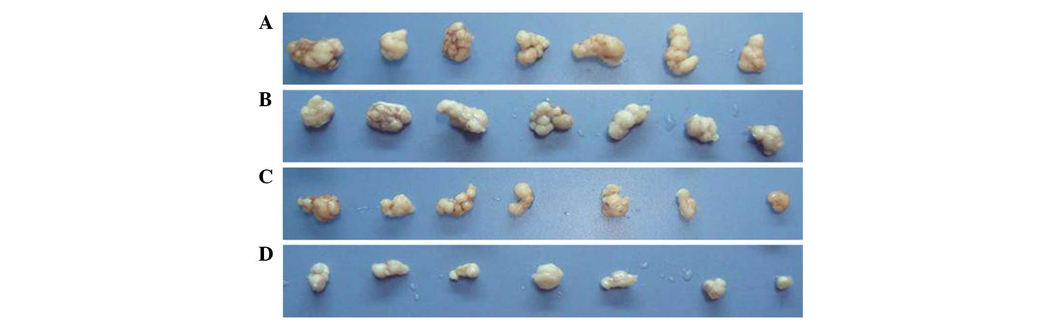
Weight change and tumor inhibition
rate
The formation of nodules was observed in the 28 nude
mice 14 days after gastric cancer cell inoculation; all grew into a
tumor. The average volumes of the tumor mass in the four groups
were not significantly different prior to the administration of the
various treatments (P>0.05). However, 15 days after the
treatment was initiated (1 day after the final day of treatment),
the average volume of the tumor mass in the 5-Fu group was less
than that in the control group, but the difference was not
significant (P>0.05). The average volumes of the tumor mass in
the celecoxib group and the combined group were significantly lower
than that in the control group (P<0.05), and the average volume
of the tumor mass in the combined treatment group was significantly
less than the volume in the 5-Fu group (P<005). Similar results
were obtained for the tumor weight. The mean tumor weight in the
5-Fu group was not statistically significant different from that in
the control group (P>0.05). The mean tumor weights in the
celecoxib and combined treatment groups were significantly lower
than that in the control group (P<0.05), and the mean tumor
weight in the combined treatment group was significantly less than
the mean weight in the 5-Fu group (P<0.05). The tumor inhibition
rates in the 5-Fu, celecoxib and combination groups were 26.36,
59.70 and 88.37%, respectively. A statistically significant
difference was identified between the combination group and the
5-Fu and celecoxib groups (Table I
and Fig. 1).
 | Table IVolume and body weight changes in each
group of xenografted nude mice following treatment. |
Table I
Volume and body weight changes in each
group of xenografted nude mice following treatment.
| Group | Tumor volume
(cm3) | Tumor weight (g) | Inhibition rate
(%) |
|---|
| Control | 0.691±0.197 | 1.287±0.274 | - |
| 5-Fu | 0.586±0.135 | 0.949±0.185 | 26.36 |
| Celecoxib | 0.255±0.035a,b | 0.523±0.146a,b | 59.70b |
| Combination | 0.101±0.031a,b | 0.153±0.023a,b | 88.37b |
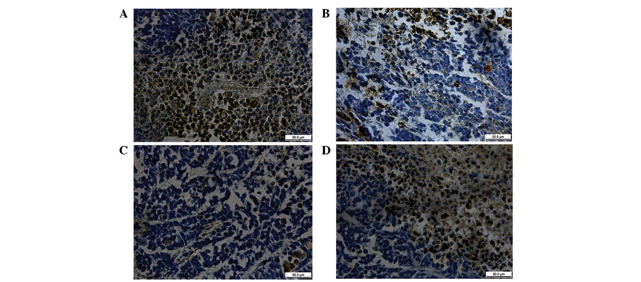
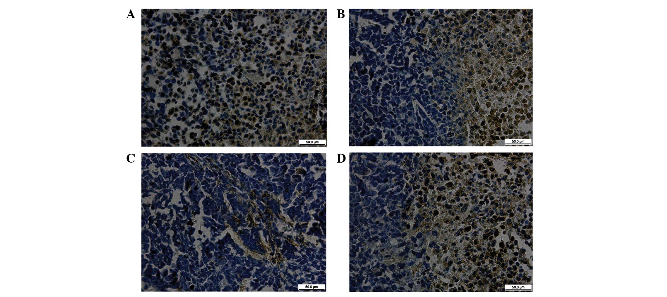
HIF-2α, ABCG2 and Oct-4 protein
expression in tumor tissue evaluated by immunohistochemical
staining
The positive expression of HIF-2α, ABCG2 and Oct-4
proteins was identified as brown granular staining. The proteins
were mainly dispersed throughout the cytoplasm and along the
nuclear membrane border in a linearly distributed manner, and a
strong positive reaction was observed in the nucleus.
For the four groups of nude mice after 14 days, the
expression levels of HIF, ABCG2 and Oct-4 proteins were highest in
the tumor tissue of the 5-Fu group, followed by the blank control
group, and the expression levels of the three proteins in the
combined and celecoxib groups were significantly reduced (Figs. 2–4).
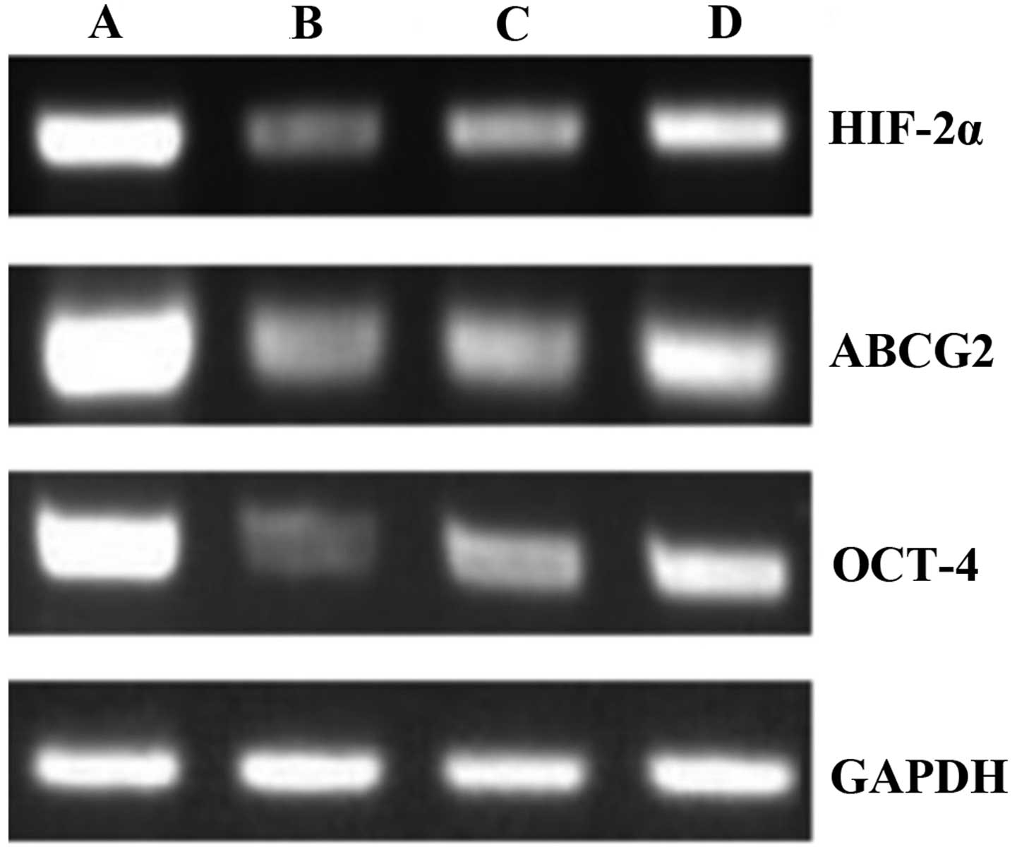
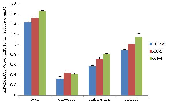
HIF-2α, ABCG2 and Oct-4 mRNA expression
in tumor tissue evaluated by RT-PCR
The RT-PCR technique was used in the four groups of
nude mice to detect the mRNA expression of HIF-2α, ABCG2 and Oct-4
in the tumor tissue after 14 days of treatment. In the control
group, which received saline every other day, HIF-2α, ABCG2 and
Oct-4 mRNA expression was observed at high levels. In the 5-Fu
group, the levels of HIF-2α, ABCG2 and Oct-4 mRNA expression were
increased by the injection of 5-Fu every other day. In the
celecoxib and combined treatment groups, the HIF-2α, ABCG2 and
Oct-4 mRNA expression levels were lower than those in the 5-Fu
group. The celecoxib and combination treatment groups showed a
significant difference from the control group when a pairwise
comparison was performed (P<0.01; Figs. 5 and 6).
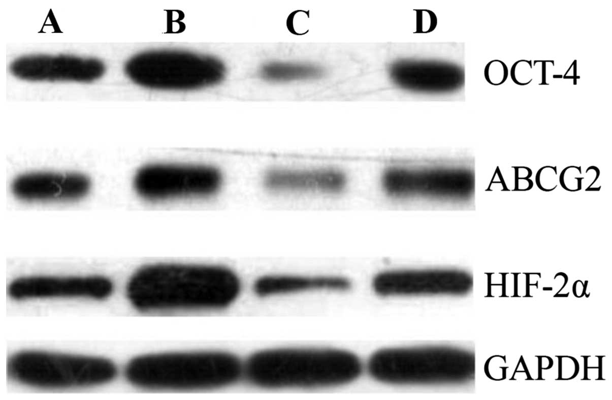
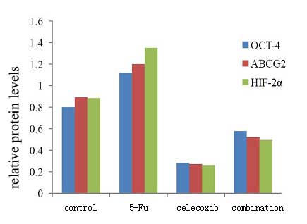
HIF-2α, ABCG2 and Oct-4 protein
expression in tumor tissue evaluated by western blotting
The western blotting technique was used to detect
the protein expression of HIF-2α, ABCG2 and Oct-4 in the tumor
tissue after 14 days of treatment. The results showed that the
HIF-2, ABCG2 and Oct-4 protein expression levels in the tumor
tissue were high in the control group, and were further increased
in the 5-Fu group. The HIF-2α, ABCG2 and Oct-4 protein expression
in the tumor tissue was significantly decreased in the celecoxib
and combination treatment groups compared with the control and 5-Fu
groups. For all four groups, a pairwise comparison was performed
(Figs. 7 and 8). The protein expression levels of
HIF-2α, ABCG2 and OCT-4 within each group were progressively lower
from the 5-Fu group (highest level), to the control group, and to
the celecoxib and combination groups (both the lowest levels; all
P<0.05).
Discussion
Gastric cancer is an disease that is seriously
harmful to human health, as it has a high morbidity and mortality.
Surgery remains the only mean possible to cure gastric cancer, but
in approximately two-thirds of cases, the patient’s condition has
reached advanced gastric cancer at the time of diagnosis (7,8). It
has a high rate of recurrence and metastasis. Chemotherapy is a
primary treatment means for gastric cancer (9). Although new anticancer drugs and
chemotherapies have been introduced, there has been no significant
progress in the effectiveness of treatment. This is primarily due
to gastric cancer cells developing multidrug resistance to
chemotherapeutic drugs, which limits the application of
chemotherapy drugs. 5-Fu is one of the most frequently selected
drugs in clinical adjuvant chemotherapy and neoadjuvant
chemotherapy. It acts as an inhibitor of thymidylate synthase, and
blocks the transformation of deoxyuridylate into deoxythymidylate,
affects DNA synthesis and leads to cell damage and death (10). Apoptosis is one of the anti-tumor
mechanisms of 5-Fu (11). In
addition, it is a cell cycle-specific drug; it can inhibit each
stage of the cell cycle, but has its optimum effects on cells in
the S phase (12). However,
resistance in cancer patients reduces the practical effect of 5-Fu.
The present study revealed that the tumor inhibition rate of 5-Fu
was only 26.36% in gastric xenografts in nude mice. When compared
with the saline-treated control group, the difference in tumor
weight was not statistically significant. The identification of
novel methods to attenuate tumor resistance to chemotherapy and to
enhance the effect of chemotherapy is necessary.
Previous studies have suggested that a very small
amount of tumor tissue has unlimited self-renewal capacity and
multi-cell proliferative potential, due to the presence of cancer
stem cells. These are considered to be the root cause of
metastasis, recurrence, drug resistance and chemotherapy resistance
(13,14). However, at present, since many
cancer stem cell markers have not yet been determined, it is not
possible to directly isolate and identify the stem cells. HIFs are
closely associated with the malignant phenotype of tumor
angiogenesis, invasion, metastasis and chemotherapy resistance
(15). For cancer stem cells, the
association with HIF-2α is closer than that with HIF-1α. HIF-2α can
regulate a variety of stem cell-related pathways to maintain the
stem cell phenotype and allow the transformation of non-stem cells
into a stem cell phenotype. ABCG2 is a member of the ABC
transporter super family, which can cause the efflux of a variety
of chemotherapy drugs. Its high level of expression has been found
to be a significant cause of multidrug resistance (16). This previous study identified that
a plurality of stem cells highly expressed ABCG2. ABCG2 is a direct
target gene of HIF-2α. The high expression of components of the
HIF-2α-ABCG2 pathway leads to MDR in cancer stem cells (17). Oct-4 is a member of the POU family
of transcription factors and totipotent or pluripotent stem cell
markers. Previous studies have suggested that Oct-4 may be closely
associated with tumor stem cells (18,19).
Covello et al (20)
reported that Oct-4 is a direct target gene of HIF-2α. Hypoxia can
activate the HIF-2α-Oct-4 pathway to maintain the tumor stem cell
phenotype. Dallas et al (21) demonstrated that compared with their
parental cells, 5-Fu-resistant colon cancer cells (HT29/5Fu-R)
highly expressed the stem cell phenotype
(CD133+/CD44+), indicating that a 5-Fu
insensitive subpopulation of cancer stem cells is the source of
resistance to chemotherapy. The preliminary results of the present
study demonstrated that under hypoxic conditions, when 5-Fu was
used to treat human gastric cancer cell lines, the expression of
cancer stem cell markers HIF-2α and ABCG2 increased (22). In the present study, following the
intraperitoneal injection of 5-Fu into gastric cancer xenografts in
nude mice, the HIF-2α, ABCG2 and Oct-4 mRNA and protein levels
increased, indicating that the gastric cancer cells were exhibiting
chemotherapy resistance to 5-Fu. This chemoresistance may be
associated with the high expression of cancer stem cell markers
HIF-2α, ABCG2 and Oct-4 and tumor stem cell promotion.
Celecoxib is a selective COX-2 inhibitor that has
anti-inflammatory and analgesic effects. Clinically, it is used for
the treatment of acute and chronic osteoarthritis and rheumatoid
arthritis. Compared with conventional NSAIDs, it has significantly
reduced gastrointestinal side-effects. Clinical and experimental
studies have shown that celecoxib also plays a role in tumor
suppression. Steinbach et al (23) performed a double-blind,
placebo-controlled clinical trial, which showed that celecoxib
inhibited the formation of familial adenomatous polyposis. Animal
experiments have shown that celecoxib has preventive and
suppressive effects on gastric cancer (24,25).
In the present study, the tumor inhibition rates in the 5-Fu,
celecoxib and combination groups were 26.36, 59.70 and 88.37%,
respectively. The inhibition rate in the combination group was
statistically significantly different from those in the 5-Fu and
celecoxib groups. This indicates that celecoxib is able to inhibit
tumor growth in vivo, specifically, in gastric cancer
transplants in nude mice. The inhibition rate in the combination
treatment group was significantly enhanced compared with that in
the 5-Fu group. Celecoxib and 5-Fu exhibited a synergistic effect
in the treatment of gastric cancer. However, the specific antitumor
mechanism of NSAIDs remains unclear. Previous cell and animal
experiments have shown that NSAIDs inhibit COX-2 activity to reduce
the synthesis of prostaglandin E2, thereby inducing tumor cell
apoptosis (26). However, Ding
et al found in a premalignant and malignant oral mucosal
cell culture model, that the potential anticancer and
apoptosis-inducing effects of celecoxib occurred via a mechanism
that was independent of COX pathways (27). Numerous other studies have
indicated that the NSAID celecoxib can promote the apoptosis of
tumor cells and achieve an antitumor effect via non-COX-2 dependent
pathways. The mechanism by which COX-2 inhibitors promote tumor
cell apoptosis has been indicated to be achieved via regulation of
the mRNA and protein expression of p21, Fas, Akt, GSK3β, FKHR,
caspase-9, bcl-2/bax, p53 and survivin genes (28–33).
The present study found that the NSAID celecoxib reduces HIF-2α,
Oct-4 and ABCG2 mRNA and protein expression in gastric cancer
tissues implanted in nude mice. This result indicates that in
addition to acting via an apoptosis-promoting pathway, celecoxib
may achieve antitumor effects by reducing the expression of the
cancer stem cell markers HIF-2α, Oct-4 and ABCG2.
In conclusion, HIF-2α, ABCG2 and Oct-4 mRNA and
protein expression levels were significantly increased in the tumor
tissues of the 5-Fu group; this may be a due to the tumor cells
having resistance to 5-Fu. However, when 5-Fu and celecoxib were
used together, compared with 5-Fu used alone, the HIF-2α, ABCG2 and
Oct-4 mRNA and protein expression levels were significantly lower,
and the difference was statistically significant. This showed that
the combined use of 5-Fu and celecoxib is able to attenuate the
resistance to chemotherapy in gastric cancer and enhance the effect
of chemotherapy by reducing the expression of HIF-2α, ABCG2 and
Oct-4 and cancer stem cells in tumor tissue xenografts in nude
mice.
Acknowledgements
This study was supported by the Science and
Technology Innovation Foundation of the Affiliated Hospital of
Weifang Medical University and by a grant from the Shandong
Province Science and Technology Program (no. 2012YD18108).
References
|
1
|
Cunningham D, Allum WH, et al: Magic Trial
Participants. Perioperative chemotherapy versus surgery alone for
resectable gastroesophageal cancer. N Engl J Med. 355:11–20. 2006.
View Article : Google Scholar : PubMed/NCBI
|
|
2
|
Longley DB, Harkin DP and Johnston PG:
5-Fluorouracil: mechanisms of action and clinical strategies. Nat
Rev Cancer. 3:330–338. 2003. View
Article : Google Scholar : PubMed/NCBI
|
|
3
|
Mathew ST, Devi SG, Prasanth VV and Vinod
B: Efficacy and safety of COX-2 inhibitors in the clinical
management of arthritis: Mini review. ISRN Pharmacol.
2011:4802912011. View Article : Google Scholar : PubMed/NCBI
|
|
4
|
Dannenberg AJ and Subbaramaiah K:
Targeting cyclooxygenase-2 in human neoplasia: rationale and
promise. Cancer Cell. 4:431–436. 2003. View Article : Google Scholar
|
|
5
|
Schönthal AH: Direct non-cyclooxygenase-2
targets of celecoxib and their potential relevance for cancer
therapy. Br J Cancer. 97:1465–1468. 2007. View Article : Google Scholar : PubMed/NCBI
|
|
6
|
Chuang HC, Kardosh A, Gaffney KJ, Petasis
NA and Schönthal AH: COX-2 inhibition is neither necessary nor
sufficient for celecoxib to suppress tumor cell proliferation and
focus formation in vitro. Mol Cancer. 7:382008. View Article : Google Scholar : PubMed/NCBI
|
|
7
|
Siegel R, Naishadham D and Jemal A: Cancer
statistics, 2013. CA Cancer J Clin. 63:11–30. 2013. View Article : Google Scholar : PubMed/NCBI
|
|
8
|
Agboola O: Adjuvant treatment in gastric
cancer. Cancer Treat Rev. 20:217–240. 1994. View Article : Google Scholar : PubMed/NCBI
|
|
9
|
Cunningham D, Allum WH, et al:
Perioperative chemotherapy versus surgery alone for resectable
gastroesophageal cancer. N Engl J Med. 355:11–20. 2006. View Article : Google Scholar : PubMed/NCBI
|
|
10
|
Longley DB, Harkin DP and Johnston PG:
5-fluorouracil: mechanisms of action and clinical strategies. Nat
Rev Cancer. 3:330–338. 2003. View
Article : Google Scholar : PubMed/NCBI
|
|
11
|
Yoshida K, Yamaguchi K, et al: Challenge
for a better combination with basic evidence. Int J Clin Oncol.
13:212–219. 2008. View Article : Google Scholar : PubMed/NCBI
|
|
12
|
Jin Mao-Lin: New progress in advanced
gastric cancer systemic chemotherapy. Waike Lilun Yushijian.
8:18–20. 2003.(In Chinese).
|
|
13
|
Reya T, Morrison SJ, Clarke MF and
Weissman IL: Stem cells, cancer, and cancer stem cells. Nature.
414:105–111. 2001. View
Article : Google Scholar : PubMed/NCBI
|
|
14
|
Jordan CT, Guzman ML and Noble M: Cancer
stem cells. N Engl J Med. 355:1253–1261. 2006. View Article : Google Scholar : PubMed/NCBI
|
|
15
|
Bertout JA, Patel SA and Simon MC: The
impact of O2 availability on human cancer. Nat Rev
Cancer. 8:967–975. 2008. View
Article : Google Scholar : PubMed/NCBI
|
|
16
|
Hu L, McArthur C and Jaffe RB: Ovarian
cancer stem-like side-population cells are tumourigenic and
chemoresistant. Br J Cancer. 102:1276–1283. 2010. View Article : Google Scholar : PubMed/NCBI
|
|
17
|
Martin CM, Ferdous A, Gallardo T, et al:
Hypoxia-inducible factor-2alpha transactivates Abcg2 and promotes
cytoprotection in cardiac side population cells. Circ Res.
102:998–1001. 2008. View Article : Google Scholar
|
|
18
|
Nichols J, Zevnik B, Anastassiadis K, et
al: Formation of pluripotent stem cells in the mammalian embryo
depends on the POU transcription factor Oct4. Cell. 95:379–391.
1998. View Article : Google Scholar : PubMed/NCBI
|
|
19
|
Tai MH, Chang CC, Kiupel M, Webster JD,
Olson LK and Trosko JE: Oct4 expression in adult human stem cells:
evidence in support of the stem cell theory of carcinogenesis.
Carcinogenesis. 26:495–502. 2005. View Article : Google Scholar
|
|
20
|
Covello KL, Kehler J, Yu H, et al:
HIF-2alpha regulates Oct-4: Effects of hypoxia on stem cell
function, embryonic development, and tumor growth. Genes Dev.
20:557–570. 2006. View Article : Google Scholar : PubMed/NCBI
|
|
21
|
Dallas NA, Xia L, Fan F, et al:
Chemoresistant colorectal cancer cells, the cancer stem cell
phenotype, and increased sensitivity to insulin-like growth
factor-I receptor inhibition. Cancer Res. 69:1951–1957. 2009.
View Article : Google Scholar : PubMed/NCBI
|
|
22
|
Zhang XQ, Feng YG and Wu MY: Effect of
5-Fu on the ratio of SP cells and expression of HIF-2α and ABCG2 in
human gastric cancer cell line SGC7901 under hypoxia. World Chinese
Journal of Digestology. 20:1813–1818. 2012.(In Chinese).
|
|
23
|
Steinbach G, Lynch PM, Phillips RK, et al:
The effect of celecoxib, a cyclooxygenase-2 inhibitor, in familial
adenomatous polyposis. N Engl J Med. 342:1946–1952. 2000.
View Article : Google Scholar : PubMed/NCBI
|
|
24
|
Kuo CH, Hu HM, Tsai PY, Wu IC, Yang SF,
Chang LL, Wang JY, Jan CM, Wang WM and Wu DC: Short-term celecoxib
intervention is a safe and effective chemopreventive for gastric
carcinogenesis based on a Mongolian gerbil model. World J
Gastroenterol. 15:4907–4914. 2009. View Article : Google Scholar : PubMed/NCBI
|
|
25
|
Rocha FT, Lourenço LG, Jucá MJ, Costa V
and Leal AT: Chemoprevention by celecoxib in reflux-induced gastric
adenocarcinoma in Wistar rats that underwent gastrojejunostomy.
Acta Cir Bras. 24:189–194. 2009. View Article : Google Scholar : PubMed/NCBI
|
|
26
|
Dandekar DS, Lopez M, Carey RI and
Lokeshwar BL: Cyclooxygenase-2 inhibitor celecoxib augments
chemotherapeutic drug-induced apoptosis by enhancing activation of
caspase-3 and-9 in prostate cancer cells. Int J Cancer.
115:484–492. 2005. View Article : Google Scholar : PubMed/NCBI
|
|
27
|
Ding H, Han C, Zhu J, Chen CS and
D’Ambrosio SM: Celecoxib derivatives induce apoptosis via the
disruption of mitochondrial membrane potential and activation of
caspase 9. Int J Cancer. 113:803–810. 2005. View Article : Google Scholar
|
|
28
|
Li Q, Peng J and Zhan GY: Effect of a
selective COX-2 inhibitor on cell proliferation and apoptosis in
human gastric cancer cell line BGC-823. Zhong Nan Da Xue Xue Bao Yi
Xue Ban. 33:1123–1128. 2008.(In Chinese).
|
|
29
|
Kim N, Kim CH, Ahn DW, et al: Anti-gastric
cancer effects of celecoxib, a selective COX-2 inhibitor, through
inhibition of Akt signaling. J Gastroenterol Hepatol. 24:480–487.
2009. View Article : Google Scholar
|
|
30
|
Wang Z, Chen H and Xia GH: Study on
celecoxib inducing gastric cancer cell apoptosis and its mechanism.
Journal of Modern Oncology. 17:416–420. 2009.(In Chinese).
|
|
31
|
Swamy MV, Herzog CR and Rao CV: Inhibition
of COX-2 in colon cancer cell lines by celecoxib increases the
nuclear localization of active p53. Cancer Res. 63:5239–5242.
2003.PubMed/NCBI
|
|
32
|
Erkinheimo TL, Lassus H, et al: Elevated
cyclooxygenase-2 expression is associated with altered expression
of p53 and SMAD4, amplification of HER-2/neu, and poor outcome in
serous ovarian carcinoma. Clin Cancer Res. 10:538–545. 2004.
View Article : Google Scholar : PubMed/NCBI
|
|
33
|
Krysan K, Merchant FH, et al:
COX-2-dependent stabilization of survivin in non-small cell lung
cancer. FASEB J. 18:206–208. 2004.
|