Introduction
Invasion and metastasis are the main characteristics
during the progression of malignant tumors, which is responsible
for the majority of cancer mortalities. Tumor dissemination may
occur through a number of pathways, among which blood vessels and
lymphatics are key components of metastatic spread. Lymph node
metastasis is an important prognostic indicator in a number of
cancer types. Numerous epithelial tumors have been characterized by
lymph node metastasis, which is often an early event in tumor
progression. Lymphatic metastasis is also a key factor associated
with tumor recurrence and prognosis. Previous studies on tumor
molecular biology have revealed that the development of a
microvascular network (angiogenesis and lymphangiogenesis) is
essential for tumor metastasis.
Vascular endothelial growth factor receptor
(VEGFR)-3 was the first cloned lymphatic marker, and is
predominantly expressed on lymphatic endothelium in adult tissues.
On binding to its ligands, VEGF-C and VEGF-D, VEGFR-3 signals for
tumor lymphangiogenesis, mediating tumor metastasis to the lymph
nodes (1,2). Therefore, the inhibition of
lymphangiogenesis is a realistic therapeutic strategy for
inhibiting tumor cell dissemination and lymphatic metastasis.
Previous studies have predominantly focused on tumor metastasis via
the blood vasculature and significant progression has been made
with regard to angiogenesis and antiangiogenesis therapy (3). However, antiangiogenesis therapy
appears to be not as efficient as predicted for the treatment of
tumor metastasis, which may be due to the networking of the blood
and lymphatic vasculatures. Blocking a single route of metastasis
is unable to inhibit the distant metastasis of tumor cells, which
may transfer between the two vasculatures (4).
In the present study, mRNA and protein expression
levels of VEGFR-3 were detected in non-small-cell lung carcinoma
(NSCLC) tissues and lymph nodes using semiquantitative reverse
transcription polymerase chain reaction (RT-PCR) and
immunohistochemisty. In addition, the microlymphatic vessel density
(MLVD) was calculated in order to analyze the correlation with
lymph node metastasis, which may be an indicator of tumor
metastasis and provide evidence for personalized therapy.
Materials and methods
Study criteria
In total, 52 patients who had been diagnosed with
primary NSCLC in Beijing Chest Hospital (Beijing, China) between
April 2006 and June 2007 were selected for the study. The patients
had not undergone any previous treatment and were aged between 29
and 77 years (mean age, 59±11 years). In total, 38 patients were
male, while 14 patients were female. The histological types of the
lung cancer tissues were classified into adenocarcinoma (20 cases),
squamous cell carcinoma (27 cases) and adenosquamous carcinoma (5
cases), according to the World Health Organization’s standards
(5). All the patients provided
informed consent. According to the postoperative pathology results,
patients with at least one lymph node metastasis were classified as
the lymph node metastasis-positive group (25 patients), while
patients without lymph node metastasis were classified into the
lymph node metastasis-negative group (27 patients). In total, 196
lymph nodes were analyzed, including 72 metastasis-positive lymph
nodes and 26 metastasis-negative lymph nodes from the lymph node
metastasis-positive group and 98 lymph nodes from the lymph node
metastasis-negative group. In the group of patients with benign
lung disease, 10 lung tissues and 8 lymph nodes were analyzed. The
study was approved by the Ethics Committee of Beijing Chest
Hospital (Beijing, China).
Reagents
TRIzol was purchased from Invitrogen Life
Technologies (Carslbad, CA, USA). PCR primers were synthesized by
Shanghai Shengwu Gongcheng Co., Ltd. (Shanghai, China), while dNTPs
and Moloney murine leukemia virus (M-MLV) reverse transcriptase
were obtained from Promega Corporation (Madison, WI, USA). RNasin
Ribonuclease Inhibitor was purchased from Huamei, while rabbit
anti-VEGFR-3 antibodies and an SP-9000 ELISA kit were purchased
from Zhongshan Jinqiao Biotechnology, Co., Ltd. (Zhongshan,
China).
RT-PCR of VEGFR-3
Tissue samples were collected within 30 min
following surgery and were stored in liquid nitrogen
immediately.
For RNA extraction, the tissues were ground in
liquid nitrogen and the RNA was extracted using TRIzol and
chloroform. The optical density at 260 and 280 nm was detected
using a UV-spectrophotometer (Shimadzu Corporation, Kyoto, Japan),
with a spectral bandwidth of 1.8–2.0. The RNA samples were also
analyzed by formaldehyde denaturing gel electrophoresis and no
degradation was detected. RNA samples were diluted to 1 μg/μl and
stored at −80°C.
For reverse transcription, 2 μg RNA template, 1 μl
oligo(dT) and RNase free H2O were placed in a
microcentrifuge tube to a final volume of 15 μl and incubated at
70°C for 5 min. The samples were centrifuged at 300 × g, 4°C for 30
sec in a microcentrifuge and then placed on ice. A 25 μl reaction
was prepared by adding the following reagents in the order listed:
5 μl 5X first strand buffer, 25 units RNasin RNase inhibitor, 200
units M-MLV reverse transcriptase, 5 μl 4X dNTP and nuclease-free
water. The reaction mixture was incubated at 42°C for 60 min. The
samples were then heated at 95°C for 5 min to inactivate the
reverse transcriptase and incubated at 0–5°C for 5 min.
The primers were designed according to the DNA
sequence of VEGFR-3 (6). The
primer sequences were as follows: VEGFR-3 upstream,
CCCACGCAGACATCAAGACG and downstream, TGCAGAACTCCACGATCACC; β-actin
upstream, TGACGGGGTCACCCACACTGTGCCCATCT and downstream,
CTAGAAGCATTTGCGGTGGACGAT GGAGGG.
The following conditions were selected for the PCR
reaction: Predenaturation at 94°C for 5 min; denaturation at 94°C
for 30 sec; annealing at 60°C for 1 min; extension at 72°C for 1
min; for 32 cycles. The final extension was conducted at 72°C for
10 min. A reaction without a template was used as the negative
control and the PCR products were separated by 1.5% agarose gel
electrophoresis (Invitrogen Life Technologies, Carlsbad, CA,
USA).
Calculating the VEGFR-3 protein
expression levels in the clinical samples and lymphatic
vessels
The study consisted of 52 lung tumor tissues
collected from NSCLC patients. Formalin-fixed lung tumor tissue
samples were embedded in paraffin and cut into 4-μm slices. The
sections were detected using an immunohistochemical
streptavidin-biotin complex kit (Beijing Solarbio Science and
Technology, Beijing, China). According to the manufacturer’s
instructions, the slides were restored using citric acid buffer (pH
6.0) under high pressure conditions. Immunohistochemical staining
was performed to analyze the expression levels of VEGFR-3 using
specific antibodies, which was followed by staining with
horseradish peroxidase-conjugated secondary antibodies. Next, the
slides were developed in diaminobenzidine and counterstained with
hematoxylin. The stained slides were dehydrated and mounted in
permount solution and visualized using a microscope.
Immunohistochemical analysis
VEGFR-3 expression was primarily localized in the
cytoplasm. The results were assessed using a semiquantitative
scoring method, with the positive staining score standard as
follows: 0 was no color (same as the background color); 1 was pale
yellow (slightly higher than the background color); 2 was brown
(significantly higher than the background color); and 3 was strong
brown. In total, 400 tumor cells were selected at a high
magnification for scoring according to the percentage of positive
cells: 0, negative; 1, <10%; 2, 11–50%; 3, 51–75%; and 4,
>75%. Positive immunohistochemical results were assessed by the
product of the positive staining score and the positive cell score:
0–2, negative (−); 3–4, weak positive (+); 5–8, moderate
positive (++); and 9–12, strong positive (+++). Overall, 4–12 was
considered to be positive, while 0–3 was considered to be negative
(7).
MLVD assay
The VEGFR-3 positive microlymphatic vessel area (hot
zone) was identified and the MLVD was counted in five high power
fields (HPFs). The mean value of the HPF was the MLVD of the
tissue. A single or cluster of endothelial cells was selected for a
vessel count. The microlymphatic vessels associated with the
muscular layer were not selected for counting (7).
Statistical analysis
SPSS software (version 13.0; SPSS, Inc., Chicago,
IL, USA) was used for statistical analysis. The VEGFR-3 mRNA
expression levels are expressed as the mean ± standard deviation
and comparisons between groups were conducted using the Student’s t
test. Immunohistochemical data were analyzed with a χ2
test. P<0.05 was considered to indicate a statistically
significant difference.
Results
Semiquantitative RT-PCR
In the lung tumor tissue, the mRNA expression levels
of VEGFR-3 were significantly higher than in the benign tissues
(0.140±0.129 vs. 0.031±0.043; t=2.598; P<0.05). In addition, the
mRNA expression levels of VEGFR-3 in the lung tumor tissue with
positive and negative lymph node metastasis exhibited no
statistically significant difference (0.139±0.137 vs. 0.142±0.123;
t=0.08; P>0.05).
The mRNA expression levels of VEGFR-3 in the lymph
nodes (98 samples) from the lymph node metastasis-negative group
and metastasis-positive lymph nodes (72 samples) from the lymph
node metastasis-positive group exhibited statistically significant
differences (0.281±0.166 vs. 0.158±0.158; t=4.849, P<0.001;
Table I). The mRNA expression of
VEGFR-3 in the metastasis-positive (72 cases) and
metastasis-negative lymph nodes (26 cases) from the lymph node
metastasis-positive group exhibited no statistically significant
difference (0.158±0.158 vs. 0.123±0.115; t=0.993; P>0.05
Table II).
 | Table IComparison of VEGFR-3 mRNA expression
levels between metastasis-positive lymph nodes and lymph nodes from
lung cancer patients without metastasis. |
Table I
Comparison of VEGFR-3 mRNA expression
levels between metastasis-positive lymph nodes and lymph nodes from
lung cancer patients without metastasis.
| Classification | Cases, n | VEGFR-3 | t-value | P-value |
|---|
| Negative LN | 98 | 0.281±0.166 | 4.849 | 0.000 |
| Positive LN | 72 | 0.158±0.158 | | |
| Table IIComparison of VEGFR-3 mRNA expression
levels between negative and positive lymph nodes from lung cancer
patients with LN metastasis. |
Table II
Comparison of VEGFR-3 mRNA expression
levels between negative and positive lymph nodes from lung cancer
patients with LN metastasis.
| Classification | Cases, n | VEGFR-3 | t-value | P-value |
|---|
| Negative LN | 26 | 0.123±0.115 | 0.993 | 0.323 |
| Positive LN | 72 | 0.158±0.158 | | |
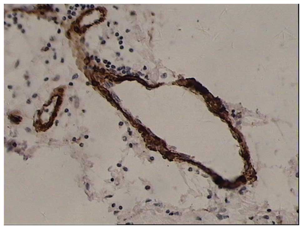
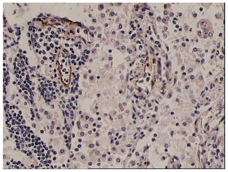
Location of VEGFR-3
Positive staining of VEGFR-3 was indicated by brown
staining and was observed in peritumoral and intratumoral lymphatic
endothelial cells and part of the cancer cell plasma. No expression
was observed in the adjacent normal bronchi and alveoli (Figs. 1–5).
Association between VEGFR-3 expression
and lymph node metastasis
VEGFR-3 expression levels were positive in 27 of 52
cases (51.9%) of NSCLC tissue, while positive expression was only
observed in one case (10%) in the control group. Statistically
significant differences were observed between the groups
(χ2=5.856; P<0.05). The expression of VEGFR-3 in
patients with lymph node metastasis (72% positive) was also
significantly higher than those without lymph node metastasis
(33.3% positive).
VEGFR-3 positive vessel count
Under a magnification of ×200, the number of VEGFR-3
positive vessels in the tumor and peritumoral tissues exhibited
statistically significant differences (9.88±3.22 vs. 3.40±1.27;
t=22.125; P<0.05). The number of VEGFR-3 positive vessels in the
peritumoral tissues of the lymph node metastasis-positive and
-negative groups also exhibited a statistically significant
difference (12.72±1.86 vs. 7.26±1.51; t=11.665; P<0.05). In
addition, the number of VEGFR-3 positive tubes was shown to
correlate with VEGFR-3 protein expression (Table III). Thus, VEGFR-3 expression is
associated with lymph node metastasis (Table IV).
| Table IIIAssociation between VEGFR-3 positive
vessels and the expression of VEGFR-3 in tumor tissue. |
Table III
Association between VEGFR-3 positive
vessels and the expression of VEGFR-3 in tumor tissue.
| Classification | Vessels, n | Peritumoral MLVD | t-value | P-value |
|---|
| VEGFR-3 (−) | 25 | 8.040±2.525 | 4.734 | 0.000 |
| Protein expression
(+) | 27 | 11.593±2.859 | | |
| Table IVAssociation between VEGFR-3 expression
in tumor tissues and lymph node metastasis. |
Table IV
Association between VEGFR-3 expression
in tumor tissues and lymph node metastasis.
| VEGFR-3
expression | | |
|---|
|
| | |
|---|
| Classification | (−) | (+) | χ2 | P-value |
|---|
| Lymph node (−) | 18 | 9 | 7.775 | 0.005 |
| Metastasis (+) | 7 | 18 | | |
Discussion
Lung cancer is one of the most common malignant
tumors, causing a severe threat to human health. Lymph node
metastasis of lung cancer directly influences clinical treatment
and patient prognosis. The survival rates for patients with lymph
node metastasis are significantly lower than those for patients
without lymph node metastasis. With the identification of
lymphangiogenesis factors (VEGF-C and VEGF-D) and lymphatic
endothelial cell specific markers, including VEGFR-3, lymphatic
vessel endothelial hyaluronan receptor-1 and prospero homeobox
protein-1, lymphangiogenesis and lymphatic metastasis have been
increasingly studied.
The first cloned molecular marker of lymphatic
endothelial cells was VEGFR-3, the gene of which is located on
5q33–q35. VEGFR-3 is essential for the initial formation of the
cardiovascular network prior to the onset of the lymphatic system.
In the early phase of embryonic development, VEGFR-3 is expressed
on developing blood endothelial cells and is required for normal
vascular development. In later development, VEGFR-3 is specifically
expressed on lymphatic vessels and regulates occurrence and growth.
However, in a number of pathological conditions, including cancer
and inflammation, VEGFR-3 has been shown to be expressed in the
endothelium of the microvasculature (8–11).
At present, whether functional tumor lymphatic
vessels exist remains controversial. In the present study,
immunohistochemical analysis of 52 lung cancer patients
demonstrated that VEGFR-3 is expressed not only in lymphatic
endothelial cells, but also in microvascular endothelial cells and
the tumor cell cytoplasm, which is in accordance with the studies
by Li et al and Peng et al (12,13).
Newly formed lymphatic capillaries in NSCLC tissues undergo
expansion, with thin walls, no significant plasma membrane
protrusions or continuous endothelial cells and in certain cases,
without endothelial cell coverage. Peritumoral tissues have a
higher number of newly formed lymphatic capillaries, exhibiting
cord-like structures with small branches and thin walls. A number
of the capillaries have a very small cavity and only appear as a
brown mass with light microscopy.
Although the expression of VEGFR-3 has been studied
extensively in a variety of tumor tissues, to the best of our
knowledge, there are no studies investigating the expression in
lymph nodes. The present study detected the expression levels of
VEGFR-3 in the lung tissue and lymph nodes using semiquantitative
RT-PCR and immunohistochemistry. The results not only confirmed the
association between VEGFR-3 and tumor lymphangiogenesis and lymph
node metastasis, but also demonstrated that high VEGFR-3 expression
levels in metastatic-negative lymph nodes can be an indicator of
lymph node metastasis.
In the present study, VEGFR-3 positive lymphatic
vessels were analyzed. With immunohistochemical staining, VEGFR-3
positive lymphatic vessels were found to predominantly exist in the
peritumoral tissue, particularly in the region between the tumor
and normal tissue, which is also referred to as the
tumor-infiltrating area. However, VEGFR-3 positive lymphatic
vessels were rarely observed within the tumor area. The number of
VEGFR-3 positive lymphatic vessels in the peritumoral tissue was
significantly higher compared with the tumor tissue. In addition,
the expression levels were higher in the NSCLC tissue with lymph
node metastasis as compared with the tissue without lymph node
metastasis (P<0.05), indicating that tumor lymphangiogenesis is
associated with tumor invasion. As the proportion of intratumoral
lymphatic vessels was small, we hypothesized that peritumoral
lymphatic vessels play a more important role in the invasion and
metastasis of lung cancer. The decreasing number of VEGFR-3
positive lymphatic vessels in tumors may be associated with the
following factors. Firstly, the lack of an original structure of
the lymphatic valves within the tumor may prevent the intake of
tissue fluid and suppress lymphatic functions. Secondly, continuous
growth of tumor cells produces mechanical force, which causes
lymphatic atrophy and non-function. Finally, tumor cells invade and
destruct the lymphatic network, thus, leave only lymphatic vessel
epithelial remnants within the tumor. With regard to the increase
in VEGFR-3 lymphatic vessels in the peritumoral tissue, we
hypothesized that tumor cells secreting VEGF-C induced the
proliferation of peritumoral lymphatic vessels via VEGFR-3, which
is located on the lymphatic endothelium, including increasing the
diameter and the number of peritumoral lymphatic vessels. Tumor
cells maintain close contact with the lymphatic endothelium, then
infiltrate into the lymphatic vessels and move to the regional
lymph nodes, proliferating there and finally resulting in lymphatic
metastasis (1).
In the present study, VEGFR-3 mRNA expression in the
lung cancer tissues exhibited no significant correlation with lymph
node metastasis. A possible reason may be that expression of
VEGFR-3 in the cytoplasm of tumor cells and small blood vessels may
not be detected by RT-PCR. Therefore, immunohistochemistry and the
MLVD counting method may be more suitable for investigating VEGFR-3
expression. The association between the expression of VEGFR-3 in
lung cancer tissues and lymph node metastasis remains controversial
(14,15), and the exact mechanism requires
further investigation.
The mRNA expression levels of VEGFR-3 were also
detected in the lymph node tissue. The VEGFR-3 mRNA expression
level in the lymph node tissue from the lymph node metastasis group
was significantly lower than that from the group without lymph node
metastasis (P<0.05; 0.158±0.158 vs. 0.281±0.166;
P<0.001).
However, no statistically significant difference was
observed in the VEGFR-3 mRNA expression level between the
metastasis -positive and -negative lymph nodes from the NSCLC
patients with lymph node metastasis (P>0.05). The result was
consistent with a previous study investigating VEGF-C mRNA
expression levels in lymph nodes (16), indicating that pathologically
negative lymph node tissue may exhibit metastasis at a molecular
level.
The present study applied semiquantitative RT-PCR
and immunohistochemisty to investigate the association between
VEGFR-3 mRNA expression and lymph node metastasis in NSCLC
patients. However, the conclusions obtained by these two methods
were not consistent since VEGFR-3 expression in the cytoplasm of
tumor cells and small blood vessels can not be detected using
RT-PCR methods. Thus, we hypothesize that VEGFR-3 should be used as
a marker of lymphatic endothelial cells to assess the number of
lymphatic vessels, which correlates with lymph node metastasis of
lung cancer. For this purpose, the immunohistochemistry method was
more appropriate compared with the semiquantitative RT-PCR
method.
References
|
1
|
Yonemura Y, Fushida S, Bando E, et al:
Lymphangiogenesis and the vascular endothelial growth factor
receptor (VEGFR)-3 in gastric cancer. Eur J Cancer. 37:918–923.
2001. View Article : Google Scholar : PubMed/NCBI
|
|
2
|
Stacker SA, Caesar C, Baldwin ME, et al:
VEGF-D promotes the metastatic spread of tumor cells via the
lymphatics. Nat Med. 7:186–191. 2001. View
Article : Google Scholar : PubMed/NCBI
|
|
3
|
Stacker SA, Stenvers K, Caesar C, et al:
Biosynthesis of vascular endothelial gowth factor involves
Proteolytic Processing which generates non-covalent homodimers. J
Biol Chem. 274:32127–32136. 1999. View Article : Google Scholar : PubMed/NCBI
|
|
4
|
Lee J, Gray A, Yuan J, et al: Vascular
endothelial growth factor related protein: a 1igand and specific
activator of tyrosine kinase receptor Flt4. Proc Natl Acad Sci USA.
93:1988–1992. 1996. View Article : Google Scholar
|
|
5
|
Brambilla E, Travis WD, Colby TV, et al:
The new World Health Organization classification of lung tumours.
Eur Respir J. 18:1059–1068. 2001. View Article : Google Scholar
|
|
6
|
Tanaka K, Kurebayashi J, Sonoo H, et al:
Expression of vascular endothelial growth factor family messenger
RNA in diseased thyroid tissues. Surg Today. 32:761–768. 2002.
View Article : Google Scholar : PubMed/NCBI
|
|
7
|
Fromowitz FB, Viola MV, Chao S, et al: Ras
p21 expression in the progression of breast cancer. Human Pathol.
18:1268–1275. 1987. View Article : Google Scholar
|
|
8
|
Partanen TA, Arola J, Saaristo A, et al:
VEGF-C and VEGF-D expression in neuroendocrine cells and their
receptor, VEGFR-3, in fenestrated blood vessels in human tissues.
FASEB J. 14:2087–2096. 2000. View Article : Google Scholar : PubMed/NCBI
|
|
9
|
Dumont DJ, Jussila L, Taipale J, et al:
Cardiovascular failure in mouse embryos deficient in VEGF
receptor-3. Science. 282:946–949. 1998. View Article : Google Scholar : PubMed/NCBI
|
|
10
|
Akagi K, Ikeda Y, Miyazaki M, et al:
Vascular endothelial growth faetor-C (VEGF-C) expression in human
colorectal cancer tissues. Br J Cancer. 83:887–891. 2000.
View Article : Google Scholar : PubMed/NCBI
|
|
11
|
Partanen TA, Alitalo K and Miettinen M:
Lack of lymphatic vascular specificity of vascular endothelial
growth factor receptor 3 in 185 vascular tumors. Cancer.
86:2406–2412. 1999. View Article : Google Scholar : PubMed/NCBI
|
|
12
|
Li Q, Dong X, Gu W, et al: Clinical
significance of co-expression of VEGF-C and VEGFR-3 in non-small
cell lung cancer. Chin Med J (Engl). 116:727–730. 2003.
|
|
13
|
Peng ZY, Xiao TH, Chen SS and Li DZ:
Mechanism of early lymphatic metastasis of non-small cell lung
cancer. Hua Nan Guo Fang Yi Xue Za Zhi. 19:30–31. 2005.(In
Chinese).
|
|
14
|
Carrillo de Santa Pau E, Arias FC, Caso
Peláez E, et al: Prognostic significance of the expression of
vascular endothelial growth factors A, B, C, and D and their
receptors R1, R2, and R3 in patients with nonsmall cell lung
cancer. Cancer. 115:1701–1712. 2009. View Article : Google Scholar : PubMed/NCBI
|
|
15
|
Takizawa H, Kondo K, Fujino H, et al: The
balance of VEGF-C and VEGFR-3 mRNA is a predictor of lymph node
metastasis in non-small cell lung cancer. Br J Cancer. 95:75–79.
2006. View Article : Google Scholar : PubMed/NCBI
|
|
16
|
Li J, Li BL, Zhang HQ, et al: Relationship
between vascular endothelial growth factor C expression level and
lymph node metastasis in non small cell lung cancer. Zhonghua Yi
Xue Za Zhi. 88:2982–2985. 2008.(In Chinese). PubMed/NCBI
|