Introduction
Contrast-induced acute kidney injury (CIAKI) is a
serious clinical complication, associated with increased use of
iodinated contrast media (CM) in diagnostic and interventional
procedures. It is now the third most important cause of
hospital-acquired acute kidney injury and accounts for 12% of all
cases (1). Although the use of
low-osmolar CM (LOCM) reduces the risk of CIAKI, the incidence of
CIAKI remains high following intravascular administration of LOCM
in high-risk patients with renal insufficiency (2).
The pathogenic mechanism of CIAKI is multifactorial,
and previous studies have shown that it is mainly associated with
oxidative stress, renal ischemia and direct nephrotoxicity
(3). In addition, a series of
endogenous vasoactive substances, including renin and angiotensin
II, have an important role in the pathogenesis of CIAKI (4). NADPH oxidase is the major source of
reactive oxygen species (ROS) and is significant in cisplatin- or
cyclosporine-induced acute kidney injury (5). In previous studies, we demonstrated
that NADPH oxidase was a pivotal contributor to oxidative stress
damage in CIAKI (6,7). These results suggest that NADPH
oxidase might be a novel target for the development of drugs
against CIAKI.
Pentoxifylline, a nonspecific phosphodiesterase
inhibitor, is a methylxanthine derivative with multiple hematologic
properties (8). It has been
demonstrated that pentoxifylline may be used as a renoprotective
agent against a number of nephrotoxic drugs, including cyclosporine
or cisplatin in clinical and animal studies (9), and a recent study performed by
Firouzi et al (10)
concluded that the prophylactic oral use of pentoxifylline may be
recommended for the prevention of CIAKI. However, the mechanism for
its protective effect has yet to be elucidated. Therefore, the aim
of the present study was to further evaluate the protective effect
of pentoxifylline on CIAKI in rats with hypercholesterolemia and
the underlying mechanisms.
Materials and methods
Reagents and animals
Low-osmolar iodinated contrast medium, iohexol
(Amersham Pharmaceutical Co., Ltd, Shanghai, China), was diluted to
300 mg I/ml with distilled water. Cholesterol and cholic acid were
obtained from DingGuo Biotechnology Co., Ltd. (Beijing, China) and
the NADPH oxidase assay kit was purchased from Jiemei Gene Medicine
Technology Co., Ltd. (Shanghai, China). The MDA and SOD detection
kits were purchased from Jiancheng Bioengineering Institute
(Nanjing, China). Pentoxifylline was obtained from Northeast
Pharmaceutical Group Shenyang No.1 Pharmaceutical Co., Ltd.
(Shenyang, China).
A total of 32 healthy male Sprague-Dawley rats,
weighing 160–180 g, with negative tests for urine protein and
glucose, were provided by the Second Xiangya Hospital Animal Center
(Changsha, China). The rats were randomly divided into the normal
diet group (NN; n=8) and high cholesterol-supplemented dietary
group (HN; 4% cholesterol and 1% cholic acid; n=24) (11). After eight weeks, the rats in the
HN group were randomly divided into three subgroups (n=8 in each
group): the high cholesterol diet group (HN), the high cholesterol
plus LOCM iohexol group (HL) and the pentoxifylline protective
group (high cholesterol plus iohexol plus pentoxifylline; HLP). All
experiments were approved by the Medical Science Animal Care
Committee of the Central South University (Changsha, Hunan,
China).
Experimental treatment
At the end of the eight weeks of feeding, the eight
rats in each group were given a tail vein injection of either
iohexol (10 ml/kg; HL and HLP groups) or an equivalent volume of
normal saline (HN and NN groups) over 2 min. The rats in the HLP
group were injected with pentoxifylline (50 mg/kg) into the
peritoneal cavity, 12 h prior to and following CM injection. Rats
from the NN, HN and HL groups were administered an equal volume of
normal saline. Urine samples and blood samples were obtained prior
to and 48 h following the injection of CM to determine the level of
serum creatinine (SCr), triglyceride (TG), cholesterol (CHOL),
urine creatinine, sodium and potassium, using an automated
biochemical analyzer (Olympus AU100, Tokyo, Japan). In addition,
creatinine clearance (Ccr), fractional excretion of sodium (FENa%)
and potassium (FEK%) were calculated as previously described
(12).
Once the blood samples were obtained, the rats were
sacrificed by cervical dislocation. The right kidney tissue was
homogenized to measure the levels of malondialdehyde (MDA),
superoxide dismutase (SOD) and nicotinamide adenine dinucleotide
phosphate-oxidase (NADPH oxidase) activity using commercial kits.
The total protein content of renal tissue was then measured using
the Coomassie brilliant blue method.
Renal tissue MDA, SOD and NADPH oxidase
assay
In brief, 200 mg renal cortex was washed in ice-cold
saline in tubes, sectioned into small pieces and homogenized in
ice-cold saline homogenization buffer in a 1:9 ratio (w:v). The
homogenate was centrifuged at 2971 × g for 10 min at 4°C. The
supernatant was separated and analyzed for total protein content
and SOD and NADPH oxidase activity. The protein concentration of
the homogenate was determined using the BCA assay method. The
results were expressed as nmmol/mg protein (MDA) and U/mg protein
(SOD and NADPH oxidase).
Renal histopathological evaluation
The upper halves of the left kidney were cut into
sections at 4 μm and stained using hematoxylin and eosin for
histopathological evaluation under a light microscope (Leica
DMI-3000B; Leica Microsystems AG, Wetzlar, Germany). Evaluations
were performed and scored semi-quantitatively in a blinded manner
using an arbitrary scale (6). The
calculation was determined at a ×200 magnification in 10 fields for
each biopsy. Tubular injuries were graded as follows: 0, no tubular
injury; 1, <25% of tubules injured; 2, between 25 and 50% of
tubules injured; 3, between 51 and 75% of tubules injured; 4,
>76% of tubules injured (7).
Statistical analysis
Statistical analyses were performed using SPSS
software, version 16.0 (SPSS, Inc., Chicago, IL, USA). The results
are expressed as the mean ± standard deviation. Data were analyzed
using one-way analysis of variance, and the two-groups comparison
among multiple samples was performed using the Fisher Least
Significant Difference test. P<0.05 was considered to indicate a
statistically significant difference.
Results
Baseline characteristics in different
groups
Body weight, serum TG, CHOL, Scr, Ccr, FENa% and
FEK% values were analyzed in the four groups at baseline. As shown
in Table I, there were no
significant differences in the baseline characteristics of Scr,
Ccr, FENa% and FEK% (P>0.05). In the rats fed a high cholesterol
diet, the levels of serum CHOL were higher compared with those in
rats fed a normal diet (P<0.05). All rats tolerated the
treatment well, and survived until the end of the experiment.
 | Table IComparison of biochemical indicators
in each group prior to contrast media injection (mean±standard
deviation; n=8). |
Table I
Comparison of biochemical indicators
in each group prior to contrast media injection (mean±standard
deviation; n=8).
| Group | BW (g) | Scr (μmol/l) | TG (mmol/l) | CHOL (mmol/l) | Ccr (ml/min) | FENa% | FEK% |
|---|
| NN | 321.26±85.27 | 32.13±2.96 | 0.32±0.17 | 1.35±0.18 | 0.41±0.05 | 1.36±0.42 | 71.24±7.08 |
| HN | 334.30±72.67 | 35.50±2.44 | 0.39±0.18 | 2.71±0.14a | 0.39±0.04 | 1.48±0.38 | 75.00±6.63 |
| HL | 317.54±86.80 | 34.61±2.49 | 0.41±0.21 | 2.86±0.22a | 0.38±0.03 | 1.56±0.31 | 74.30±6.96 |
| HLP | 321.68±79.46 | 34.92±2.73 | 0.48±0.20 | 2.62±0.25a | 0.38±0.02 | 1.61±0.47 | 73.84±5.84 |
Effect of pentoxifylline on renal
function parameters
In CM-treated groups, rats showed marked increases
in Scr, FENa% and FEK% (P<0.01), and a decline in Ccr
(P<0.01) following iohexol administration, compared with those
in control animals. However, the CM-treated rats that underwent
treatment with pentoxifylline showed significantly decreased values
of Scr, FENa% and FEK% compared with those in the rats treated with
CM alone (P<0.05; Table
II).
| Table IIChanges in renal function indicators
in each group 48 h following contrast media injection
(mean±standard deviation; n=8). |
Table II
Changes in renal function indicators
in each group 48 h following contrast media injection
(mean±standard deviation; n=8).
| Group | Scr (μmol/l) | Ccr (ml/min/100
g) | FENa% | FEK% |
|---|
| NN | 32.95±2.14 | 0.42±0.04 | 1.28±0.25 | 70.61±5.09 |
| HN | 35.62±1.81 | 0.40±0.03 | 1.55±0.41 | 75.29±4.13 |
| HL | 46.69±2.91a | 0.29±0.02a | 3.95±0.15a | 98.25±4.08a |
| HLP | 36.16±2.06b | 0.39±0.03b | 1.78±0.16b | 77.12±5.97b |
Effect of pentoxifylline on renal
oxidative stress parameters and NADPH oxidase activity
As shown in Table
III, in CM-treated rats, a trend towards higher values of renal
MDA levels and NADPH oxidase activity was observed compared with
control animals (P<0.01), while the renal SOD activity was lower
in the CM-treated rats than in the control rats (P<0.01).
However, in the rats treated with pentoxifylline and iohexol, the
renal NADPH oxidase activity and MDA levels were significantly
lower compared with those in the rats treated with iohexol alone
(P<0.01), while a significant increase in the activity of renal
SOD was observed (P<0.01).
| Table IIIComparison of renal oxidative stress
parameters following contrast media injection (mean±standard
deviation; n=8). |
Table III
Comparison of renal oxidative stress
parameters following contrast media injection (mean±standard
deviation; n=8).
| Group | MDA (nmmol/mg
protein) | SOD (U/mg
protein) | NADPH oxidase (U/mg
protein) |
|---|
| NN | 3.43±0.47 | 413.03±23.28 | 14.95±5.12 |
| HN | 4.21±0.75 | 394.67±43.62 | 21.26±8.26a |
| HL | 8.46±0.92a,b | 317.01±47.36a,b | 82.42±31.18a,b |
| HLP | 5.27±0.48c | 422.32±41.50c | 26.59±7.56c |
Histomorphological comparison of renal
injury
As shown in Fig. 1,
tubular epithelial cell shedding and basement membrane nudity,
vacuolar degeneration of tubular epithelial cells, protein cast,
tubular dilation, loss of tubular brush border, and necrosis of
partial tubular epithelial cells was observed in CM-injected rats.
However, in the pentoxifylline-injected rats, only renal tubular
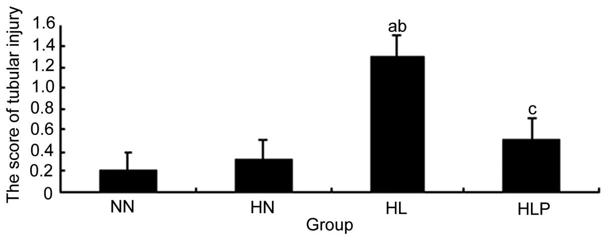
epithelial cell degeneration was observed. As shown in Fig. 2, light microscopic examination and
semi-quantitative analysis demonstrated that the tubular
pathological scores in the HL group were significantly higher
compared with those of the control animals (P<0.01).
Pretreatment with pentoxifylline was observed to significantly
attenuate the development of these lesions, as the tubular
pathological scores of the rats treated with iohexol alone were
significantly higher compared with those of iohexol-treated rats
that were pretreated with pentoxifylline (P<0.01).
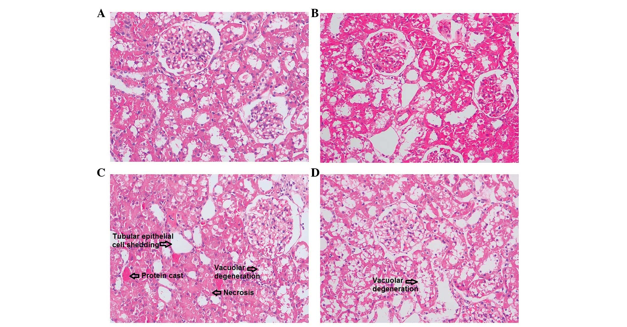 | Figure 1Representative renal
histomorphological changes in the (A) NN, normal diet group, (B)
HN, high cholesterol diet group, (C) HL, high cholesterol plus
low-osmolar contrast media iohexol group and (D) HLP, high
cholesterol plus iohexol plus pentoxifylline group. (C) In
iohexol-injected rats, tubular epithelial cell shedding and
basement membrane nudity, vacuolar degeneration of tubular
epithelial cells, protein cast, tubular dilation, loss of tubular
brush border, and necrosis of partial tubular epithelial cells were
observed. However, in (D) the rats treated with pentoxifylline and
iohexol, only renal tubular epithelial cell degeneration was
observed. Hematoxylin and eosin staining, original magnification,
×200. |
Discussion
In the present study, it was investigated whether
pentoxifylline has protective effect against CIAKI. Pentoxifylline
is known to have antioxidant properties. It is also cost-effective,
readily available and is currently being used clinically for
chronic kidney disease (13). The
results from the present study have further demonstrated that
pentoxifylline may also protect against CIAKI in rats with
hypercholesterolemia, which may be due to an antioxidant
action.
The pathological mechanism of CIAKI remains unclear.
In a previous study we demonstrated that high-osmolality CM induces
cultured human renal tubular epithelial cell apoptosis, while LOCM
does not induce tubular epithelial cell apoptosis (14). In addition, clinical studies have
demonstrated a reduction in contrast nephropathy with the
introduction of LOCM (15).
However, the cytotoxicity of radiocontrast agents has been shown to
correlate with not only hyperosmolality, but also iodine
concentration (14). The osmotic
pressures of high-osmolality CM (>1,500 mOsm/kg) and LOCM
(550–850 mOsm/kg) are higher than those of plasma (280–310
mOsm/kg). LOCM is an important risk factor for radiocontrast
nephrotoxicity, and in the present study, a CIAKI model was
successfully established using the LOCM iohexol in rats with
hypercholesterolemia. This was consistent with the study by Yang
et al (12), which observed
that long-term hypercholesterolemia diet and LOCM appeared to be
risk factors for CIAKI (12).
A recent study demonstrated that oxidative stress
has an important role in the pathogenesis of CIAKI (3). NADPH oxidases represent a class of
hetero-oligomeric enzymes whose primary function is the generation
of ROS. In addition, NADPH oxidase has been shown to have an
important role in cisplatin or cyclosporine-induced acute kidney
injury (5). Furthermore, a
recently study by Ahmad et al (16) demonstrated that the NADPH-oxidase
inhibitor apocynin attenuates the degree of contrast-induced
nephropathy in diabetic rats (16). The present study demonstrated that
in the iohexol-injected rats, the levels of Scr, renal tissue MDA
content and NADPH oxidase activity were significantly increased. In
addition, in the iohexol-treated animals compared with the control
animals, the tubular pathological injury scores were found to be
higher, while the Ccr and renal SOD activity decreased
significantly. Furthermore, it was demonstrated that NADPH oxidase
and oxidative stress appear to have an important role in CIAKI.
There is currently no accepted method for the
prevention of CIAKI, apart from the importance of hydration and
avoidance of hypovolemia prior to exposure to CM (17). Studies have shown that the
formation of ROS is increased in the kidney following the
administration of CM, which has an important role in the
development of CIAKI (18). In
accordance with previous studies, the results from the present
study showed that iohexol administration significantly increased
renal oxidative stress, suggesting a protective effect of ROS
scavenging in CIAKI.
Pentoxifylline, an inhibitor of phosphodiesterase,
was first considered in the treatment of peripheral vascular
diseases (19). It is known to
have several pharmacological effects, including inhibition of free
radical production, stimulation of the biosynthesis of renal
vasodilator prostaglandins, and improvement in oxygen delivery to
ischemic tissues (20).
Additionally, it has been shown that pentoxifylline is able to
inhibit the formation of free radicals by inhibiting NADPH oxidase
activity in neutrophils (21). The
results from animal and clinical studies suggest that
pentoxifylline may prevent renal cell injuries induced by glycerol,
cisplatin or cyclosporine (9,22). A
recent study by Firouzi et al (10) indicated that the prophylactic oral
use of pentoxifylline may be recommended for CIAKI prevention;
however, the mechanisms for this protective effect have yet to be
elucidated.
The results from the present study showed that
pretreatment with pentoxifylline prior to iohexol infusion may
reduce the SCr and the renal MDA content and NADPH oxidase
activity. In the pentoxifylline treatment group, the tubular
pathological changes associated with iohexol were found to be
reduced, while the levels of renal SOD activity were increased.
This indicates that pentoxifylline has a protective effect against
the nephrotoxicity induced by iohexol in renal tissue, due to an
antioxidant action.
There are several potential limitations of the
present study that should be addressed. Firstly, pentoxifylline has
been found to have novel biochemical effects, including the
alleviation of renal vasoconstriction (23), reduction of oxidative stress and
modulation of inflammatory responses (24). These properties may also be used to
prevent CIAKI as proposed by Roozbeh et al (8). The main limitation of this study is
that the mechanism of the anti-oxidation effects of pentoxifylline
was analyzed only in CIAKI. Secondly, only tubular injury scores
were measured to indicate the degree of tubular injury, and TUNEL
immunostaining and analysis of urine tubular injury biomarkers (for
example, NGAL) was not performed. However, despite these
limitations, the results from the present study remain valid.
In conclusion, the results from the present study
demonstrated that pentoxifylline is able to protect the renal
tissue from the nephrotoxicity induced by CM in rats with
hypercholesterolemia due to an antioxidant action. However, further
clinical trials in patients with CIAKI are required in order to
determine its exact role.
Acknowledgements
This study was supported by a grant from the
Scientific Foundation Of Hunan Province, China (2010FJ6008,
2008JT3005).
References
|
1
|
Itoh Y, Yano T, Sendo T and Oishi R:
Clinical and experimental evidence for prevention of acute renal
failure induced by radiographic contrast media. J Pharmacol Sci.
97:473–488. 2005. View Article : Google Scholar : PubMed/NCBI
|
|
2
|
Aspelin P, Aubry P, Fransson SG, Strasser
R, Willenbrock R and Berg KJ: NEPHRIC Study Investigators:
Nephrotoxic effects in high-risk patients undergoing angiography. N
Engl J Med. 348:491–499. 2003. View Article : Google Scholar : PubMed/NCBI
|
|
3
|
Seeliger E, Sendeski M, Rihal CS and
Persson PB: Contrast-induced kidney injury: mechanisms, risk
factors, and prevention. Eur Heart J. 33:2007–2015. 2012.
View Article : Google Scholar : PubMed/NCBI
|
|
4
|
Russo D, Minutolo R, Cianciaruso B, Memoli
B, Conte G and De Nicola L: Early effects of contrast media on
renal hemodynamics and tubular function in chronic renal failure. J
Am Soc Nephrol. 6:1451–1458. 1995.PubMed/NCBI
|
|
5
|
Perianayagam MC, Liangos O, Kolyada AY, et
al: NADPH oxidase p22phox and catalase gene variants are associated
with biomarkers of oxidative stress and adverse outcomes in acute
renal failure. J Am Soc Nephrol. 18:255–263. 2007. View Article : Google Scholar
|
|
6
|
Duan SB, Liu GL, Chen GC, Wang P, Pan P
and Xu XQ: Aged rats are susceptible to nephrotoxicity induced by
iodinated contrast media. Ren Fail. 35:150–154. 2013. View Article : Google Scholar
|
|
7
|
Duan SB, Yang SK, Zhou QY, et al:
Mitochondria-targeted peptides prevent on contrast-induced acute
kidney injury in the rats with hypercholesterolemia. Ren Fail.
35:1124–1129. 2013. View Article : Google Scholar : PubMed/NCBI
|
|
8
|
Roozbeh J, Hamidian Jahromi A, Sharifian
M, Pakfetrat M and Afshariani R: Protective effect of
pentoxifylline on contrast induced nephropathy. Saudi J Kidney Dis
Transpl. 19:985–986. 2008.PubMed/NCBI
|
|
9
|
Nasiri-Toosi Z, Dashti-Khavidaki S,
Khalili H and Lessan-Pezeshki M: A review of the potential
protective effects of pentoxifylline against drug-induced
nephrotoxicity. Eur J Clin Pharmacol. 69:1057–1073. 2013.
View Article : Google Scholar
|
|
10
|
Firouzi A, Eshraghi A, Shakerian F, et al:
Efficacy of pentoxifylline in prevention of contrast-induced
nephropathy in angioplasty patients. Int Urol Nephrol.
44:1145–1149. 2012. View Article : Google Scholar
|
|
11
|
Yang D, Lin S, Yang D, Wei L and Shang W:
Effects of short- and long-term hypercholesterolemia on
contrast-induced acute kidney injury. Am J Nephrol. 35:80–89. 2012.
View Article : Google Scholar
|
|
12
|
Yang DW, Jia RH, Yang DP, Ding GH and
Huang CX: Dietary hypercholesterolemia aggravates contrast
media-induced nephropathy. Chin Med J (Engl). 117:542–546.
2004.
|
|
13
|
Lin SL, Chiang WC, Chen YM, Lai CF, Tsai
TJ and Hsieh BS: The renoprotective potential of pentoxifylline in
chronic kidney disease. J Chin Med Assoc. 68:99–105. 2005.
View Article : Google Scholar : PubMed/NCBI
|
|
14
|
Duan S, Zhou X, Liu F, et al: Comparative
cytotoxicity of high-osmolar and low-osmolar contrast media on HKCs
in vitro. J Nephrol. 19:717–724. 2006.PubMed/NCBI
|
|
15
|
Rudnick MR and Goldfarb S: Pathogenesis of
contrast-induced nephropathy: experimental and clinical
observations with an emphasis on the role of osmolality. Rev
Cardiovasc Med. 4(Suppl 5): S28–S33. 2003.PubMed/NCBI
|
|
16
|
Ahmad A, Mondello S, Di Paola R, et al:
Protective effect of apocynin, a NADPH-oxidase inhibitor, against
contrast-induced nephropathy in the diabetic rats: a comparison
with N-acetylcysteine. Eur J Pharmacol. 674:397–406. 2012.
View Article : Google Scholar
|
|
17
|
Benko A, Fraser-Hill M, Magner P, et al:
Canadian Association of Radiologists: consensus guidelines for the
prevention of contrast-induced nephropathy. Can Assoc Radiol J.
58:79–87. 2007.PubMed/NCBI
|
|
18
|
Quintavalle C, Brenca M, De Micco F, et
al: In vivo and in vitro assessment of pathways involved in
contrast media-induced renal cells apoptosis. Cell Death Dis.
2:e1552011. View Article : Google Scholar : PubMed/NCBI
|
|
19
|
Samlaska CP and Winfield EA:
Pentoxifylline. J Am Acad Dermatol. 30:603–621. 1994. View Article : Google Scholar : PubMed/NCBI
|
|
20
|
Dávila-Esqueda ME and Martínez-Morales F:
Pentoxifylline diminishes the oxidative damage to renal tissue
induced by streptozotocin in the rat. Exp Diabesity Res. 5:245–251.
2004. View Article : Google Scholar
|
|
21
|
Costantini TW, Deree J, Peterson CY, et
al: Pentoxifylline modulates p47phox activation and downregulates
neutrophil oxidative burst through PKA-dependent and -independent
mechanisms. Immunopharmacol Immunotoxicol. 32:82–91. 2010.
View Article : Google Scholar
|
|
22
|
Savic V, Vlahovic P, Djordjevic V,
Mitic-Zlatkovic M, Avramovic V and Stefanovic V: Nephroprotective
effects of pentoxifylline in experimental myoglobinuric acute renal
failure. Pathol Biol (Paris). 50:599–607. 2002. View Article : Google Scholar
|
|
23
|
Tyagi P, Sharma P, Sharma BC, Puri AS,
Kumar A and Sarin SK: Prevention of hepatorenal syndrome in
patients with cirrhosis and ascites: a pilot randomized control
trial between pentoxifylline and placebo. Eur J Gastroenterol
Hepatol. 23:210–217. 2011. View Article : Google Scholar : PubMed/NCBI
|
|
24
|
de Moura FJ, Leal PP, de Souza Furtado R,
Muniz-Junqueira MI and Veiga JP: Pentoxifylline prevents the
meglumine antimonate-induced renal toxicity in rats, but not that
induced by the inorganic antimony pentachloride. Toxicology.
243:66–74. 2008. View Article : Google Scholar
|