Introduction
Bone marrow (BM)-derived mesenchymal stem cells
(MSCs) are becoming a target for use in cell and gene therapy due
to the fact that they possess several therapeutic advantages. MSCs
can contribute to wound healing and tissue regeneration through
their multiple differentiation capacity, immune modulation
properties and ability to promote the neovascularization of
ischemic tissues (1–4). Clinical and preclinical trials based
on stem cell therapies by use of MSCs have reported that these
cells can be beneficial in the treatment of several conditions,
such as renal failure, myocardial infarction and bone and cartilage
diseases (4–6). A great majority of these trials are
based on the allogeneic or autologous transplantation of MSCs
isolated from healthy donors or patients. This therapeutic approach
requires the obtainment of high numbers of MSCs, which can be
accomplished by the isolation of autologous MSCs and further
expansion in vitro; however, studies have shown that culture
expansion of MSCs can cause them to gradually lose their early
progenitor properties, such as multipotency, proliferation
potential and bone-forming efficiency (7). Efforts are therefore being made to
find substances that can enhance the natural egress of MSCs from
the BM into the periphery, which would facilitate the harvest of
higher numbers of MSCs and increase their proliferation in
vitro in order to shorten the expansion process. Eventually, in
this way, the changes in the therapeutic properties of MSCs would
be minimized prior to their clinical application.
A study by Hong et al (8), showed that substance P (SP), an
11-amino acid neuropeptide involved in pain perception, induced the
mobilization of cluster of differentiation (CD) 29-positive MSCs
from the BM into the periphery, which were found to participate in
wound repair. In addition, the same study revealed that SP enhanced
the proliferation of human MSCs in vitro, most likely
through the activation of the extracellular signal-regulated
kinase-1/2 (ERK-1/2) and T-cell factor/lymphoid enhancer-binding
factor/β-catenin pathways (8).
Furthermore, in a previous study our data showed an increase in the
fibroblast-colony forming units (CFU-F) inside the BM two days
after the injection of SP in mice, suggesting that the MSC
population increases inside the BM in response to SP. Furthermore,
the data demonstrated the ability of SP to modulate other essential
characteristics of MSCs, such as their differentiation potential
(9); however, the mechanisms
involved in the SP-mediated mobilization and expansion of MSCs have
yet to be elucidated.
Based on the aforementioned findings, SP could be
considered as one of the substances that could be tested for its
use to improve the isolation and in vitro expansion
processes of MSCs in the development of stem cell therapies by use
of these cells. In order to evaluate this possibility and to search
for stable cell lines that could permit further investigation into
the mechanisms involved in the SP-mediated effects on MSCs, the
present study was designed to test the effects of SP on the
proliferation and migration potential of two mouse BM-derived
MSC-like cell lines, OP9 and ST2.
Materials and methods
Cell lines and culture
The OP9 cell line was purchased from the American
Type Culture Collection (#CRL-2749; Manassas, VA, USA) and
maintained in α-Minimum Essential Medium (1X, without
ribonucleosides and deoxyribonucleosides; Gibco-BRL, Carlsbad, CA,
USA) containing 10% heat-inactivated fetal bovine serum (FBS;
Gibco-BRL), 2 mmol/l L-glutamine (Gibco-BRL), 1.5 g/l sodium
bicarbonate (Gibco-BRL), 100 U/ml penicillin and 100 μg/ml
streptomycin (P/S; Gibco-BRL). The ST2 cell line was obtained from
the Riken Bioresource Center Cell Bank (Tsukuba, Japan) and
maintained in RPMI-1640 (Gibco-BRL) supplemented with 10% FBS and
1% P/S. The two cell lines were incubated at 37°C in a humidified
atmosphere containing 5% CO2. At 80% confluence, the
cells were harvested using 0.25% trypsin/EDTA (Gibco-BRL) and
sub-cultured at a ratio of 1:3 to 1:4. The media were changed every
3–4 days. Only cells in passages five to eight were used for the
experiments.
Cell counting
SP was obtained from EMD Millipore (#05-23-0600; San
Diego, CA, USA) and was prepared with 5% acetic acid
(Sigma-Aldrich, St. Louis, MO, USA). To evaluate the cytotoxicity
of SP and its effect on the proliferation of the OP9 and ST2 cell
lines, cell counting was performed. Cultured OP9 or ST2 cells were
seeded in six-well plates at a density of 1.3×10⁴ or
5×10³ cells/well, respectively. After 24 h of
incubation, the media were switched to control media (growth media
containing 5% acetic acid (22 μm), a solvent of SP) or media
containing 0.1, 1, 10, 100 or 300 nM SP for 72 h. In the case of
the serum-free condition experiment, the cells were incubated for
18–24 h with serum-free growth media, following which the cells
were treated with SP for 72 h. The cells were collected, stained
with trypan blue and mounted on a Marienfeld-Superior hemocytometer
(Paul Marienfeld GmbH & Co. KG, Lauda-Königshofen, Germany) for
cell counting. The number of alive and dead cells was counted.
Three replicates were analyzed for each concentration of SP
tested.
5-Bromo-2′-deoxyuridine (BrdU)
incorporation assay
OP9 or ST2 cells were seeded on fibronectin-coated
coverslips (1 μg/ml) in 24-well plates at a density of
2.7×10³ cells/well or in six-well plates at a density of
5×10³ cells/well, respectively. The cells were incubated
for 24 h before the treatment with SP. The cells were treated with
control media (growth media containing the appropriate amount of
solvent of SP) or media containing 1, 10 or 100 nM SP for 48 h. In
the case of the serum-free condition experiment, the cells were
serum-starved for 18–24 h, following which the cells were treated
with SP for 48 h. The OP9 and ST2 cells were treated with 20 μM
BrdU (Sigma-Aldrich) for the last 12 or 6 h of incubation,
respectively. For the antagonist experiment, the OP9 cells were
pretreated for 30 min with the neurokinin-1 (NK-1) receptor
antagonist RP67580 (1 μM) (Tocris Bioscience, Bristol, UK). The
cells were then treated with 10 nM SP in the presence or absence of
RP67580 (1 μM) for 48 h. Following immunocytochemical staining for
BrdU, images were captured using a Leica fluorescence microscope
(Leica Microsystems GmbH, Wetzlar, Germany) and the number of total
and BrdU-positive cells was counted.
Immunocytochemistry
For immunofluorescence staining, the cells were
fixed on coverslips with 4% paraformaldehyde in phosphate-buffered
saline (PBS) for 10 min on ice. Following permeabilization with
0.2% Triton X-100 (USB Corp., Cleveland, OH, USA) and blocking
solution treatment (5% non-fat milk in PBS with 0.1% Triton X-100)
for 30 min at room temperature, the cells were incubated with mouse
monoclonal anti-BrdU primary antibody (1:20, #11-170-376-001; Roche
Diagnostics GmbH, Mannheim, Germany) for 1.5 h at room temperature.
Subsequent to being washed three times with 1% non-fat milk in PBS
with 0.1% Triton X-100, the cells were treated with Alexa 488
anti-mouse immunoglobulin G1 secondary antibody (Invitrogen Life
Technologies, Carlsbad, CA, USA) for 45 min at room temperature.
When necessary, actin was stained with phalloidin (Invitrogen Life
Technologies). Finally, the samples were mounted using
ProLong® Gold Antifade mounting solution with DAPI
(Invitrogen Life Technologies) and left to dry overnight prior to
observation. The samples were then examined using a fluorescence
microscope (Leica Microsystems Gmbh).
RNA isolation and reverse
transcription-quantitative polymerase chain reaction (RT-qPCR)
analysis
Total RNA was extracted from cultured ST2 or OP9
cells using TRIzol™ reagent (Invitrogen Life Technologies)
according to the manufacturer’s instructions. Samples of 5 μg total
RNA were used for single strand cDNA synthesis using Superscript
First-Strand cDNA Synthesis System (Invitrogen Life Technologies)
according to the manufacturer’s instructions. After reverse
transcription, RNA was degraded by Escherichia coli RNase H
(Invitrogen Life Technologies). qPCR was performed in 20 μl
reaction buffer containing 1.5 U i-Taq DNA polymerase (iNtRON
Biotechnology, Seongnam, Korea), 1X PCR buffer (iNtRON), 2.5 mM
each of dATP, dCTP, dGTP and dTTP (iNtRON) and 10 pM of each
specific primer (NK-1: F 5′-TGGACTCTGATCTCTTCCCCAACA-3′ and R
5′-GGACCCAGATGACAAAGATGACCA-3′). Primers were purchased from Cosmo
Genetech Co., Ltd. (Seoul, Korea). PCR products were
electrophoretically separated on 1.5% (w/v) agarose gels
(M.biotech, Inc., Hanam, Korea) and visualized after staining with
RedSafe nucleic acid staining solution (iNtRON).
Western blotting
Cells were treated with the correct amount of
solvent of SP (5% acetic acid) or 10 nM SP for 0 or 24 h under
normal medium conditions. To obtain the cell lysate, the cells were
rinsed twice with ice-cold PBS and incubated with 400 μl 2X sodium
dodecyl sulfate (SDS) loading buffer [100 mM Tris-Cl (pH 6.8), 4%
(w/v) SDS, 0.2% (w/v) bromophenol blue, 20% glycerol and 200 mM
β-mercaptoethanol] for 5 min at room temperature. The cell lysate
was collected and denatured at 92°C for 10 min. Protein samples
were subjected to 10% SDS-polyacrylamide gel electrophoresis. The
separated proteins were transferred onto nitrocellulose membranes
(Whatman; GE Healthcare Life Sciences, Munich, Germany). Subsequent
to being blocked with 5% non-fat milk in 20 mM Tris-buffer
containing 0.1% Tween-20 (TBS-T), the membranes were incubated with
mouse monoclonal anti-cyclin D1 primary antibody (DCS-3, 1:800,
#sc-20044; Santa Cruz Biotechnology, Inc., Santa Cruz, CA, USA)
diluted with TBS-T buffer containing 5% non-fat milk overnight at
4°C. The membranes were then incubated with horseradish
peroxidase-conjugated secondary antibodies at room temperature for
30 min. Target proteins were visualized using enhanced
chemiluminescence detection (EMD Millipore). Band densities were
measured using ImageJ software (National Institutes of Health
(NIH), Bethesda, MD, USA). If required, membrane stripping was
performed using Thermo Scientific™ Restore™ Western Blot Stripping
Buffer (Thermo Fisher Scientific, Inc., Waltham, MA, USA) for 15
min at room temperature. The membranes were re-blotted by use of
mouse monoclonal anti-α-tubulin antibody (1:10,000, #T5618;
Sigma-Aldrich).
Wound healing migration assay
ST2 cells were seeded on six-well plates and
incubated until complete confluence was reached. The media were
switched to serum-free growth media for 18 h. The confluent
monolayer was then wounded using a 200-μl yellow pipette tip and
washed twice with PBS to remove the floating cells. Treatment with
10, 100 and 300 nM SP was performed for 9 h. If necessary,
pretreatment with the NK-1 receptor antagonist RP67580 (10 μM)
(Tocris Bioscience) was applied for 30 min. Following incubation,
the cells were fixed with 4% paraformaldehyde in PBS for 10 min at
room temperature and stained with crystal violet solution (1%
solution; Sigma-Aldrich) for 30 min. Cell culture images were
captured using a light microscope and the wound area was measured
with ImageJ software (NIH).
Statistical analysis
Quantitative data are presented as the mean ±
standard error of mean or standard deviation. An unpaired Student’s
t-test was applied to evaluate differences between two groups.
P<0.05 was considered to indicate a statistically significant
difference. All statistical analyses were performed using GraphPad
version 5.01 (GraphPad Software, Inc., La Jolla, CA, USA).
Results
SP induces the mobilization of ST2
cells
Due to the as yet unexplained difficulty in
isolating and culturing murine MSCs, several stable MSC-like cell
lines derived from mouse BM have been established that permit the
in vitro study of the properties of MSCs. One of these cell
lines, ST2, has been shown to possess the characteristic trilineage
differentiation potential of MSCs along with their capacity to
support the growth and differentiation of early B-lineage and T
cells (10–13).
MSCs have been found to be able to mobilize in
response to several signals from the BM into the periphery in order
to contribute to the processes of wound healing and tissue repair
(8). This results in an increase
in the number of circulating MSCs, which could then be harvested
and further expanded for their use in cell therapy. A previous
study showed that SP induces the mobilization of MSCs in
vivo; however, the mechanisms involved in this SP-mediated
mobilization of MSCs have yet to be elucidated (8). To further analyze the effects of SP
on the mobilization of MSCs, the effects of SP on the
murine-derived stromal cell line ST2 were examined in vitro.
The expression of the NK-1 receptor in ST2 cells was confirmed
using RT-qPCR. No cytotoxic effect on the ST2 cells was observed
following treatment with several concentrations of SP for 72 h
(Fig. 1). A wound healing
migration assay was subsequently performed in order to investigate
the effect of SP on the mobilization of ST2 cells. It was found
that SP induced the mobilization of ST2 cells at 9 h of SP
treatment following the wound induction. This was reflected by a
reduced wound area in the SP-treated groups compared with that in
the untreated control group (Con) [Con, 0.86±0.01, n=68; 10 nM SP,
0.81±0.01 (P<0.0001 vs. Con), n=45; 100 nM SP, 0.78±0.006
(P<0.0148 vs. Con), n=70; 300 nM SP, 0.72±0.02 (P<0.0001 vs.
Con), n=17] (Fig. 2A and B).
Treatment with the NK-1 receptor antagonist RP67580 inhibited the
migration induced by SP in the ST2 cells [Con, 662,300±14,930,
n=26; SP, 565,100±103 (P<0.0001 vs. Con), n=35; SP-RP67580,
681,100±22,460 (P<0.0001 vs. SP), n=14; RP67580, 662,500±25,530,
n=15] (Fig. 2C). To exclude the
possibility that the reduction in the wound area in response to SP
could be due to an enhanced proliferation of the cells, the effect
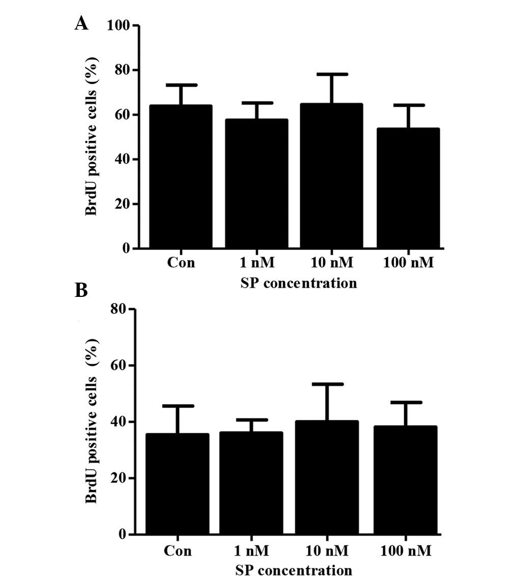
of SP on the proliferation of ST2 cells was evaluated. No increase
in the proliferation of this cell line was found in response to SP
under normal or serum-free culture conditions following treatment
with several concentrations of SP for 48 h (Fig. 3A and B). These data show that SP
can induce the migration of ST2 cells and suggest that this cell
line could be used in order to study the mechanisms involved in the
SP-mediated mobilization of MSCs.
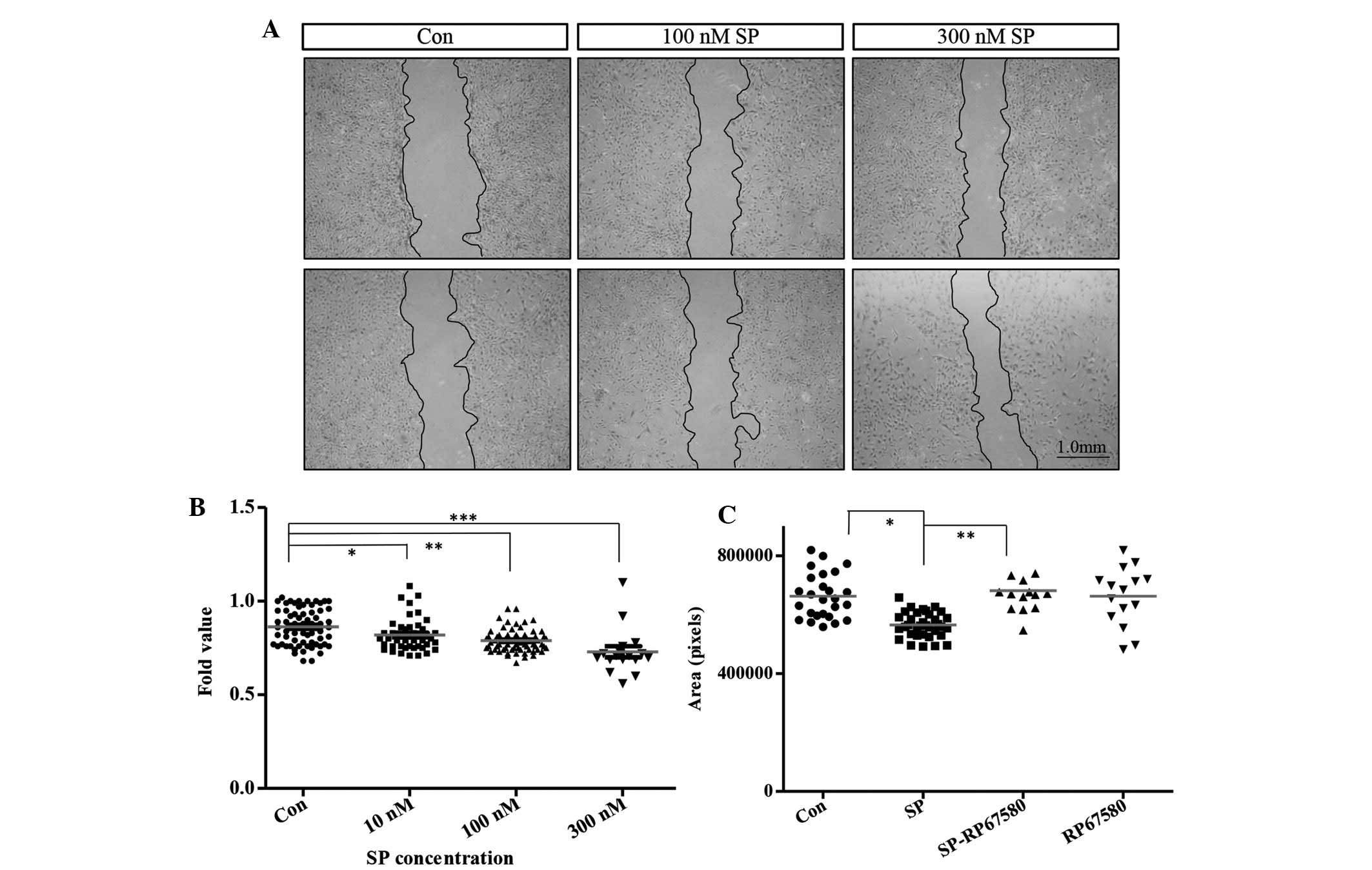 | Figure 2SP induces the mobilization of ST2
cells. (A) Representative images (scale bar, 1.0 mm) and (B) wound
area measurement 9 h after SP treatment (10, 100 and 300 nM). Three
independent experiments were performed and data are shown as the
mean ± standard error of mean. Con (0 nM SP), n=68; 10 nM SP, n=45;
100 nM SP, n=70; 300 nM SP, n=17. *P<0.0001,
**P<0.0148 and ***P<0.0001. (C) Wound
area measurement 9 h after 300 nM SP treatment with or without 10
μM RP67580. Data are shown as the mean ± standard deviation. Con (0
nM SP), n=26; SP, n=35; SP-RP67580, n=14; RP67580, n=15.
*P<0.0001 and **P<0.0001. Con, control;
SP, substance P. |
SP increases the proliferation of OP9
cells under normal serum culture conditions
OP9, another important cell line that is often used
to study the characteristics of MSCs, was established from newborn
calvaria of the (C57BL/6 × C3H) F2-op/op mouse. By
examining the immunophenotype, triple-differentiation capacity and
immunological and migration features of these cells, they were
found to be identical to standard MSCs. This cell line has been
used in co-culture systems with mouse embryonic stem cells where
they were found to support hematopoiesis (14).
In a previous study, our data suggested that SP
increased the number of MSCs inside the BM in vivo;
therefore, an aim of the present study was to evaluate whether SP
had the same effect on the proliferation of MSCs in vitro
(9). Since no effect of SP was
observed on the proliferation of ST2 cells, a decision was made to
evaluate the effect of SP on another mouse-derived MSC line,
knowing that established cell lines from the same cell type often
present different characteristics due to their distinct origin. The
effect of SP on the proliferation of the mouse-derived MSC line OP9
was therefore examined (Fig. 4).
By performing RT-qPCR analysis the expression of the NK-1 receptor
in the OP9 cells was confirmed (data not shown). No cytotoxic
effect of SP on the OP9 cells was observed at any of the
concentrations tested after 48 h of treatment (Fig. 4A). An increase in the proliferation
of OP9 cells, which was reflected by higher numbers of
BrdU-incorporating cells, was observed in the SP-treated groups
compared with the control group after 72 h of treatment with
different concentrations of SP; however, this increase was
significant only at a concentration of 10 nM SP (Con, 50.6±3.6,
n=5; 10 nM SP, 75.6±4.2, n=5; P<0.0021) (Fig. 4C and D). The specificity of this
effect was then confirmed by treatment with the NK-1 receptor
antagonist RP67580. It was observed that the effect of SP on the
proliferation of OP9 cells was inhibited in the group treated with
RP67580 in combination with SP compared with the group treated with
SP only (SP, 74.5±1.6, n=4; SP-RP67580, 61.8±4.0, n=4, P<0.042)
(Fig. 4E). It was of note that the
effect of SP on the proliferation of OP9 cells was not observed in
serum-free culture conditions, even after 72 h of treatment with
increasing concentrations of SP (Fig.
4B). In combination, these data show that SP increases the
proliferation of OP9 cells through the interaction with its
receptor NK-1, and that this effect may require the presence of
certain serum component(s).
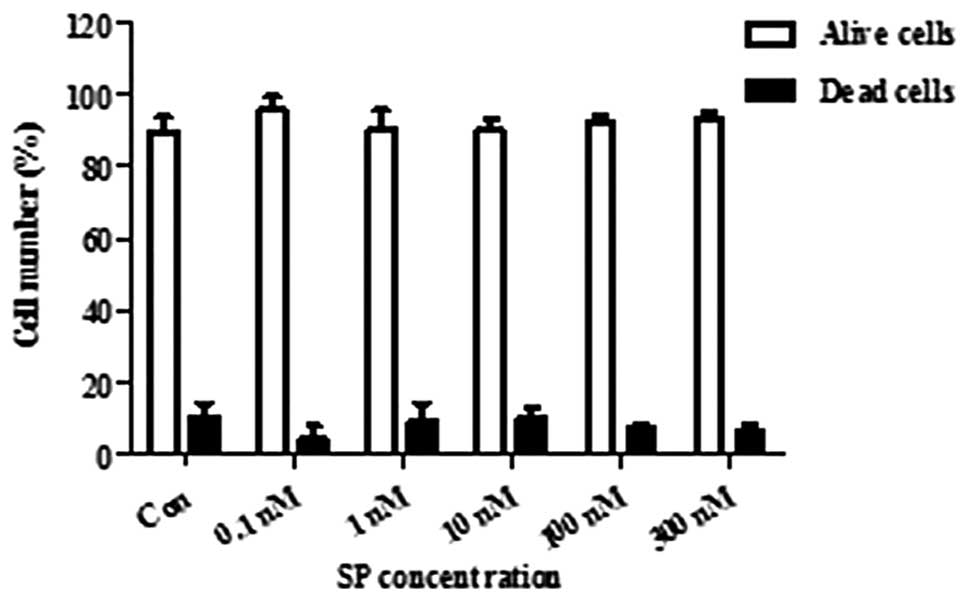
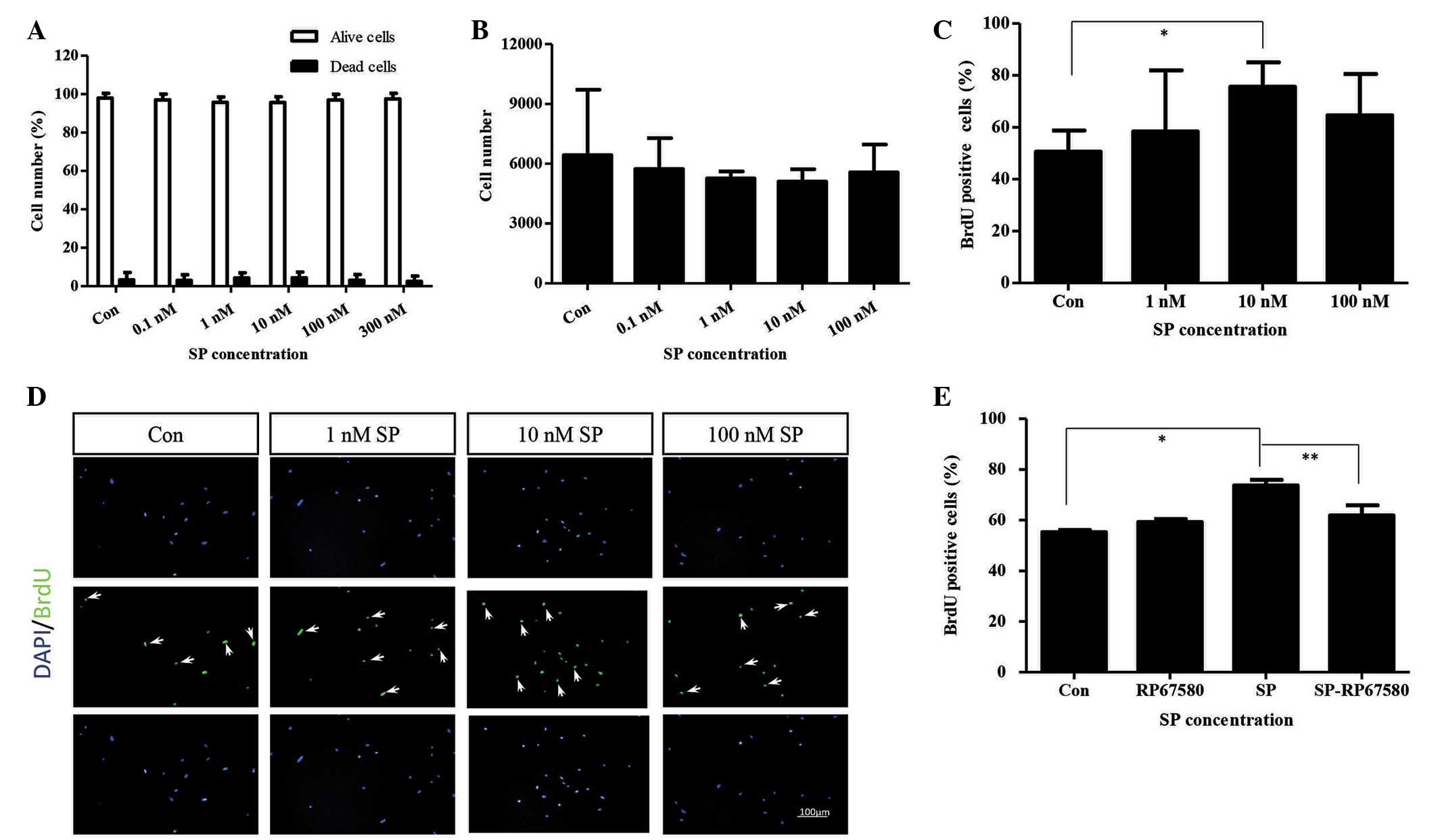 | Figure 4SP enhances the proliferation of OP9
cells. (A) Percentages of dead and alive cells 72 h after SP
treatment (0.1, 1, 10, 100 and 300 nM). Cell viability was assessed
on the basis of the exclusion of trypan blue-stained cells. Data
are presented as the mean ± standard deviation (n=3 per group). (B)
Numbers of OP9 cells 72 h after SP treatment (0.1, 1, 10 and 100
nM) under serum-free condition. Data are shown as the mean ±
standard deviation (n=3 per group). (C) Percentages and (D)
representative images of BrdU-positive cells 48 h after SP
treatment (1, 10 and 100 nM) under normal serum conditions. Cells
were treated with 20 μM BrdU for the last 12 h. Data are shown as
the mean ± standard deviation. (n=5 per group).
*P<0.0021. Arrows indicate BrdU-positive cells.
Nuclei were stained in blue with DAPI and BrdU was stained in green
(scale bar, 100 μm). (E) Percentage of BrdU-positive cells 48 h
after 10 nM SP treatment with or without 1 μM RP67580. Cells were
treated with 20 μM. BrdU for the last 12 h. Data are shown as the
mean ± standard error of mean from four independent experiments
(n=4 per group). *P<0.0002 and
**P<0.042. Con, control; SP, substance P; BrdU,
5-bromo-2′-deoxyuridine. |
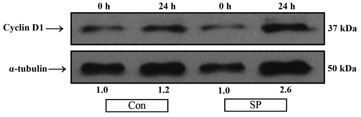
SP increases the levels of cyclin D1 in
OP9 cells
A key event in the activation of the cell cycle by
proliferative signaling pathways is the activation of cyclin D1. An
increased expression level of this protein is required for the
G1- to S-phase transition of the cell cycle (15). In the present study it was
evaluated whether the levels of cyclin D1 were altered by SP
treatment in the OP9 cells. An increase in the levels of this
protein was observed after 24 h of treatment in the SP-treated
cells compared with the untreated control cells (Fig. 5). These data show that SP increases
the levels of cyclin D1 in OP9 cells, suggesting that SP may
promote the proliferation of OP9 cells by inducing the transition
from G1- to S-phase of the cell cycle; however, further
studies are required in order to prove this hypothesis and to
evaluate the upstream signals involved in the SP-induced
proliferation of OP9 cells.
Discussion
The present study showed the ability of the
neurotransmitter SP to enhance the migration potential of the mouse
BM-derived MSC-like cell line ST2. SP was also demonstrated to
increase the proliferation of another mouse BM-derived MSC-like
cell line, OP9, under normal serum culture conditions. Furthermore,
SP increased the level of cyclin D1 protein in the OP9 cells. The
effects of SP on the proliferation and migration potential of the
above-mentioned cell lines resulted from the interaction of SP with
its receptor NK-1, which is expressed by these cells.
As more therapeutic properties and clinical
applications of MSCs are being identified, there is an increasing
requirement to improve the techniques to harvest endogenous
BM-derived MSCs and to further expand them in vitro. This is
important to contribute to the progress of the development of stem
cell therapies based on the use of autologous or allogeneic
BM-derived MSCs, as this would ultimately shorten the culture
expansion process, which has been proven to influence the innate
therapeutic characteristics of MSCs as early as the first passage
culture (7,16–18).
Reducing such changes could determine the successful application of
MSCs under different therapeutic settings where the most innate
stem cell properties are required. Alternatively, the success of
the application of MSCs in certain therapeutic treatments could
depend predominantly on the number of cells required to obtain a
positive effect. At least three different approaches could be
evaluated in the matter of improving the usage of MSCs as stem cell
therapy: i) To increase the proliferation of endogenous MSCs in
vivo; ii) to enhance the migration of MCSs from the BM into the
periphery; and iii) to induce the proliferation of harvested
endogenous MSCs in vitro. Accordingly, there is currently a
search for substances that could stimulate the naturally occurring
process of proliferation and liberation of endogenous reparative
MSCs in the organism and/or alternatively induce higher expansion
rates of MSCs in vitro (19).
The BM has been shown to be abundantly innervated
with sensory nerves that, in addition to conducting information
about different stimuli that could have the potential to cause
tissue damage, also secrete a variety of neurotransmitters, such as
SP and the calcitonin gene-related peptide, which could modulate
the characteristics of the BM-derived stem cells (20). In a previous study we found that SP
increased the CFU-F inside the BM two days after the injection of 5
nmol/kg SP in mice, suggesting that SP could increase the MSC
population inside the BM and, in this way, allow the isolation of
more MSCs from patients or donors (9). Further studies are required to
correctly identify whether these cells are true MSCs, since the
CFU-F in the BM comprises diverse cell populations.
Substances that can induce the mobilization of MSCs
from the BM into the periphery to facilitate the harvest of higher
numbers of endogenous MSCs are a current research focus; however,
despite the fact that several substances, such as
granulocyte-colony stimulating factor and AMD3100, have been
suggested to induce an increase in the number of circulating MSCs,
there is still no adequate protocol or drug combination that can
give high yields of mobilized endogenous MSCs. This shows the
necessity of expanding these cells in vitro in order to
reach sufficient cell numbers for each treatment (19,21,22).
A study by Hong et al (8) proposed that SP could be a suitable
stem cell mobilizer that could induce the mobilization of MSCs from
the BM into the periphery. In the study it was shown that SP
treatment caused an increase in the number of circulating
CD29-positive MSCs, which were found to participate in wound
repair; however, the mechanism for the SP-induced mobilization of
MSCs remains unknown. This motivated us to search for a stable cell
line that could permit the study of the mechanisms involved in the
SP-mediated mobilization of MSCs. For this reason, the effect of SP
on the migration potential of the ST2 cell line was evaluated in
the present study. It was found that SP increased the migration
potential of the ST2 cells 9 h after treatment in a
concentration-dependent manner without affecting their
proliferation potential. Furthermore, treatment with the NK-1
receptor antagonist RP67580 inhibited the migration induced by SP
in the ST2 cells. These data support the findings obtained in the
study by Hong et al (8). In
our previous study, it was observed that SP increased the mRNA
levels of N-cadherin and stromal cell-derived factor 1 inside the
BM one day after the injection of 5 nmol/kg SP into mice,
suggesting that these molecules could be involved in the mechanism
by which SP induces the mobilization of MSCs (9); however, further studies are required
to identify the role of these two molecules in the SP-mediated
mobilization of MSCs and the migration of ST2 cells. In
combination, these data show that SP enhances the migration
potential of the mouse-derived MSC-like cell line ST2 and suggest
that this cell line is suitable to investigate the mechanism(s)
involved in the SP-mediated mobilization of MSCs.
A previous study demonstrated that SP induced the
proliferation of human MSCs in vitro (8), as well as the activation of the
ERK-1/2 pathway and an increase in the nuclear translocation of
β-catenin (8); however, the
question still remains of whether these pathways are involved in
the mechanism(s) underlying the SP-mediated proliferation of MSCs.
Having found that SP induced the proliferation of mouse MSCs in
vivo (9), we subsequently
evaluated whether SP could exert the same effects on the mouse
BM-derived MSC-like cell line OP9 in vitro in order to
evaluate the ability of SP to improve the in vitro expansion
of MSCs and to examine the possibility of using this cell line to
identify the mechanism(s) involved in the proliferation mediated by
SP in MSCs. It was found that SP induced the proliferation of OP9
cells under normal serum culture conditions at an optimal
concentration of 10 nM. An increase in the protein levels of cyclin
D1 in the OP9 cells following treatment with 10 nM SP was also
observed. Cyclin D1 is proposed to act as an active switch in the
regulation of continued cell cycle progression, and high levels of
this protein are known to be necessary for cells to progress from
the G1- to the S-phase of the cell cycle (15,23).
The results of the present study suggest that SP
could have the potential to induce OP9 cells to undergo a
G1-/S-phase transition, hence inducing them to
proliferate; however, further investigations are required to
elucidate the effects of SP on the cell cycle in OP9 cells and to
identify the upstream signals involved in the SP-mediated
proliferation of OP9 cells. In this matter, the first candidates to
be evaluated would be the mitogen-activated protein kinase/ERK and
the Wingless/β-catenin pathways. It is of note that SP did not have
any effect on the proliferation of OP9 cells under serum-free
conditions, which suggests that SP may require the presence of
certain serum component(s) to execute its stimulating effect. We
did not performed any further studies in this matter, but it would
be interesting to determine whether SP is also dependent upon the
collaboration of other molecules to induce its effects on MSCs
in vivo. In conclusion, this study presents evidence that SP
could be considered for use in the development of stem cell
therapies based on MSCs as a substance that could facilitate the
harvest of high numbers of endogenous MSCs from patients and/or
donors and increase the in vitro expansion rates of
endogenous MSCs prior to their use in stem cell therapy.
Acknowledgements
This study was supported by the Korean Health
Technology R&D Project, Ministry of Health and Welfare,
Republic of Korea (no. HI13C1479), the Basic Science Research
Program through the National Research Foundation of Korea (NRF)
funded by the Ministry of Education (no. NRF-2012R1A1A2042265) and
the Bio and Medical Technology Development Program of the NRF
funded by the Ministry of Science, ICT and Future Planning (no.
NRF-2012M3A9C6050485).
References
|
1
|
Lotfinegad P, Shamsasenjan K,
Movassaghpour A, Majidi J and Baradaran B: Immunomodulatory nature
and site specific affinity of mesenchymal stem cells: A hope in
cell therapy. Adv Pharm Bull. 4:5–13. 2014.PubMed/NCBI
|
|
2
|
Tang YL, Zhao Q, Zhang YC, et al:
Autologous mesenchymal stem cell transplantation induce VEGF and
neovascularization in ischemic myocardium. Regul Pept. 117:3–10.
2004. View Article : Google Scholar
|
|
3
|
Aggarwal S and Pittenger MF: Human
mesenchymal stem cells modulate allogeneic immune cell responses.
Blood. 105:1815–1822. 2005. View Article : Google Scholar
|
|
4
|
Herrera MB, Bussolati B, Bruno S, Fonsato
V, Romanazzi GM and Camussi G: Mesenchymal stem cells contribute to
the renal repair of acute tubular epithelial injury. Int J Mol Med.
14:1035–1041. 2004.PubMed/NCBI
|
|
5
|
Shake JG, Gruber PJ, Baumgartner WA, et
al: Mesenchymal stem cell implantation in a swine myocardial
infarct model: Engraftment and functional effects. Ann Thorac Surg.
73:1919–1925. 2002. View Article : Google Scholar : PubMed/NCBI
|
|
6
|
Kim N and Cho SG: Clinical applications of
mesenchymal stem cells. Korean J Intern Med. 28:387–402. 2013.
View Article : Google Scholar : PubMed/NCBI
|
|
7
|
Banfi A, Muraglia A, Dozin B,
Mastrogiacomo M, Cancedda R and Quarto R: Proliferation kinetics
and differentiation potential of ex vivo expanded human bone marrow
stromal cells: Implications for their use in cell therapy. Exp
Hematol. 28:707–715. 2000. View Article : Google Scholar : PubMed/NCBI
|
|
8
|
Hong HS, Lee J, Lee E, et al: A new role
of substance P as an injury-inducible messenger for mobilization of
CD29(+) stromal-like cells. Nat Med. 15:425–435. 2009. View Article : Google Scholar : PubMed/NCBI
|
|
9
|
Dubon MJ, Byeon Y, Jung N, Son Y and Park
KS: Substance P modulates properties of bone marrow-derived
mesenchymal stem cells. Tissue Eng Regen Med. 11:217–223. 2014.
View Article : Google Scholar
|
|
10
|
Ogawa M, Nishikawa S, Ikuta K, et al: B
cell ontogeny in murine embryo studied by a culture system with the
monolayer of a stromal cell clone, ST2: B cell progenitor develops
first in the embryonal body rather than in the yolk sac. EMBO J.
7:1337–1343. 1988.PubMed/NCBI
|
|
11
|
Tong J, Kishi H, Matsuda T and Muraguchi
A: A bone marrow-derived stroma cell line, ST2, can support the
differentiation of fetal thymocytes from the CD4+
CD8+ double negative to the CD4+
CD8+ double positive differentiation stage in vitro.
Immunology. 97:672–678. 1999. View Article : Google Scholar : PubMed/NCBI
|
|
12
|
Trentin JJ: Determination of bone marrow
stem cell differentiation by stromal hemopoietic inductive
microenvironments (HIM). Am J Pathol. 65:621–628. 1971.PubMed/NCBI
|
|
13
|
Mercier FE, Ragu C and Scadden DT: The
bone marrow at the crossroads of blood and immunity. Nat Rev
Immunol. 12:49–60. 2011. View
Article : Google Scholar : PubMed/NCBI
|
|
14
|
Gao J, Yan XL, Li R, et al:
Characterization of OP9 as authentic mesenchymal stem cell line. J
Genet Genomics. 37:475–482. 2010. View Article : Google Scholar : PubMed/NCBI
|
|
15
|
Stacey DW: Cyclin D1 serves as a cell
cycle regulatory switch in actively proliferating cells. Curr Opin
Cell Biol. 15:158–163. 2003. View Article : Google Scholar : PubMed/NCBI
|
|
16
|
Bernardo ME, Cometa AM, Pagliara D, et al:
Ex vivo expansion of mesenchymal stromal cells. Best Pract Res Clin
Haematol. 24:73–81. 2011. View Article : Google Scholar : PubMed/NCBI
|
|
17
|
Fossett E and Khan WS: Optimising human
mesenchymal stem cell numbers for clinical application: A
literature review. Stem Cells Int. 2012:4652592012. View Article : Google Scholar : PubMed/NCBI
|
|
18
|
Bentivegna A, Miloso M, Riva G, et al: DNA
methylation changes during in vitro propagation of human
mesenchymal stem cells: Implications for their genomic stability?
Stem Cells Int. 2013:1924252013. View Article : Google Scholar : PubMed/NCBI
|
|
19
|
Pelus LM: Peripheral blood stem cell
mobilization: New regimens, new cells, where do we stand. Curr Opin
Hematol. 15:285–292. 2008. View Article : Google Scholar : PubMed/NCBI
|
|
20
|
Offley SC, Guo TZ, Wei T, et al:
Capsaicin-sensitive sensory neurons contribute to the maintenance
of trabecular bone integrity. J Bone Miner Res. 20:257–267. 2005.
View Article : Google Scholar : PubMed/NCBI
|
|
21
|
Deng J, Zou ZM, Zhou TL, et al: Bone
marrow mesenchymal stem cells can be mobilized into peripheral
blood by G-CSF in vivo and integrate into traumatically injured
cerebral tissue. Neurol Sci. 32:641–651. 2011. View Article : Google Scholar : PubMed/NCBI
|
|
22
|
Pitchford SC, Furze RC, Jones CP, Wengner
AM and Rankin SM: Differential mobilization of subsets of
progenitor cells from the bone marrow. Cell Stem Cell. 4:62–72.
2009. View Article : Google Scholar : PubMed/NCBI
|
|
23
|
Hitomi M and Stacey DW: Cellular ras and
cyclin D1 are required during different cell cycle periods in
cycling NIH 3T3 cells. Mol Cell Biol. 19:4623–4632. 1999.PubMed/NCBI
|