Introduction
Cryptorchidism is a common congenital abnormality
with high incidence and prevalence (1). The prevalence varies with apparent
geographical differences (2). In
general, the incidence rate is 30.3% for preterm infants, 4% for
newborns and 0.8% for one-year-old babies. The condition also
represents one of the major causes of male infertility. The
infertility rate of patients with bilateral cryptorchidism may be
as high as 50–100%, whereas the infertility rate of patients with
unilateral cryptorchidism is 30–60% (3). The reason for this may be that the
incidence rate of patients with bilateral cryptorchidism is very
low, and with the development of society, the majority of people
select the surgery in childhood (4).
The dysfunction of the contralateral testis could be an important
cause of male infertility with unilateral cryptorchidism symptoms
(4); however, the mechanism
associated with the damage has not been fully elucidated. Foresta
et al (5) proposed that
unilateral cryptorchidism is a congenital disease and that the
damage to the contralateral testicle is the ultimate result of this
inherent disease. Vasquez et al (6) demonstrated that the concentrations of
follicle stimulating hormone, luteinizing hormone and testosterone
were associated with the sperm density of patients with unilateral
cryptorchidism, and that damage to the contralateral testicular was
caused by endocrine abnormalities. It is additionally believed that
the blood-testis barrier in unilateral cryptorchidism could be the
cause of autoimmune reactions or allergic orchitis, which could
then cause contralateral testis damage (7,8).
The genitofemoral nerve (GFN) originates in the
first and second lumbar plexus, cycles through the psoas muscle and
then forms the reproductive and groin branches. The reproductive
branch is a sensory-motor hybrid neuron that enters the inguinal
canal at the inner ring and descends along the inguinal canal. The
fibers are widely distributed in the cremaster muscle, gubernaculum
and testicular hydrocele (9). By
contrast, the groin branch enters the testicular vascular system
directly and is the most important afferent nerve supplying the
testis (10). In the present study,
the nerve distribution of the contralateral testis, including the
areas of calcitonin gene-related peptide (CGRP)-positive cells and
certain associated factors, was observed, and the mechanism
underlying the impairment of the contralateral testes in unilateral
cryptorchidism was explored in experimental rats.
Materials and methods
Establishment of the experimental
model
Thirty-six male Sprague Dawley rats (weight, 120–180
g) were randomly assigned to the control (group A), left unilateral
cryptorchidism (group B) and left unilateral cryptorchidism with
division of the left GFN (group C) groups (n=12/group) A ventral
midline incision was performed in group C following anesthesia and
the GFN was separated from the leading edge of the left psoas
muscle. The ipsilateral testicular gubernaculum was cut off, the
testis was fixed to the posterior abdominal wall with 4-0 sutures
(without damage) and the abdomen was closed in two layers. The rats
from group B were treated in the same way but the GFN was kept
intact. The operation was finished following the opening of the
abdominal wall of the rats. For the rats in group A, no additional
treatment was applied following the closure of the abdominal wall
and the rats were raised as normal. All rats were sacrificed by
rapid cervical dislocation subsequent to a further 100 days of
feeding, and the contralateral testis from each rat was collected
for further measurement. The weights of the testes were determined.
The housing of the rats and the procedures involving the
experimental animals were in accordance with the Guide for the Care
and Use of Laboratory Animals (eighth edition, 2011). All animal
experiments were approved by the Animal Care and Use Committee of
Wuhan University (Wuhan, China).
Immunohistochemistry to analyze the
CGRP-positive cells of the contralateral testis
Conventional specimen slices were prepared and the
immunohistochemical staining method was applied. Following
deparaffinization and dehydration, the sections underwent 0.5%
potassium citrate (Wuhan Boster Biological Technology Co., Ltd.,
Wuhan, China) antigen retrieval at 100°C for 15 min. The sections
were then blocked with 5–10% goat serum and incubated at 37°C for
20 min. The primary antibody (rabbit polyclonal anti-CGRP; cat. no.
BA0204; Wuhan Boster Biological Technology Co., Ltd.) was
subsequently added at a dilution of 1:100 and the samples were
incubated at 37°C for a further 15 h. The samples were then washed
with phosphate-buffered saline (PBS) for 2 min for a total of three
times, incubated with secondary antibody (biotin-labeled goat
anti-rabbit immunoglobulin G; Beijing Zhongshan Biotechnology Co.,
Ltd., Beijing, China) at 37°C in a water bath for 2 h and re-washed
with PBS for 2 min for a total of 3 times. Following treatment with
3,3′-diaminobenzidine solution, the sections were flushed,
counterstained with hematoxylin, washed with water, dehydrated,
cleared, mounted on slides and observed under the microscope. Ten
immunohistochemical staining slices were randomly selected from
each group and the average luminosity, area and positive rate of
CGRP-positive cells were detected with an automatic image analyzer
(HPIAS-2000 image analysis software; Tongji Qiangping Image
Engineering Co., Wuhan, China).
Determination of malondialdehyde (MDA)
content in samples
The MDA content in the samples was determined using
a chemical colorimetric method and an MDA assay kit (Nanjing
Jiancheng Institute of Biology, Nanjing, China). The assay was
performed in accordance with the manufacturer's instructions.
Detection of apoptosis
Testicular germ cell apoptosis was detected using
the terminal deoxynucleotidyl-transferase-mediated dUTP nick end
labeling method and an apoptosis detection kit (Wuhan Boster
Biological Technology Co., Ltd. in accordance with the
manufacturer's instructions. Thirty seminiferous tubule sections
were selected from each slice and the percentage of positive cells
was calculated to give the apoptosis index (AI).
Ultrastructural changes of testicular
Sertoli cells
The ultrastructural changes of testicular Sertoli
cells were observed using transmission electron microscopy (TEM;
JEM100CXII; JEOL Ltd., Beijing, China).
Statistical analysis
Values are presented as the mean ± standard
deviation. All data analysis was performed with SPSS 17.0
statistical software (SPSS, Inc., Chicago, IL, USA). The data were
analyzed using the Student's t-test for comparisons among groups.
P<0.05 was considered to indicate a statistically significant
difference.
Results
Weight, AI and MDA
Compared with the results for group A, the weight of
the contralateral testis in group B was significantly reduced and
the AI and levels of MDA were significantly increased (P<0.01).
Compared with the results for group B, the weight of the
contralateral testis in group C was significantly increased. The
MDA levels and AI in group C were significantly reduced compared
with those in group B (P<0.01), but remained higher than those
in group A (P<0.05) (Table
I).
 | Table I.Changes in the weight, MDA levels and
AI of the contralateral testis in groups A, B and C. |
Table I.
Changes in the weight, MDA levels and
AI of the contralateral testis in groups A, B and C.
| Group | Weight (mg) | MDA (nmol/mg) | AI (%) |
|---|
| A | 746.33±95.22 | 2.41±0.50 | 6.48±1.64 |
| B |
504.71±50.10a |
7.24±1.19a |
17.24±3.36a |
| C |
642.28±69.51b |
3.63±0.72b |
8.06±2.07b |
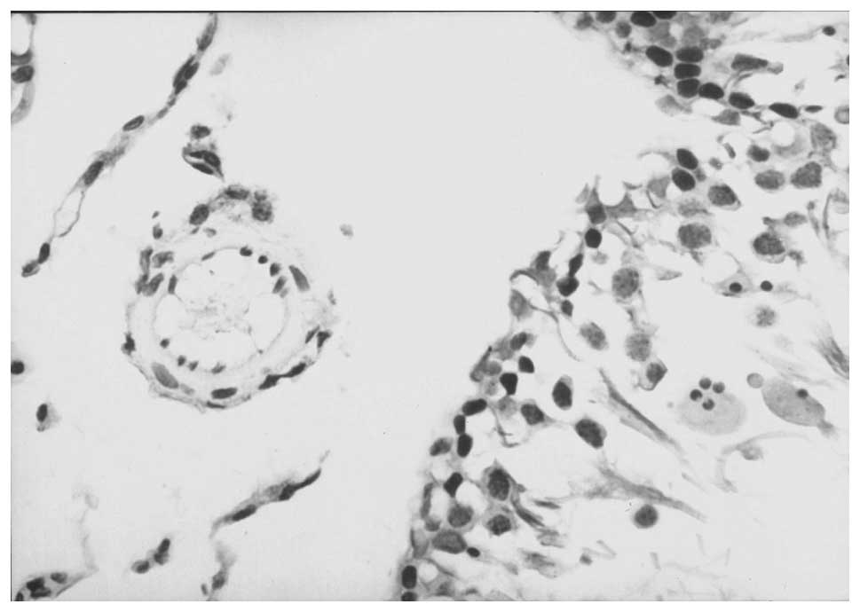
Immunohistochemistry
Immunohistochemistry showed that there were numerous
nerve fibers rich in CGRP-positive cells in the interstitial tissue
of the testis in group A. The brown immunoreactive substances in
the cells exhibited a granular distribution. The staining also
revealed intensive agglomeration, with positive fibers distributed
along the perivascular nerves belonging to the peripheral component
of the sympathetic nervous system. The vascular smooth muscle was
thick and lined by endothelial cells (Fig 1). By contrast, the seminiferous
epithelium in group B was thin and the number of CGRP-positive
cells was significantly reduced (P<0.01). In addition, the
vascular wall was thin and the diameter was narrow. The neural
image analysis results of the CGRP-positive cells are shown in
Table II. Compared with group A,
the CGRP-positive nerve distribution and content in group B was
significantly reduced (P<0.01); however, compared with group B,
the CGRP-positive nerve distribution and content in group C was
significantly increased (P<0.01).
| Table II.Neural image analysis results of the
CGRP-positive cells in the contralateral testis in groups A, B and
C. |
Table II.
Neural image analysis results of the
CGRP-positive cells in the contralateral testis in groups A, B and
C.
| Group | Average area
(cm2) | Average luminosity
(A) | Positive rate
(%) |
|---|
| A | 6.80±1.05 | 0.48±0.18 | 2.22±0.69 |
| B |
2.11±0.65a | 0.21±0.90 |
0.73±0.22a |
| C | 4.76±0.82 |
0.34±0.13b |
1.49±0.38b |
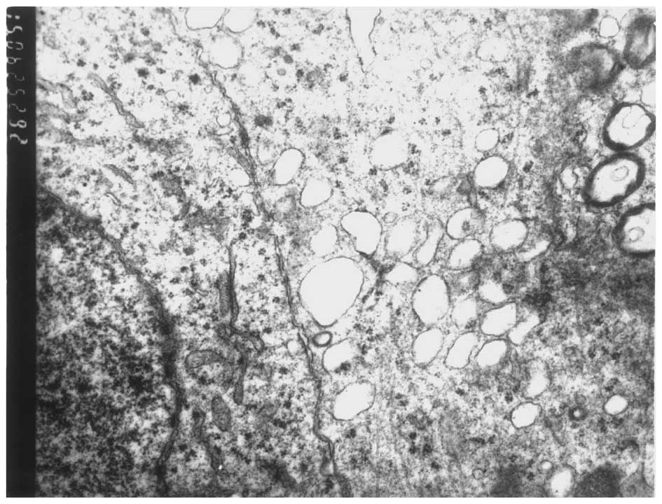
TEM
Observation with TEM showed that the Sertoli cells
of group B were in the basement membrane of the seminiferous
tubule. The cells exhibited swollen mitochondria with fewer cristae
and smooth endoplasmic reticulum expansion, which suggested the
existence of vacuolar changes. Additionally, the number of
lysosomes was increased (Fig. 2).
Few changes were observed in the Sertoli cells in groups A and
C.
Discussion
Incomplete descent of the testes is the most common
genital anomaly in newborn males (2). The dysfunction of the contralateral
testis could be an important cause of male infertility with
unilateral cryptorchidism symptoms. The present experimental
results showed that the seminiferous epithelium was thinner in rats
with unilateral cryptorchidism, while the MDA content was increased
and there was a higher proportion of germ cells undergoing
apoptosis. A positive association existed between the MDA content
and the level of germ cell apoptosis. The swelling of the
mitochondria and smooth endoplasmic reticulum of the Sertoli cells
due to lysosomal accumulation indicated the existence of
contralateral testis damage, although the potential mechanism
underlying the damage is not entirely clear.
The testis and epididymis efferent nerves are highly
affected by visceral sympathetic nerves, which are derived from the
periarterial nerve plexuses, and both the GFN and lumbar
sympathetic trunk have traffic branches. In the present study, the
GFN of the cryptorchid side was removed, which reduced or prevented
the degeneration of the contralateral testis and demonstrated that
nerve conduction in general, and through the GFN in particular,
plays an important role in the mechanism underlying contralateral
testicular damage. The present results were consistent with the
conclusions of Patkowski et al (11).
CGRP is a single polypeptide chain consisting of 37
amino acids that is transcribed following alternative splicing of
the calcitonin gene-encoded mRNA, and is widely found in the
central and peripheral nervous systems. A large number of nerve
cells containing CGRP are found in the normal testis, and these
exhibit sympathetic characteristics (12,13).
Numerous studies (14,15) have shown that CGRP is the strongest
neural active peptide that dilates the blood vessels of the body.
Following the binding of CGRP with the corresponding target
receptor, adenylate cyclase is activated to play a role similar to
that of Ca2+ antagonists, by reducing the intracellular
free C2+ levels and relaxing the smooth muscle of the
blood vessels. CGRP can additionally antagonize the release of
endothelin and angiotensin II by activating the cyclic adenosine
monophosphate/protein kinase pathway with prostacyclin. A decrease
in the concentration of CGRP can not only reduce the normal
vasodilatory effect but also reduce the inhibitory effect of
endothelin and angiotensin II. As a result, the vascular lumen
narrows and changes occur in the hemodynamics, leading to a
reduction in the perfusion of the tissue. Ischemia, hypoxia and
oxygen free radicals in the cells within the tissue also increase.
CGRP can additionally reduce the leakage of intracellular proteins
and enzymes, promote DNA synthesis, stabilize the target organ
membrane and regulate cell function by reducing Ca2+
overload, thus exerting a direct protective effect on cells
(16).
MDA is a metabolite produced by the attack of oxygen
free radicals on polyunsaturated fatty acids, and changes in the
MDA content reflect the changes in oxygen free radical levels
within the cells. An increase in MDA indicates that the
intracellular oxygen free radical and lipid peroxidation levels
have also increased, leading to a promotion of cell apoptosis.
The results of the present study showed that
unilateral cryptorchidism could cause spermatogenic damage of the
contralateral testis, and the increases in the level of germ cell
apoptosis could be due to the reduction in the CGRP released by
contralateral nerves. With continuous stimulation from ectopic
sources, such as intra-abdominal high temperature, levels of
contralateral CGRP undergo a marked reduction, as GFN fluxes to the
sympathetic center; however, a decrease in the levels of CGRP can
also cause a decrease in the vasodilation capacity, resulting in
increased levels of intracellular oxygen free radicals and lipid
peroxidation. The reduction in CGRP can simultaneously cause a
further decline in cytoprotection and promote the degeneration and
apoptosis of testicular cells. The removal of one side of the GFN,
i.e. the removal of the afferent nerve, can alleviate the damage to
the contralateral testis. The present results therefore showed that
the surgery of cryptorchidism or the removal of part of the GFN in
the early clinical stages could help protect the contralateral
testis from being further damaged.
The nerve conduction mechanism underlying the
contralateral testicular damage in unilateral cryptorchidism is a
complex process. Questions regarding whether other
neurotransmitters besides CGRP are also involved, and whether
exogenous CGRP has a protective effect on the contralateral testis,
warrant further study.
References
|
1
|
Kumar V, Misro MM and Datta K:
Simultaneous accumulation of hyaluronan binding protein 1
(HABP1/p32/gC1qR) and apoptotic induction of germ cells in
cryptorchid testis. J Androl. 33:114–121. 2012. View Article : Google Scholar : PubMed/NCBI
|
|
2
|
Kollin C and Ritzén EM: Cryptorchidism: a
clinical perspective. Pediatr Endocrinol Rev. 11(2): 240–250.
2014.PubMed/NCBI
|
|
3
|
Lee PA, Bellinger MF, Songer NJ, O'Leary
L, Fishbough R and LaPorte R: An epidemiologic study of patemity
after cryptorchidism; intial results. Eur J Pediatr. 152(2):
S25–S27. 1993. View Article : Google Scholar : PubMed/NCBI
|
|
4
|
Huff DS, Fenig DM and Canning DA: Abnormal
germ cell development in cryptorchidism. Horm Res. 55:11–17. 2001.
View Article : Google Scholar : PubMed/NCBI
|
|
5
|
Foresta C, Ferlin A, Garolla A, Milani C,
Oliva G and Rossato M: Functional and cytologic features of the
contralateral testis in cryptorchidism. Fertil Steril. 66:624–629.
1996.PubMed/NCBI
|
|
6
|
Vasquez JM, Ben-Num L, Greenblatt RB, et
al: Correlation between follicle-stimulating hormone, luteinizing
hormone, prolactin, and testerone with sperm cell concentration and
motility. Obstet Gynecol. 67:86–89. 1986.PubMed/NCBI
|
|
7
|
Stewart RJ, Boyd S, Brown S and Toner PG:
The blood-testis barrier in experiment unilateral cryptorchidism. J
Pathol. 160:51–55. 1990. View Article : Google Scholar : PubMed/NCBI
|
|
8
|
Lenzi A, Geandini L, Lombardo F, et al:
Unilateral cryptorchidism corrected in prepubertal age: evaluation
of sperm parameters, hormones, and antisperm antibodies in adult
age. Fertil Steril. 67:943–948. 1997. View Article : Google Scholar : PubMed/NCBI
|
|
9
|
Zempoalteca R, Martínez-Gómez M, Hudson R,
Cruz Y and Lucio RA: An anatomical and electrophysiological study
of the genitofemoral nerve and some of its targets in the male rat.
J Anat. 201:493–505. 2002. View Article : Google Scholar : PubMed/NCBI
|
|
10
|
Kar S, Gibson SJ and Polak JM: Origins and
projections of peptide-immunoreactive nerves in the male rat
genitofemoral nerve. Brain Res. 512:229–237. 1990. View Article : Google Scholar : PubMed/NCBI
|
|
11
|
Patkowski D, Czernik KJ and Jelen M:
Division of the genitofemoral nerve in unilateral cryptorchid rats.
J Pediatr Surg. 29:832–835. 1994. View Article : Google Scholar : PubMed/NCBI
|
|
12
|
Morris HR, Panico M, Etienne T, et al:
Isolation and characterization of human calcitonin gene-related
peptide. Nature. 308:746–748. 1984. View
Article : Google Scholar : PubMed/NCBI
|
|
13
|
Rasmussen TN, Bersani M, Schmidt P, et al:
Isolation and molecular characterization of porcine calcitonin
gene-related peptide (GGRP) and its endocrine effects in the
porcine pancreas. Pancreas. 16:195–204. 1998. View Article : Google Scholar : PubMed/NCBI
|
|
14
|
Mimaki Y, Kawasaki H, Okazaki M, et al:
Involvement of calcitonin gene-related peptide (CGRP) receptors in
insulin-induced vasodilatation in mesenteric resistance blood
vessels of rats. Br J Pharmacol. 123:1684–1690. 1998. View Article : Google Scholar : PubMed/NCBI
|
|
15
|
Benemei S, Nicoletti P, Capone JA and
Geppetti P: Pain pharmacology in migraine: Focus on CGRP and CGRP
receptors. Neurol Sci. 28:S89–S93. 2007. View Article : Google Scholar : PubMed/NCBI
|
|
16
|
Ren YS, Ma TG, Wang and Yu SQ: Membrane
fluidity changes in myocardial cells following severe hypoxia and
simulated reperfusion and effects of calcitonin gene-related
peptide. Med Sci Res. 21:627–628. 1993.
|