Introduction
Gastric cancer is one of the most common clinical
malignancies. The condition has a high incidence in China and can
have a serious impact on human health (1). The pathogenesis of gastric cancer is
currently believed to include multiple steps: Chronic atrophic
gastritis, intestinal metaplasia of the gastric mucosa, atypical
hyperplasia of the gastric mucosa and the eventual development of
gastric cancer (2–4). These steps (chronic atrophic gastritis,
intestinal metaplasia of the gastric mucosa and atypical
hyperplasia of the gastric mucosa) are known as gastric
precancerous lesions. Gastric precancerous lesions play an
important cohesive role in the development of tumors (5). Helicobacter pylori (Hp) is one
of the main causative agents of chronic active gastritis. Recent
studies have shown that Hp infection is an independent risk factor
for intestinal metaplasia, which suggests that it may play an
important role in the occurrence and development of gastric
precancerous lesions (6–8).
The mechanism underlying the Hp-induced occurrence
and development of gastric precancerous lesions has yet to be fully
elucidated. Cyclooxygenase-2 (COX-2) is the rate-limiting enzyme in
the synthesis of prostaglandins, and its expression level is low or
non-existent in normal tissues (9).
Studies have shown that COX-2 exhibits high expression in numerous
tumor tissues, which may be closely associated with tumorigenesis
and metastasis (10,11). Inducible nitric oxide synthase (iNOS)
is the main observation index of the inflammatory reaction, and
high expression levels of the enzyme are indicative that an
inflammatory reaction is ongoing (12). The aim of the present study was to
analyze the effect of Hp infection on the expression levels of
COX-2 and iNOS in gastric precancerous lesion tissue and in a cell
line in vitro, and to explore the clinical significance of
COX-2 and iNOS in the development of Hp-induced gastric
precancerous lesions.
Subjects and methods
General data
A total of 114 patients, who were diagnosed with
gastric precancerous lesions by gastroscopy and pathological
examination in the Henan University Huaihe Hospital (Kaifeng,
China) between June 2010 and 2013, were included in this study.
Pathological diagnosis was classified according to the diagnostic
criteria formulated by the National Chronic Gastritis Conference
(13). Among these patients, 57 were
confirmed to be Hp-positive by Warthin-Starry silver staining and
the rapid urease test, and they were classified into the
observation group; the remaining 57 patients were Hp-negative and
were referred to as the control group. The observation group
comprised 32 males and 25 females who were aged 38–75 years (mean
age, 51.4±10.2 years); there were 25 cases of chronic atrophic
gastritis, 20 cases of intestinal metaplasia of the gastric mucosa
and 12 cases of atypical hyperplasia of the gastric mucosa. In the
control group, there were 35 males and 22 females (age range, 39–73
years; mean age, 50.1±10.9 years); among these patients, there were
22 cases of chronic atrophic gastritis, 21 cases of intestinal
metaplasia of the gastric mucosa and 14 cases of gastric mucosal
atypical hyperplasia. No significant differences were found in the
age, gender, pathological type or other indicators between the two
groups (P>0.05). This study was conducted in accordance with the
Declaration of Helsinki and with approval from the Ethics Committee
of Henan University Huaihe Hospital. Written informed consent was
obtained from all participants.
Collection of clinical specimens
All patients were examined by gastroscopy. Part of
the obtained tissue was used for the extraction of total RNA, and
part of tissue was used for the detection of COX-2 and iNOS
levels.
Cell infection experiment in
vitro
Normal human gastric mucosal GES-1 cells (American
Type Culture Collection, Manassas, VA, USA) were cultured to the
exponential growth phase in Dulbecco's modified Eagle's medium with
10% fetal bovine serum (Gibco-BRL, Grand Island, NY, USA), and then
digested with 0.25% pancreatin. The cell concentration was adjusted
to 4×10⁵/ml, and the cells were inoculated in a 12-well
plate with 1 ml per hole. Following 24 h of culture, when the cells
were completely attached to the wall, 1×10⁵ CFU Hp
(Qicheng Industrial Co., Ltd, Shanghai, China) was added to the
12-well plate. Equal quantities of PBS were added to the control
group wells. The cells were placed in a 37°C CO2
incubator for 6, 12, 18 and 24 h, and then dissolved in TRIzol®
(Life Technologies, Carlsbad, CA, USA) for the extraction of total
RNA. The expression of COX-2 and iNOS was analyzed.
Reverse transcription-quantitative
polymerase chain reaction (RT-qPCR)
Biopsy tissue or HP-treated cells were placed in 1
ml TRIzol solution and 200 µl chloroform was added to the
homogenate. The homogenate was agitated, mixed and placed on ice
for 15 min to create a layered solution, which was then centrifuged
at 15,000 × g for 15 min. The supernatant was transferred to 500 µl
isopropanol, mixed, placed on ice for 15 min and further
centrifuged at 15,000 × g for 10 min for RNA precipitation. The
precipitate was then washed twice with pre-cooled 75% ethanol,
prior to being dissolved in double-distilled water treated with
diethylpyrocarbonate. The concentration of the sample was
determined, and RNA was transcribed into cDNA using an RT kit
(Takara, Dalian, China) to create a template for the PCR.
According to the COX-2 and iNOS mRNA sequences from
GenBank (http://www.ncbi.nlm.nih.gov/genbank/), the primers
were designed as follows: COX2 forward, 5′-GAA TCA TTC ACC AGG CAA
ATTG-3′ and reverse, 5′-TCT GTA CTG CGG GTG GAA CA-3′; iNOS
forward, 5′-ACA ACA AAT TCA GGT ACG CTG TG-3′ and reverse, 5′-TCT
GAT CAA TGT CAT GAG CAA AGG-3′; β-actin forward, 5′-GCG GGA AAT CGT
GCG TGAC-3′ and reverse, 5′-CGT CAT ACT CCT GCT TGC TG-3′.
Following the dilution of primer, the conditions for specificity
and the annealing temperature were optimized The reaction system
was prepared as follows: 10 µl 2X SYBR Green qPCR Master Mix
(Takara), 1 µl upstream primer and 1 µl downstream primer (10
µmol/1), and 1 µl cDNA. The double-distilled water was supplemented
to a final volume of 20 µl. Following centrifugation at 1,800 × g,
the reaction mixture was gathered at the bottom of the tube. PCR
was performed according to the following reaction conditions:
Predegeneration at 95°C for 30 sec; degeneration at 95°C for 3 sec;
and annealing and extension at 60°C for 30 sec for 25 cycles. The
standard curve was constructed. Data were then directly read from
the ABI® 7500 real-time PCR instrument (Applied Biosystems, Milan,
Italy).
Western blot analysis
A total of 100 µl cell lysate was obtained from the
gastric mucosa tissue in two groups of patients, and 1 µl protease
inhibitor was added. The homogenate was then placed on ice and left
to stand for 30 min, prior to centrifugation at 15,000 × g for 15
min. The supernatant was removed and the concentration of protein
was measured. Sample buffer (4X) was added and boiled using boiling
water for 30 min. The sample was added and centrifugation was
performed following the addition of sample buffer at 15,000 × g for
10 min. Electrophoresis was performed using 12% SDS-PAGE at an
initial voltage of 80 V. The voltage was adjusted to 120 V once the
bromophenol blue had entered the gel. When the electrophoresis was
complete, the sample was transferred to a polyvinylidene difluoride
membrane. Blocking was performed using 5% skimmed milk powder, and
then the primary monocolonal mouse antibody against COX-2 (1:100;
#sc-29411; Santa Cruz Biotechnology, Inc., Santa Cruz, CA, USA) was
added for overnight incubation. The membrane was then washed with
PBS-Tween 20 (PBST) three times. The horseradish-peroxidase
conjugated goat anti-rat secondary antibody (ZSGB-BIO, Beijing,
China) was then added for 1 h of incubation at room temperature,
prior to three further washes with PBST. The ECL luminous liquid
(ZSGB-BIO) was added, the signal was detected and images were
captured. β-actin was considered as a reference protein. The band
intensity of the target protein was analyzed using Tocan 430 gray
calculation software (Tocan Biotechnology Co., Shanghai, China),
and the relative expression level of the target protein was
calculated.
Analysis of iNOS activity
The activity of iNOS was analyzed using the Griess
reaction principle. In brief, a small quantity of gastric mucosa
was homogenized and then centrifuged at 15,000 × g for 5 min. The
supernatant was removed and an equal volume of Griess reaction
liquid (Wako Pure Chemical Industries Ltd., Osaka, Japan) was added
and mixed fully, and the mixture was then left to stand at room
temperature for 15 min. The optical density was analyzed at a
wavelength of 540 nm by microplate reader. The NO level in the
supernatant was calculated according to the results of standard
curve to reflect the biological activity of iNOS.
Observation indexes
The mRNA and protein levels of COX-2 and iNOS in the
gastric mucosa samples from the control and observation groups were
analyzed. Furthermore, the changes in the mRNA and protein levels
of COX-2 and iNOS in the GES-1 cells were examined at different
time-points following treatment by Hp.
Statistical analysis
All data were analyzed using SPSS 13.0 statistical
software (SPSS Inc., Chicago, IL, USA). The measurement data are
presented as the mean ± standard deviation and were compared using
the Student's t-test, P<0.05 was considered to indicate a
statistically significant difference.
Results
Comparison of COX-2 and iNOS mRNA
levels in the gastric mucosa of the two groups
An RT-qPCR method was utilized to determine the
COX-2 and iNOS levels in the gastric mucosa. The stability of the
system was high, and the repeatability and melting curve were good.
The results for the analysis of COX-2 and iNOS mRNA in the two
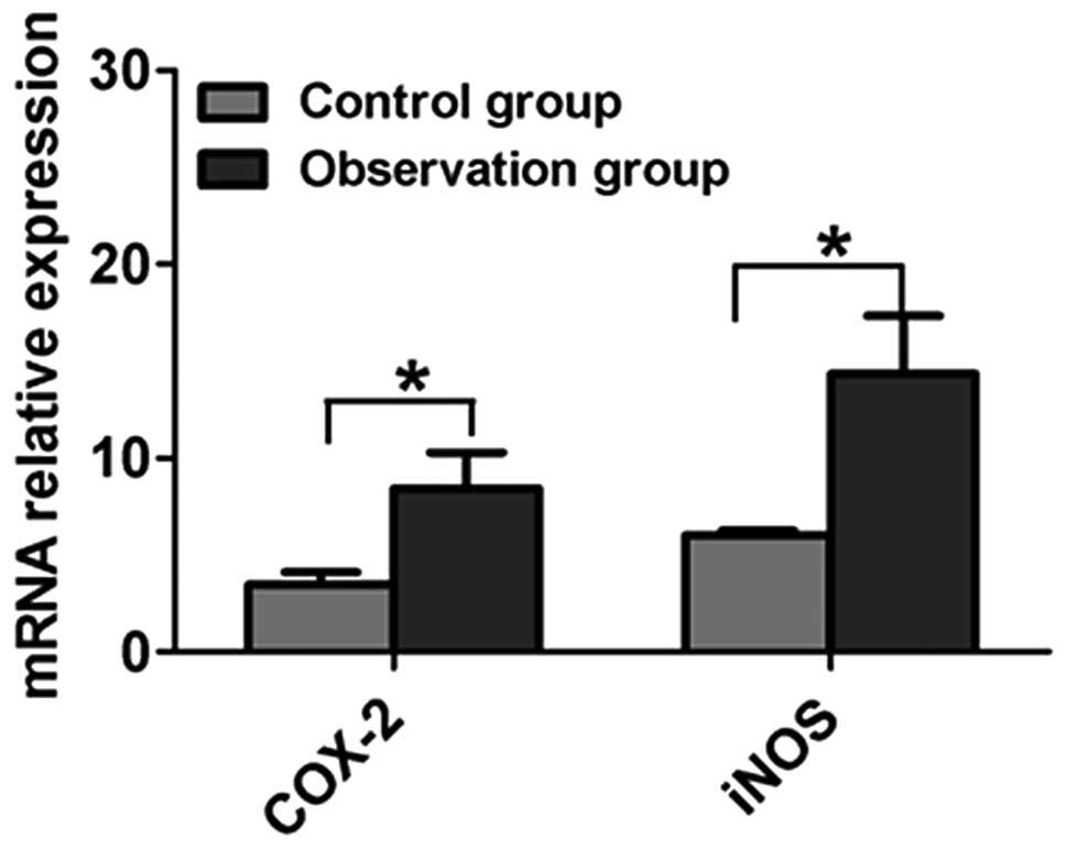
gastric mucosa groups are shown in Fig.
1. Compared with the control group, the COX-2 and iNOS mRNA
levels in the gastric mucosa of the patients in the observation
group were significantly increased, and the difference was
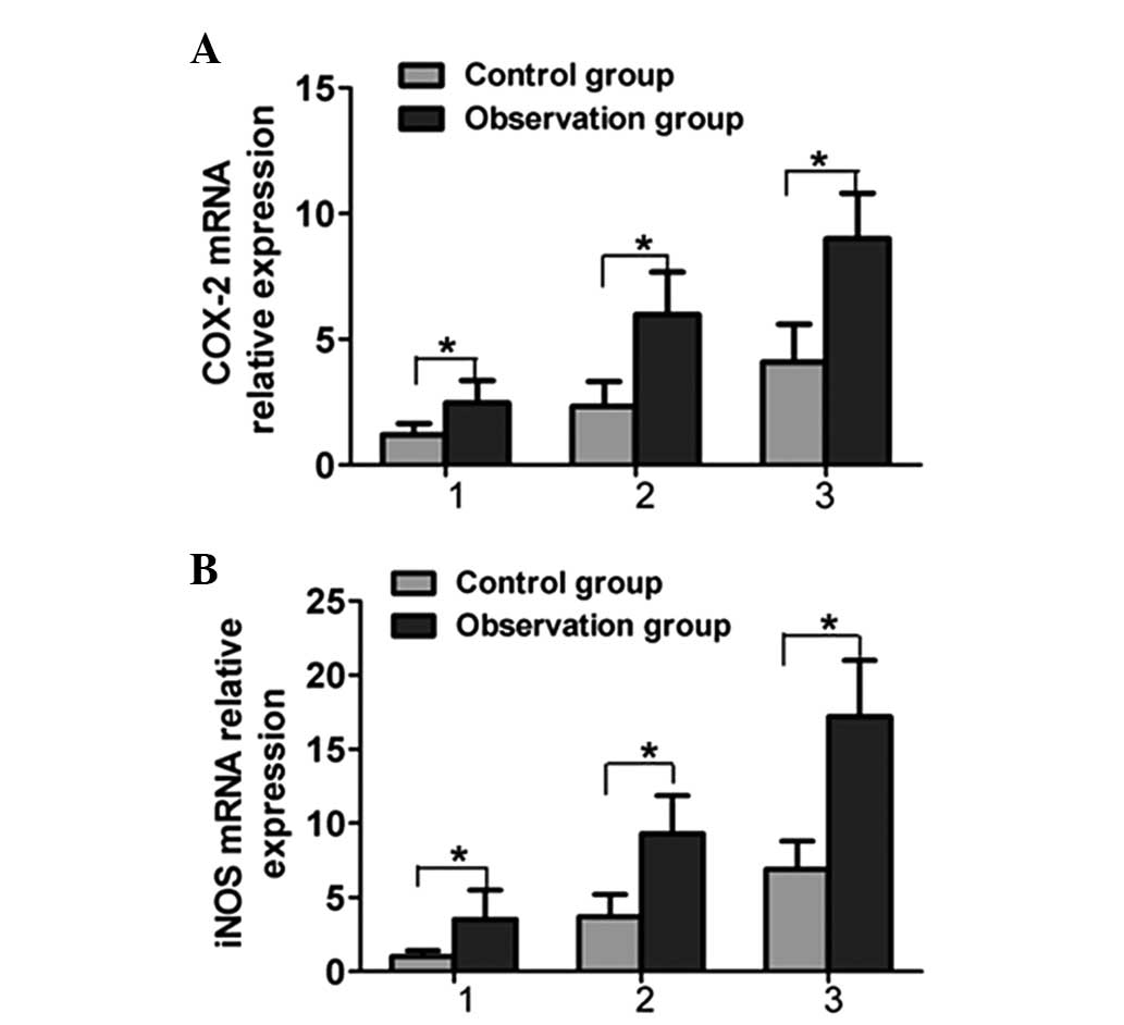
statistically significant (P<0.05) (Fig. 2). The COX-2 and iNOS mRNA levels in
the gastric mucosa of the patients in the observation group with
different pathological types were significantly higher than those
in the control group (P<0.05).
Comparison of COX-2 and iNOS protein
levels in the gastric mucosa of the two groups
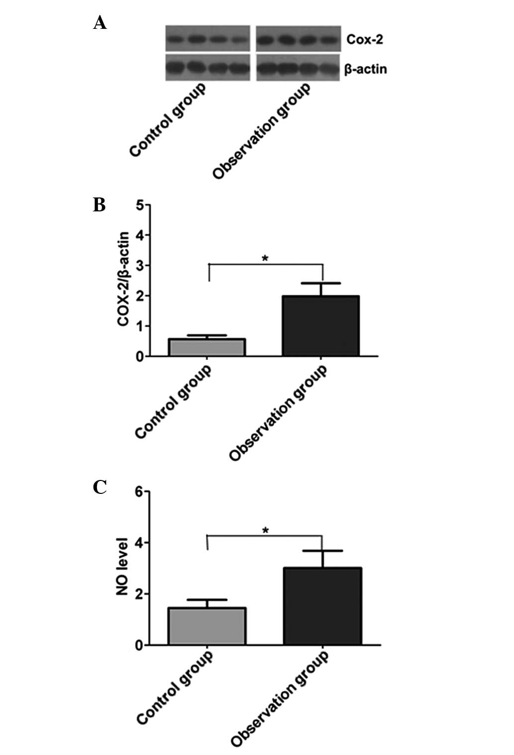
Western blotting was used to analyze the changes in
COX-2 protein expression in the gastric mucosa in the two groups,
and quantitative correction was performed through the internal
reference protein, β-actin (Fig.
3A). The expression level of COX-2 in the observation group was
significantly higher than that in the control group (P<0.05).
The NO (the reaction product of iNOS) level in the gastric mucosa
was determined by the Griess reaction; the level of NO in the
gastric mucosa of the observation group was significantly higher
than that in the control group (P<0.05) (Fig. 3B).
Effect of Hp on COX-2 and iNOS mRNA
expression in gastric mucosa cells
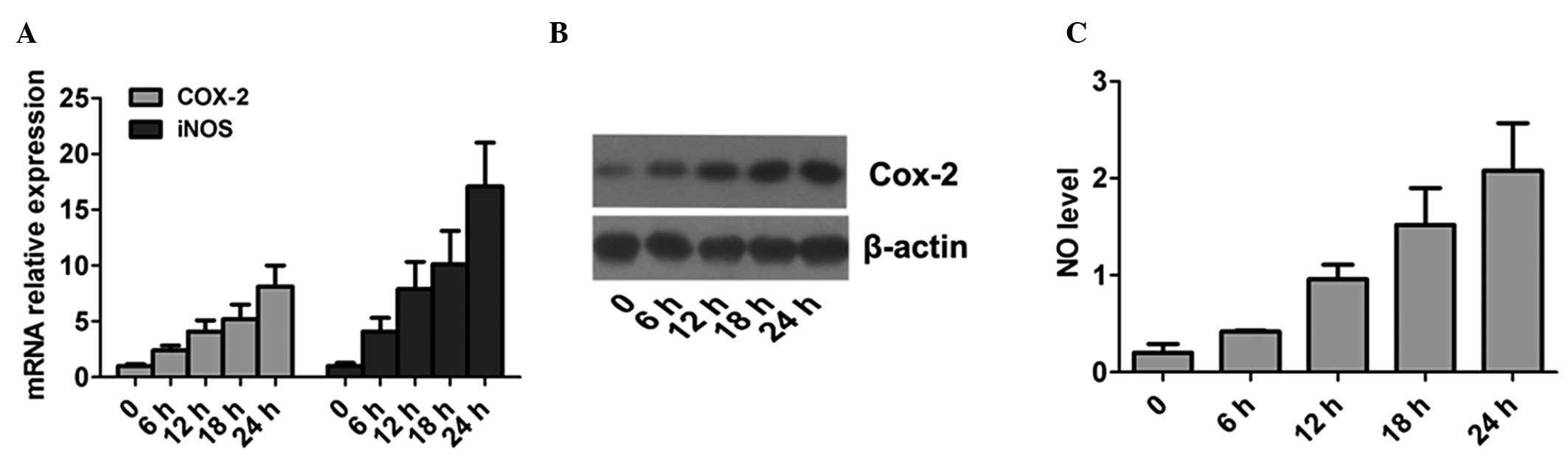
The infection process of Hp was simulated using
normal gastric mucosa cells in vitro, and the changes in
cellular COX-2 and iNOS mRNA were analyzed (Fig. 4). Compared with the negative control
group, the expression levels of COX-2 and iNOS mRNA in the cells
stimulated by Hp were significantly increased (P<0.05), and the
changes showed a time-dependent manner. This suggested that Hp
infection could stimulate cells to produce higher levels of COX-2
and iNOS.
Discussion
Gastric precancerous lesions are a type of
histopathological change in the gastric mucosa and are closely
associated with gastric cancer. The formation of precancerous
lesions is an important stage in the transformation process from
normal gastric mucosa to gastric carcinoma, including dysplasia and
intestinal metaplasia. Studies have shown that the infection of the
normal gastric mucosa with Hp can lead to chronic atrophic
gastritis, intestinal metaplasia and dysplasia; however the
molecular mechanism underlying these Hp-induced processes is still
not completely understood at present (14,15).
COX-2 is an essential enzyme for the synthesis of prostaglandin,
and is also the key rate-limiting enzyme in the initial step of
prostaglandin synthesis. COX-2 is generally produced when the body
suffers stimulation, and is involved in the inflammatory response
(16). Studies have found an
abnormal expression of COX-2 protein in several types of tumor
tissues, which suggests that it may be involved in the
tumorigenesis and the development and metastasis of cancer
(17,18). iNOS is generally produced by
macrophages or monocytes and is the main sign of an inflammatory
reaction. iNOS can aggravate the injury to the gastric mucosa. By
promoting the imbalance between the proliferation and apoptosis of
epithelial cells and promoting tumor angiogenesis, iNOS is involved
in the occurrence of gastric cancer and gastric lesions. iNOS
production is the early main event in the development of gastric
cancer (19,20).
In the present study, RT-qPCR was initially utilized
to analyze the mRNA levels of COX-2 and iNOS. The results showed
that the gastric mucosal mRNA levels of COX-2 and iNOS were
increased significantly in the patients with Hp-positive gastric
precancerous lesions compared with those in the patients with
Hp-negative lesions. This indicated that Hp stimulation resulted in
abnormal transcription of COX-2 and iNOS mRNA in the gastric mucosa
of patients. The expression levels of COX-2 and iNOS protein were
analyzed using western blotting. The analysis showed that the
expression level of COX-2 protein in Hp-positive gastric cancer
tissues was significantly higher than that in Hp-negative patients.
Since NO is the main product of iNOS, measurement of NO was used to
reflect the expression level of iNOS. The results showed that the
NO level in Hp-positive gastric cancer tissues was significantly
higher than that in the Hp-negative gastric cancer tissues. These
results suggested that Hp infection may stimulate the gastric
mucosa, resulting in an abnormal activation of COX-2 and iNOS
transcription and translation. COX-2 and iNOS are the main
inflammatory indexes, which indicated that Hp infection aggravated
inflammation levels in the gastric mucosa. The present study showed
that chronic inflammation was closely associated with
tumorigenesis.
In order to explain the universality of this
phenomenon, Hp was used to infect normal human gastric mucosal
cells in vitro. The result showed that, with the extension
of the infection time, the expression levels of COX-2 and iNOS
increased significantly compared with the levels in unstimulated
cells. This result was consistent with the detection results for
the clinical sample. In addition, it was found that a correlation
existed between the expression levels of COX2 and iNOS in gastric
cancer and the pathological types of gastric precancerous lesions.
The expression levels of COX-2 and iNOS increased significantly in
the gastric mucosal tissues of severe pathological types,
suggesting that serious chronic inflammation in the gastric mucosa
was associated with a higher chance of gastric cancer.
In conclusion, the levels of COX-2 and iNOS were
increased significantly in the mucosa of patients with gastric
precancerous lesions infected by Hp. The expression of COX-2 and
iNOS was correlated with the pathological grading in patients;
therefore, anti-Hp therapy should be performed to reduce the level
of inflammation in the gastric mucosa and reverse the development
of the disease.
References
|
1
|
Siegel R, Naishadham D and Jemal A: Cancer
statistics, 2012. CA Cancer J Clin. 62:10–29. 2012. View Article : Google Scholar : PubMed/NCBI
|
|
2
|
Falt P, Hanousek M, Kundrátová E and Urban
O: Precancerous conditions and lesions of the stomach. Klin Onkol.
26:Suppl. S22–S28. 2013.[(In Czech)]. View Article : Google Scholar : PubMed/NCBI
|
|
3
|
Rugge M, Capelle LG, Cappellesso R, Nitti
D and Kuipers EJ: Precancerous lesions in the stomach: from biology
to clinical patient management. Best Pract Res Clin Gastroenterol.
27:205–223. 2013. View Article : Google Scholar : PubMed/NCBI
|
|
4
|
Zorzetto V, Maddalo G, Basso D and
Farinati F: Immunotherapy for gastric premalignant lesions and
cancer. Immunotherapy. 4:587–599. 2012. View Article : Google Scholar : PubMed/NCBI
|
|
5
|
Watari J and Miwa H: Precancerous lesions
of the stomach. Nihon Shokakibyo Gakkai Zasshi. 107:1759–1769.
2010.[(In Japanese)]. PubMed/NCBI
|
|
6
|
Rizzato C, Kato I, Plummer M, et al: Risk
of advanced gastric precancerous lesions in Helicobacter pylori
infected subjects is influenced by ABO blood group and cagA status.
Int J Cancer. 133:315–322. 2013. View Article : Google Scholar : PubMed/NCBI
|
|
7
|
Kato I, Canzian F, Plummer M, et al:
Polymorphisms in genes related to bacterial
lipopolysaccharide/peptidoglycan signaling and gastric precancerous
lesions in a population at high risk for gastric cancer. Dig Dis
Sci. 52:254–261. 2007. View Article : Google Scholar : PubMed/NCBI
|
|
8
|
Deng X, Liu ZW, Wu FS, Li LH and Liang J:
A clinical study of weining granules in the treatment of gastric
precancerous lesions. J Tradit Chin Med. 32:164–172. 2012.
View Article : Google Scholar : PubMed/NCBI
|
|
9
|
Greenhough A, Smartt HJ, Moore AE, Roberts
HR, Williams AC, Paraskeva C and Kaidi A: The COX-2/PGE2 pathway:
key roles in the hallmarks of cancer and adaptation to the tumour
microenvironment. Carcinogenesis. 30:377–386. 2009. View Article : Google Scholar : PubMed/NCBI
|
|
10
|
Chell S, Kaidi A, Williams AC and
Paraskeva C: Mediators of PGE2 synthesis and signalling downstream
of COX-2 represent potential targets for the prevention/treatment
of colorectal cancer. Biochim Biophys Acta. 1766:104–119.
2006.PubMed/NCBI
|
|
11
|
Chu AJ, Chou TH and Chen BD: Prevention of
colorectal cancer using COX-2 inhibitors: basic science and
clinical applications. Front Biosci. 9:2697–2713. 2004. View Article : Google Scholar : PubMed/NCBI
|
|
12
|
Janakiram NB and Rao CV: iNOS-selective
inhibitors for cancer prevention: promise and progress. Future Med
Chem. 4:2193–2204. 2012. View Article : Google Scholar : PubMed/NCBI
|
|
13
|
Digestive Disease Branch of Chinese
Medical Association: National seminar consensus on chronic
gastritis. Zhong Hua Xiao Hua Za Zhi. 20:199–201. 2000.[(In
Chinese)].
|
|
14
|
Correa P and Houghton J: Carcinogenesis of
Helicobacter pylori. Gastroenterology. 133:659–672. 2007.
View Article : Google Scholar : PubMed/NCBI
|
|
15
|
Kato I, van Doorn LJ, Canzian F, et al:
Host-bacterial interaction in the development of gastric
precancerous lesions in a high risk population for gastric cancer
in Venezuela. Int J Cancer. 119:1666–1671. 2006. View Article : Google Scholar : PubMed/NCBI
|
|
16
|
Zhang J, Luo J, Ni J, et al: MMP-7 is
upregulated by COX-2 and promotes proliferation and invasion of
lung adenocarcinoma cells. Eur J Histochem. 58:22622014. View Article : Google Scholar : PubMed/NCBI
|
|
17
|
Jana D, Sarkar DK, Ganguly S, et al: Role
of cyclooxygenase 2 (COX-2) in prognosis of breast cancer. Indian J
Surg Oncol. 5:59–65. 2014. View Article : Google Scholar : PubMed/NCBI
|
|
18
|
Kim JM, Kim JS, Jung HC, Song IS and Kim
CY: Up-regulation of inducible nitric oxide synthase and nitric
oxide in Helicobacter pylori-infected human gastric epithelial
cells: possible role of interferon-gamma in polarized nitric oxide
secretion. Helicobacter. 7:116–128. 2002. View Article : Google Scholar : PubMed/NCBI
|
|
19
|
Li JB, Li XM and Zhan W: Expression of
VEGF and iNOS protein in gastric cancer and precancerous lesions.
Yi Bao Yu Fen Zi Mian Yi Za Zhi. 27:561–562. 2011.[(In
Chinese)].
|
|
20
|
Szlachcic A, Krzysiek-Maczka G, Pajdo R,
et al: The impact of asymmetric dimethylarginine (ADAMA), the
endogenous nitric oxide (NO) synthase inhibitor, to the
pathogenesis of gastric mucosal damage. Curr Pharm Des. 19:90–97.
2013. View Article : Google Scholar : PubMed/NCBI
|