Introduction
Hypoglycemia in nondiabetic patients is not a common
clinical problem and can be a diagnostic and therapeutic challenge
(1). Persistent hyperinsulinemic
hypoglycemia (PHH) is a functional disorder caused by aberrant
insulin release by pancreatic β cells (2). Nesidioblastosis is the major cause of
PHH in infants and children, but in adults it is usually a
consequence of a solitary insulinoma. Nesidioblastosis has been
reported infrequently in adults (1–3). It
should be noted that in pediatric patients nesidioblastosis may be
classified histologically as either diffuse or focal, but only
diffuse lesions have been reported in adult patients with
histologically-confirmed nesidioblastosis. A single case of
suspected focal nesidioblastosis in an adult was reported by
McElroy et al in 2010 (4),
but was not confirmed histologically. Due to the lack of evidence,
most physicians do not take a possibility of focal
nesidioblastotosis into account when confronted with an adult
patient with PHH‥
The present study focused on the case of a
62-year-old man with a 3-year history of intermittent episodes of
symptomatic hypoglycemia. A 72-h fasting test, elevated levels of
insulin and C-peptide, concomitant with decreased blood glucose
levels and imaging, led to the discovery of a nodule in the
pancreatic tail. The pancreatic corpus and tail were enucleated
laparoscopically and the presence of focal nesidioblastosis was
confirmed histologically. We propose that focal nesidioblastosis
should be taken into consideration when confronted with PHH, even
in middle-aged patients.
Case report
Case presentation
A 62-year-old man with a body mass index of 26.99
presented with a 3-year history of intermittent episodes of
dizziness, weakness and sweating, which were apparently associated
with work load and subsided upon food intake. Previous clinical
evaluations had not included blood glucose measurements. The
patient had no history of diabetes, pancreatic diseases, von
Hippel-Lindau or multiple endocrine neoplasia syndromes and denied
using insulin or medications associated with hypoglycemia.
Based on the clinical presentation of the patient, a
72-h fasting test was performed to address the possibility of
endogenous hyperinsulinemic hypoglycemia, but was discontinued
after 61 h due to complaints of fatigue and dizziness. During the
hypoglycemic episodes, laboratory tests revealed that the glucose,
insulin and C-peptide levels were 48 mg/dl (normal range, 70–100),
162.20 pmol/l (normal range, 17.0–173.0) and 1.394 nmol/l (normal
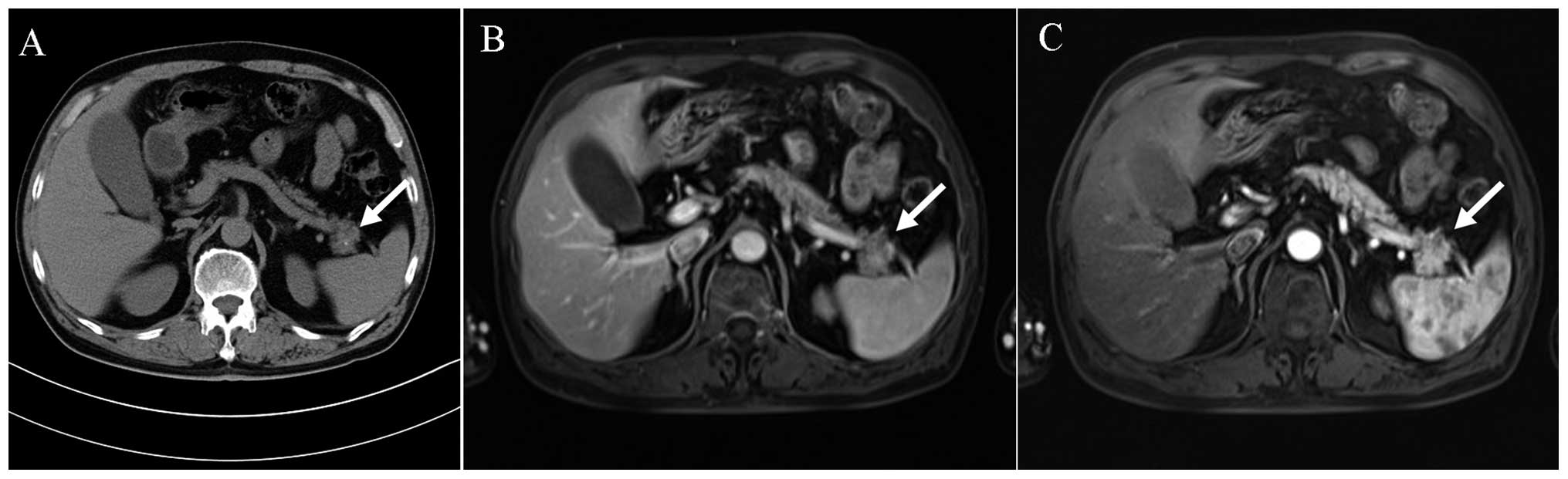
range, 0.37–1.47), respectively. A nodule in the pancreatic tail
was observed on abdominal computed tomography (CT) (Fig. 1A), which prompted further examination
using other imaging modalities. Magnetic resonance imaging (MRI)
revealed the presence of an unevenly enhanced lesion in the
pancreatic tail, measuring 2.2 cm in diameter (Fig. 1B and C). The patient underwent
laparoscopic enucleation of the pancreatic body and tail following
an initial diagnosis of a distal pancreatic tumor.
Investigations
The collected pancreatic specimen measured
~3.5×2.5×1.5 cm. This specimen was fixed in formalin, and
hematoxylin and eosin-stained serial sections were prepared for
microscopic examination by an expert pathologist.
Immunohistochemical staining was also performed, using rabbit
monoclonal antibodies against synaptophysin (1:1; #RMA-0537) and
chromogranin A (1:1; #MAB-0202), and mouse monoclonal antibodies
against neuron-specific enolase (1:1; #MAB-0584) and Ki-67 (1:1;
#Kit-0005-2) purchased from Fuzhou Maixin Biotechnology Development
Co., Ltd. (Fuzhou, China). In addition, rabbit polyclonal antibody
against insulin Rβ (1:100; #sc-711) was purchased from Santa Cruz
Biotechnology Inc., Dallas, TX, USA).
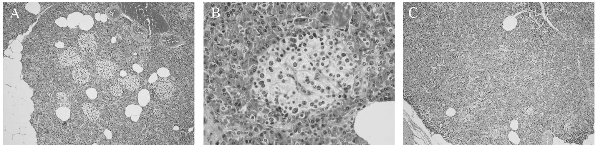
The pancreatic tissue had a normal appearance; the
cut surface was light brown and soft. The microscopic architecture
of the exocrine pancreas and the histology of the pancreatic ducts
and epithelium were normal. A significant abnormality was noted in
the endocrine pancreas, which contained substantially more than the
normal number of islets (Fig. 2A).
The endocrine islets also contained multiple enlarged cells with
enlarged nucleoli and abundant clear cytoplasm that was granulated
and slightly eosinophilic (Fig. 2B).
The hyperplastic nature of the islets decreased substantially from
the center of the lesion toward the perimeter (Fig. 2A and C), and therefore it was
concluded that the lesion was characterized by the presence of
hyperplastic islets.
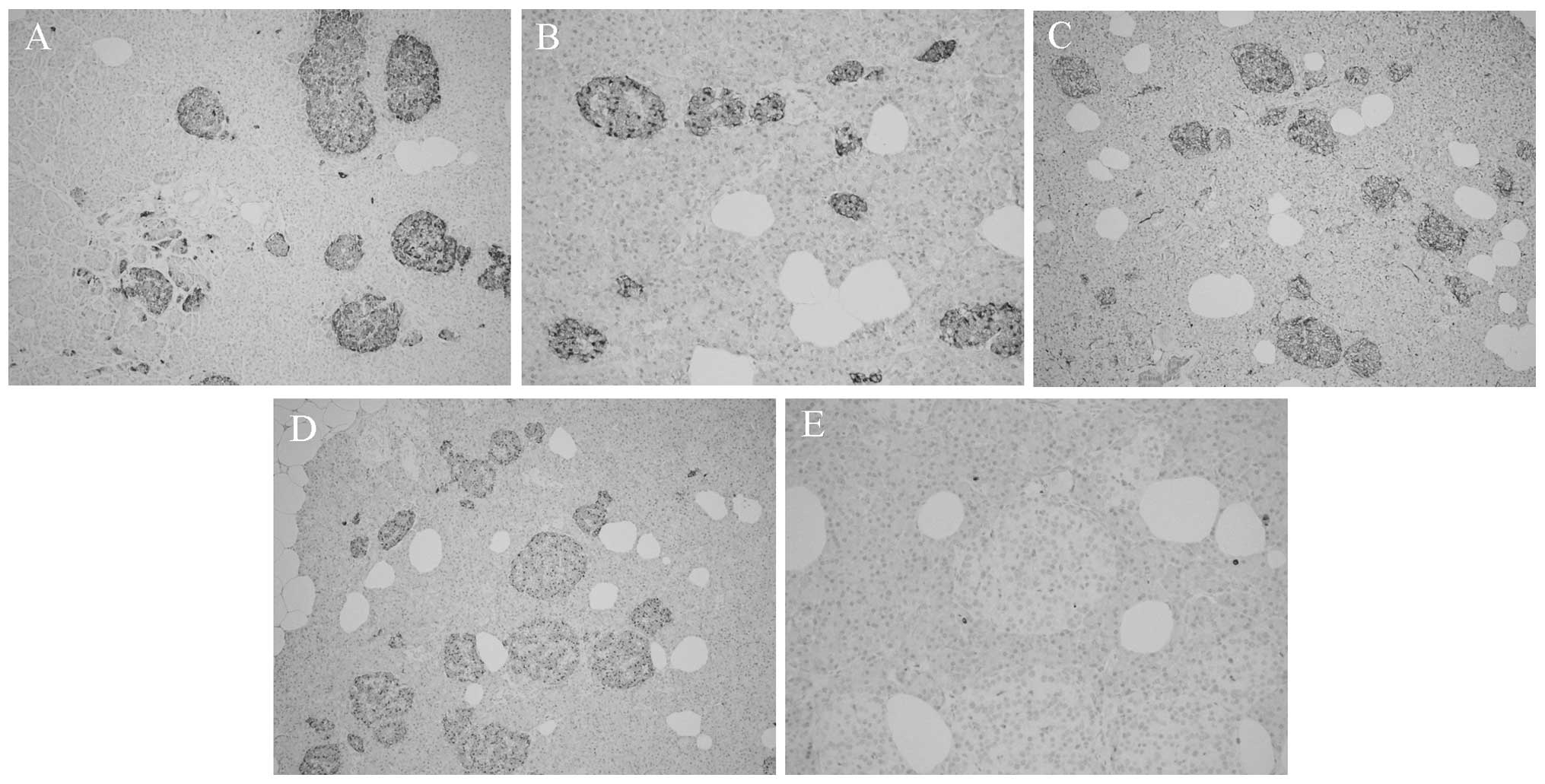
Immunohistochemical staining showed islet cells that
were positive for synaptophysin (Fig.
3A), chromogranin A (Fig. 3B)
and neuron-specific enolase (Fig.
3C). Insulin staining revealed enlarged, insulin-secreting β
cells (Fig. 3D). Ki-67 staining was
nearly absent in the islets (Fig.
3E). The pathological findings were consistent with a diagnosis
of adult nesidioblastosis (3) and
had the histological characteristics of a focal lesion.
Postoperatively, the hypoglycemic symptoms of the patient
disappeared, with fasting glucose ranging from 120 to 144
mg/dl.
Discussion
Nesidioblastosis is the most common cause of PHH of
infancy, but is rarely encountered in adults. The term
nesidioblastosis was coined by Laidlaw (5) in 1938 to describe the neoformation of
endocrine islets from pancreatic duct epithelium. The first adult
case was reported in 1975 (6), and
nesidioblastosis is currently estimated to be the cause of PHH in
0.5–7% of adult patients (4,7).
The cause of adult nesidioblastosis is unknown, but
it may be genetically induced, as in congenital hyperinsulinism in
neonates, or be a response to metabolic and hormonal changes, such
as those following gastric bypass surgery and weight loss (3). A paradoxical elevation of glucagon-like
peptide 1 reported following gastric bypass surgery (8) may influence islet cell neogenesis and
apoptosis, thus producing islet hyperplasia and PHH (9).
Preoperative diagnosis of adult nesidioblastosis is
challenging, as there are no defining clinical symptoms or history
and no highly specific functional tests (10). The occurrence of postprandial
hypoglycemia may indicate adult nesidioblastosis (3,5) if
insulinoma or factitious hypoglycemia can be excluded. In addition,
a selective arterial calcium stimulation test may indicate
hyperactive β-cell activity and provide guidance for the
localization of the disease and direct resection of the appropriate
pancreatic regions (11). A final
diagnosis of adult nesidioblastosis is best established
postoperatively by histological examination (3,7,10,11).
Diagnostic criteria of adult nesidioblastosis proposed by Klöppel
et al (3) involve exclusion
of an insulinoma and various histological characteristics.
The definitive treatment of adult nesidioblastosis
is surgical, but the resection volumes remain undefined (10). Subtotal (75 to 90%) pancreatectomy is
generally considered to be the more suitable choice, but recurrent
hypoglycemia and diabetes mellitus are frequent postoperative
complications (11).
In the present case, the histological findings met
the diagnostic criteria for adult nesidioblastosis (3) and the gradient observed in islet
hyperplasia, progressing from the center to the periphery of the
lesion, was consistent with the diagnosis of focal
nesidioblastosis.
Despite the fact that postprandial hypoglycemia has
been reported in adult nesidioblastosis, this was not evident in
the present case, in which the disease episodes appeared to be due
to increased physical activity or physical work stress. Insulin and
C-peptide were in the upper half of the normal range but laboratory
testing did not provide a definitive indication of
nesidioblastosis. Imaging techniques are not generally helpful for
indicating preoperative localization of adult nesidioblastosis
(10), but in this patient both CT
and MRI revealed the location of an exophytic lesion in the
pancreas. The intraoperative localization of the lesion was
consistent with the imaging and with the previous case report of
suspected (but histologically unconfirmed) focal nesidioblastosis
(4). Since positive imaging was also
apparent in the previous case, we propose that imaging may assist
in the localization of focal nesidioblastosis and its distinction
from diffuse nesidioblastosis.
The patient of the present study developed diabetes
mellitus following the resection of the pancreatic tissue. In the
aforementioned case of suspected focal nesidioblastosis, euglycemia
was restored following simple enucleation of the lesion, which
comprised <5% of the total pancreatic mass (4). In patients with focal nesidioblastosis,
partial pancreatectomy would be expected to control the
hypoglycemia and result in a good long-term outcome.
In conclusion, focal nesidioblastosis is rare in
adults and can be difficult to diagnose preoperatively. The present
case report, however, suggests that physicians should consider
focal nesidioblastosis when confronted with patients with PHH,
since early detection may prevent unnecessary full resection of the
pancreatic corpus and tail. Imaging can be useful, particularly
when combined with moderately high levels of insulin following
fasting; patients exhibiting these characteristics should be
considered for partial pancreatectomy.
References
|
1
|
Ng CL: Hypoglycaemia in nondiabetic
patients - an evidence. Aust Fam Physician. 39:399–404.
2010.PubMed/NCBI
|
|
2
|
Anlauf M, Wieben D, Perren A, et al:
Persistent hyperinsulinemic hypoglycemia in 15 adults with diffuse
nesidioblastosis: Diagnostic criteria, incidence and
characterization of beta-cell changes. Am J Surg Pathol.
29:524–533. 2005. View Article : Google Scholar : PubMed/NCBI
|
|
3
|
Klöppel G, Anlauf M, Raffel A, et al:
Adult diffuse nesidioblastosis: Genetically or environmentally
induced. Hum Pathol. 39:3–8. 2008. View Article : Google Scholar : PubMed/NCBI
|
|
4
|
McElroy MK, Lowy AM and Weidner N: Case
report: Focal nesidioblastosis (‘nesidioblastoma’) in an adult. Hum
Pathol. 41:447–451. 2010. View Article : Google Scholar : PubMed/NCBI
|
|
5
|
Laidlaw GF: Nesidioblastoma, the islet
tumor of the pancreas. Am J Pathol. 14:125–134. 1938.PubMed/NCBI
|
|
6
|
Sandler R, Horwitz DL, Rubenstein AH and
Kuzuya H: Hypoglycemia and endogenous hyperinsulinism complicating
diabetes mellitus. Application of the C-peptide assay to diagnosis
and therapy. Am J Med. 59:730–736. 1975. View Article : Google Scholar : PubMed/NCBI
|
|
7
|
Kaczirek K and Niederle B:
Nesidioblastosis: An old term and a new understanding. World J
Surg. 28:1227–1230. 2004. View Article : Google Scholar : PubMed/NCBI
|
|
8
|
Patti ME, McMahon G, Mun EC, et al: Severe
hypoglycaemia post-gastric bypass requiring partial pancreatectomy:
Evidence for inappropriate insulin secretion and pancreatic islet
hyperplasia. Diabetologia. 48:2236–2240. 2005. View Article : Google Scholar : PubMed/NCBI
|
|
9
|
Drucker DJ: Enhancing incretin action for
the treatment of type 2 diabetes. Diabetes Care. 26:2929–2940.
2003. View Article : Google Scholar : PubMed/NCBI
|
|
10
|
Raffel A, Krausch MM, Anlauf M, et al:
Diffuse nesidioblastosis as a cause of hyperinsulinemic
hypoglycemia in adults: A diagnostic and therapeutic challenge.
Surgery. 141:179–186. 2007. View Article : Google Scholar : PubMed/NCBI
|
|
11
|
Kenney B, Tormey CA, Qin L, et al: Adult
nesidioblastosis. Clinicopathologic correlation between
pre-operative selective arterial calcium stimulation studies and
post-operative pathologic findings. JOP. 9:504–511. 2008.PubMed/NCBI
|