Introduction
In a number of countries, a mifepristone plus
misoprostol regimen is the most frequently used medical method for
pregnancy termination in early gestation, with efficacy and safety
demonstrated for early abortion. However, abnormal uterine bleeding
is among the major limitations in such a regimen, and can lead to
endometritis or infertility (1–3).
Sheng-Hua-Tang, a well-known traditional Chinese
medicine (TCM) compound prescription, has been widely used during
the postpartum period in Chinese communities for a number of years
(4,5). A previous study demonstrated that use
of Sheng-Hua-Tang during the first month of the postpartum period
may have a positive effect on female health-related quality of life
(6). In addition, Shen-Hua-Tang
administration was shown to significantly correlate with an
anteverted uterus, and was effective for uterine contraction
(7). The Wujia Shenghua capsule
(WSC), which comprises Radix et Caulis Acanthopanacis Senticosi,
Radix Angelicae Sinensis, Rhizoma Chuanxiong, Semen Persicae, Radix
Glycyrrhizae and Rhizoma Zingiberis Preparata, is derived from
Sheng-Hua-Tang, and has been shown to have beneficial effects in
Chinese females following medically-induced abortions. These
beneficial effects include a reduction in the bleeding volume,
shortening the bleeding duration, promoting the blood flow and
resolving the blood stasis (8).
However, limited information from basic studies is available with
regard to the mechanisms underlying the effects of WSC on abnormal
uterine bleeding following medically-induced abortion.
Thus, the aim of the present study was to
investigate the effect of WSC on abnormal uterine bleeding
following a medically-induced abortion in rats, and also provide
evidence for the mechanism of action.
Materials and methods
Animals
A total of 32 eight-week-old female Wistar rats
(weight, 180–220 g) were obtained from the Laboratory Animal Center
of Heilongjiang University of Chinese Medicine (Harbin, China). The
experimental protocols were conducted in accordance with the
Guidelines for Animal Experiments (US Government Principle for the
Utilization and Care of Vertebrate Animals Used in Testing,
Research and Training) (9), and were
approved by the Ethical Committee of Heilongjiang University of
Chinese Medicine (no. SCXK Hei2008004).
WSC and reagents
WSC was purchased from Duoduo Pharmaceutical Co.,
Ltd. (Jiamusi, China); the enzyme-linked immunosorbent assay
(ELISA) kit for estradiol (E2) was obtained from Beijing
Yuande Biomedical Engineering Co., Ltd. (Beijing, China); the
radioimmunoassay (RIA) kit for laminin (LM) was purchased from
Beijing North Institute of Biotechnology (Beijing, China); the
ELISA kit for fibronectin (FN), rabbit polycolonal antibodies
against LM (BA1033) and FN (BA1049), and the streptavidin-biotin
complex (SABC) and 3,3′-diaminobenzidine (DAB) kits were all
purchased from Boster Biological Technology, Ltd. (Wuhan, China);
rabbit polycolonal antibodies against ERα (SC542), anti-ERβ
(SC8974) and anti-PR (SC538) were purchased from Beijing Zhongshan
Golden Bridge Biotechnology Co., Ltd. (Beijing, China); the
mifepristone and misoprostol tablets were obtained from Beijing
Zizhu Pharmaceutical Co., Ltd. (Beijing, China).
Model replication and groups
Female Wistar rats were mated with male rats of
proven fertility in the evening, at a ratio of 2:1, as previously
reported (10). If sperm was found
in the vaginal smears the following morning, that day was
considered as day 1 of gestation. On day 7 of gestation, 32 rats
were randomly divided into four groups, which included the normal
(group N), pregnant control (group P), model (group M) and
WSC-treated (group W) groups, with eight rats in each group.
Abortions were induced in each rat from groups M and W using 8.3
mg/kg mifepristone, followed by 100 µg/kg misoprostol 10 hours
later, which were applied via intragastic administration. Cotton
balls of the same weight, which were hemi-wrapped with plastic
films, were subsequently placed into the vagina of the rats to
verify the success of the abortion and to prevent blood leakage.
From day 8 of gestation, the rats in groups N, P and M were treated
with 1 ml per 100 g body weight distilled water each day for seven
days, whereas the rats in group W were treated with 1 ml per 100 g
body weight WSC suspension at a concentration of 0.25 g/ml per day
for seven days.
Uterine index measurement and
hematoxylin-eosin staining
On day 14 of gestation, all the rats were
anesthetized with ether and visceral organs were removed. The
unilateral uterine specimens were weighed. The uterine indexes were
subsequently calculated by dividing the uterine weight with the
body weight of each rat. The specimens were fixed in 4%
formaldehyde for 24–48 h, embedded in paraffin, serially sectioned
and restained with hematoxylin-eosin. Following incubation at 37°C
and sealing with neutral gum, the uterine shape was investigated
under a microscope (CX21; Olympus Corporation, Tokyo, Japan).
Duration and volume of uterine
bleeding measurements
From day 8 of gestation, a new cotton ball was
replaced in the vagina at an interval of 6 h from 6:00, until no
blood stain was observed. The duration of uterine bleeding was
recorded and the cotton balls were stored at −20°C for the
determination of the uterine bleeding volume. On day 14 of
gestation, a 0.02-ml blood sample was collected from the tail vein
using a hemoglobin pipet, added to 4 ml (V1) NaOH
solution (5%) and mixed. The cotton balls from each rat were placed
in a beaker, soaked and washed with the appropriate amount of NaOH
solution that was adjusted to the quantity of bleeding. The washed
solution was then preserved in an additional beaker. The process
was repeated once or twice as required, and the total volume
(V2) of NaOH solution used was recorded. Following
complete mixing of the washed solution, a 5-ml sample of the
mixture was filtrated using a filtrator (Beijing Bomei Glass Co.,
Ltd., Beijing, China). The absorbance (A) values of the filtered
extract and the 4-ml NaOH solution containing the tail vein blood
were recorded at 546 nm using an ultraviolet-visible light detector
(Helios Gamma; Thermo Fisher Scientific, Waltham, MA, USA), with a
5% NaOH solution used as the blank control. The volume of uterine
bleeding was calculated using the following equation: Volume (ml) =
tail vein blood (0.02 ml) × (A of filtered extract ×
V2)/(A of tail vein blood × V1).
Determination of the serum
E2, FN and LM levels
Blood samples were collected from the femoral artery
of the rats on days 1, 8 and 14 of gestation. Following
centrifugation at 600 × g for 10 min, the serum was aspirated and
stored at −20°C. The E2 level was analyzed with an ELISA
kit, according to the manufacturer's instructions. For the
measurement of FN and LM levels, plasma samples were collected on
day 14 of gestation and analyzed by ELISA and RIA, respectively,
according to the manufacturer's instructions.
Immunohistochemistry
Paraffin-embedded tissues prepared for
hematoxylin-eosin staining were also used in the
immunohistochemistry assay. Tissue sections were blocked with 5%
bovine serum albumin at 37°C for 30 min, and incubated with primary
rabbit polyclonal antibodies targeted against ER, PR, FN and LM at
4°C overnight. The samples were subsequently incubated with a
biotin-conjugated secondary antibody (BST06K11A; Beijing Zhongshan
Golden Bridge Biotechnology Co., Ltd.). Staining was developed by
incubation of the sections with the SABC and visualized using DAB.
All the slides were counterstained with hematoxylin, and images
were captured using an inverted microscope (magnification, x100)
(IX71-21PH; Olympus Corporation). Immunoreactive spots from five
randomly selected fields in each section were further selected for
analysis and quantified using Image-Pro Plus 6.0 software (Media
Cybernetics, Inc., Rockville, MD, USA).
Statistical analysis
The data are expressed as the mean ± standard
deviation. Statistical analyses were conducted using SPSS version
17.0 (SPSS Inc., Chicago, IL, USA). The statistical significance of
differences was evaluated using the Mann-Whitney U-test, where
P<0.05 was considered to indicate a statistically significant
difference.
Results
Effect of WSC on the uterine index and
shape
As shown in Table I,
the uterine indexes in groups P and M were significantly higher
compared with that observed in group N (P<0.01); whereas the
uterine index in group W was notably lower when compared with group
M (P<0.05).
 | Table I.Effect of WSC on the uterine weight
and uterine index. |
Table I.
Effect of WSC on the uterine weight
and uterine index.
| Group | Uterine weight
(g) | Uterine index |
|---|
| N | 0.301±0.093 | 0.0017±0.0007 |
| P | 2.328±1.073 |
0.0098±0.0051a |
| M | 1.312±0.787 |
0.0058±0.0032b |
| W | 0.505±0.1776 |
0.0021±0.0008c |
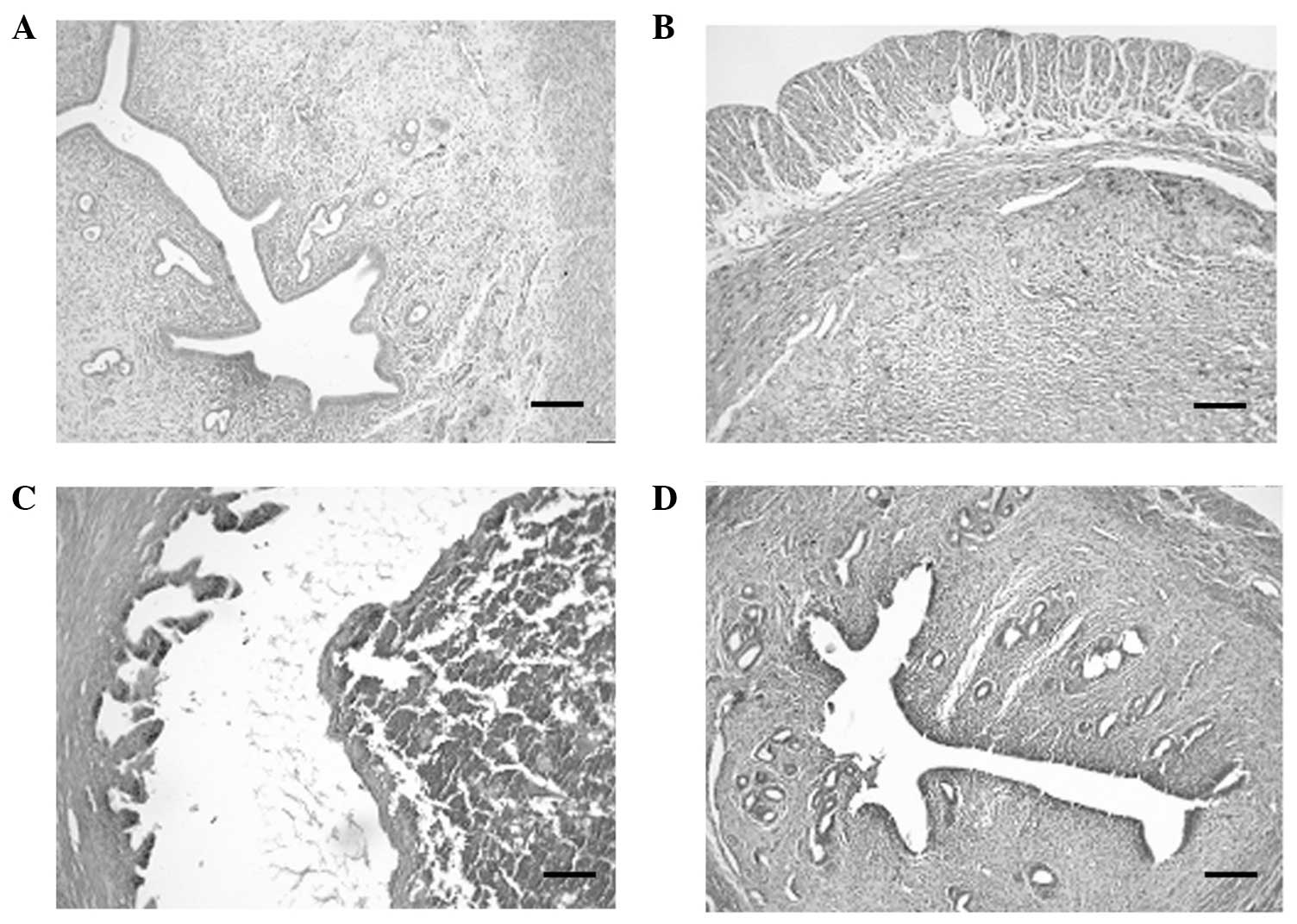
The pathological results obtained from the paraffin
sections of the uterine tissue revealed that there were a number of
trophoblastic cells observed in group P when compared with the
normal uterine shape in group N. However, in group M, there was a
predominance of decidual cells and few trophocytes, in addition to
evidence of extravasated blood. In group W, there were few decidual
cells observed and no evidence of extravasated blood (Fig. 1).
Effect of WSC on the duration and
volume of uterine bleeding
No evidence of uterine bleeding was observed in
groups N and P. The duration of uterine bleeding was prolonged and
the volume of uterine bleeding increased significantly in group M
when compared with group P (P<0.01); whereas the duration of
uterine bleeding was reduced and the volume of uterine bleeding
decreased significantly in group W when compared with group M
(P<0.01 and P<0.05, respectively; Table II).
| Table II.Effect of WSC on the duration and
volume of uterine bleeding. |
Table II.
Effect of WSC on the duration and
volume of uterine bleeding.
| Group | Volume of uterine
bleeding (ml) | Duration of uterine
bleeding (h) |
|---|
| N | 0.05±0.02 |
0±0 |
| P | 0.06±0.04 |
0±0 |
| M |
0.40±0.15a |
124.4±22.0a |
| W |
0.22±0.16b |
71.3±10.4c |
Effect of WSC on the serum
E2 level
Table III shows the
E2 level in the rats from the various groups. No
statistically significant changes were observed in the
E2 level on days 1, 8 and 14 of gestation in group N.
However, in group P, the E2 level exhibited an
increasing trend following the experimental days. In addition, the
E2 level in group M increased initially at day 8 of
gestation and then decreased by day 14 of gestation. The serum
E2 level in group W was significantly elevated when
compared with that in group M on day 14 of gestation
(P<0.01).
| Table III.Effect of WSC on the serum
E2 level. |
Table III.
Effect of WSC on the serum
E2 level.
|
|
| E2
(pg/ml) |
|---|
|
|
|
|
|---|
| Group | Day 1 | Day 8 | Day 14 |
|---|
| N | 15.27±4.20 | 14.98±7.14 | 15.63±5.36 |
| P | 15.52±3.90 | 17.18±4.14 | 18.23±4.87 |
| M | 15.44±3.25 | 33.03±9.62 |
22.47±6.37a |
| W | 15.45±3.02 | 34.89±5.72 |
39.57±5.19b,c |
Effect of WSC on the plasma levels of
FN and LM
As shown in Table
IV, the plasma levels of FN and LM in group M were
significantly elevated when compared with group P (P<0.01 and
P<0.05, respectively). However, the levels of FN and LM in group
W were markedly lower than that observed in group M (P<0.01 and
P<0.05, respectively).
| Table IV.Effect of WSC on the plasma levels of
FN and LM. |
Table IV.
Effect of WSC on the plasma levels of
FN and LM.
| Group | FN (pg/ml) | LM (ng/ml) |
|---|
| N | 0.05±0.01 | 12.44±4.32 |
| P | 0.06±0.01 | 13.57±6.52 |
| M |
2.19±1.49b |
20.45±7.47a |
| W |
0.08±0.02d |
15.82±8.97c |
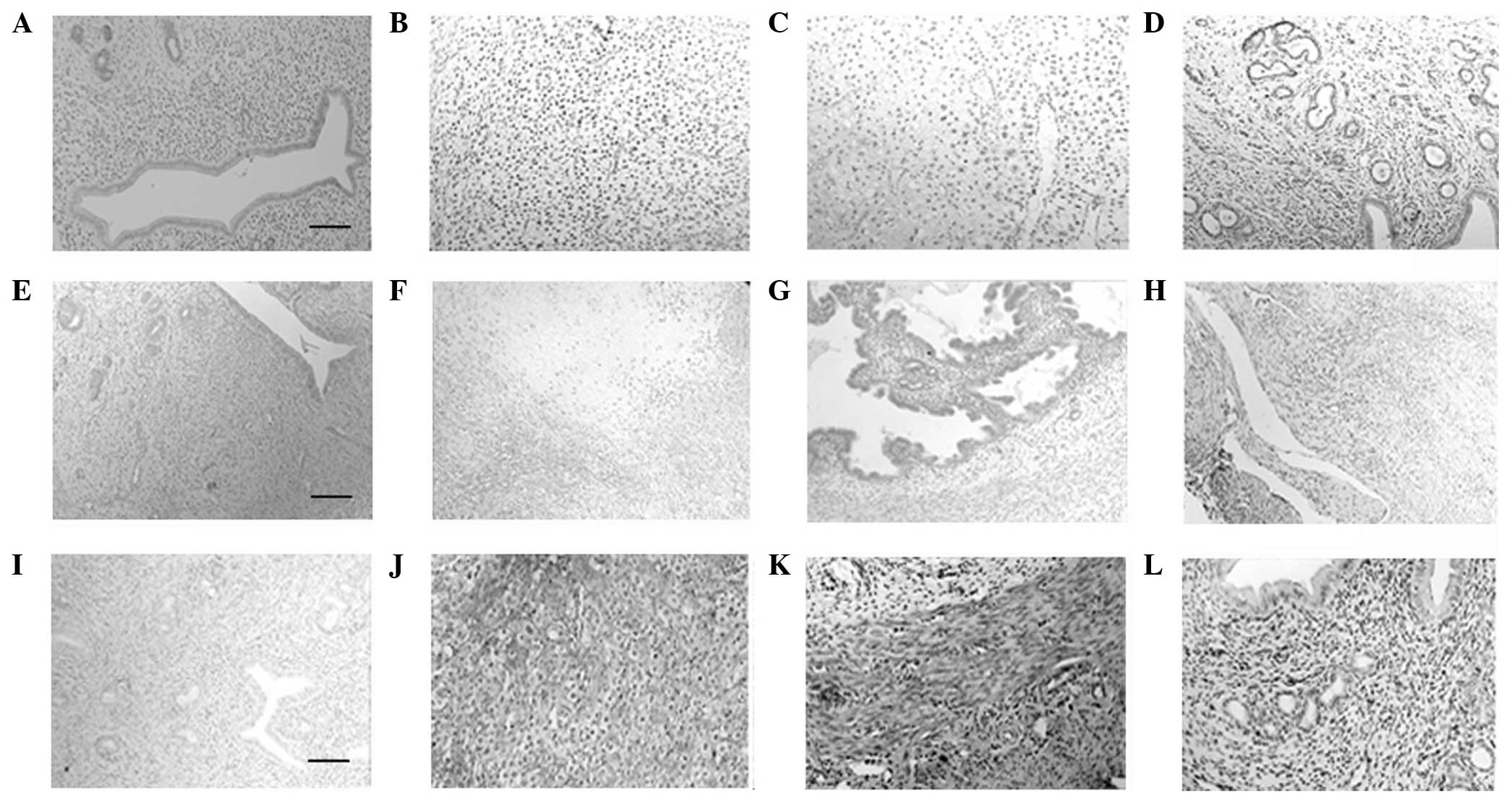
Effect of WSC on the expression levels
of ER, PR and FN in the uterine tissues
Immunohistochemistry staining results showing the
expression of the ER, PR and FN in the uterine tissues are shown in
Fig. 2. Since no evident expression
of LM was detected in the uterine tissue (data not shown), LM
expression was not analyzed with immunohistochemistry. The ERα and
PR were predominately expressed in the endometrial glandular
epithelial cells, simple columnar epithelial cells and were also
detected in the smooth muscle cells of the uterine endometrium. No
statistically significant differences were observed between the
optical density (OD) values for the ERα and PR in groups P and N.
However, the OD values of the ERα and PR were significantly
decreased in group M when compared with group P (P<0.01 and
P<0.05, respectively), whereas the OD values of the ERα and PR
were significantly increased in group W when compared with group M
(P<0.01 and P<0.05, respectively). FN was primarily expressed
in the myometrial and endometrial interstitial cells and cells of
the decidual tissues. No statistically significant difference was
observed between the OD value of FN in groups P and N. The OD value
of FN was significantly increased in group M when compared with
group P (P<0.05), whereas the OD value of FN was significantly
decreased in group W when compared with group M (P<0.01;
Table V).
| Table V.Effect of WSC on the expression of
the ER, PR and FN in the uterine tissue (OD values). |
Table V.
Effect of WSC on the expression of
the ER, PR and FN in the uterine tissue (OD values).
| Group | ERα | PR | FN |
|---|
| N | 0.091±0.024 | 0.076±0.022 | 0.084±0.011 |
| P | 0.095±0.019 | 0.065±0.012 | 0.104±0.020 |
| M |
0.036±0.025b |
0.043±0.023a |
0.251±0.069a |
| W |
0.208±0.072d |
0.095±0.035c |
0.091±0.033d |
Effect of WSC on the hemodynamic
indexes
As shown in Table
VI, no statistically significant differences were observed in
the hemodynamic indexes when comparing groups P and N. The plasma
viscosity, the whole blood viscosity at shear rates of 1 mPa•s and
5 mPa•s, the whole blood relative index (low shear) and the whole
blood reduced viscosity (low shear) were significantly increased in
group M when compared with group P; whereas the plasma viscosity,
the whole blood viscosity at shear rates of 1 s−1 and 5
s−1, the whole blood relative index (low shear) and the
whole blood reduced viscosity (low shear) were significantly
decreased in group W when compared with group M.
| Table VI.Effect of WSC on the hemodynamic
indexes. |
Table VI.
Effect of WSC on the hemodynamic
indexes.
| Hemodynamic
index | Group N | Group P | Group M | Group W |
|---|
| Whole blood
viscosity (1/s, mPa•s) | 17.92±3.20 | 18.79±4.36 |
26.78±4.91a |
20.73±4.71c |
| Whole blood
viscosity (5/s, mPa•s) | 8.88±1.06 | 8.19±1.34 |
10.71±1.38a |
9.02±1.51c |
| Whole blood
viscosity (30/s, mPa•s) | 5.01±0.45 | 5.02±0.69 | 5.59±0.42 | 5.13±0.57 |
| Whole blood
viscosity (200/s, mPa•s) | 3.85±0.29 | 3.75±0.33 | 3.92±0.17 | 3.81±0.29 |
| Plasma viscosity
(mPa•s) | 1.51±0.31 | 1.52±0.14 |
1.95±0.36b |
1.73±0.12c |
| Erythrocyte
sedimentation rate (mm/h) | 23.71±1.70 | 24.00±4.19 | 23.16±5.45 | 23.25±3.94 |
| Hematocrit
(l/l) | 0.31±0.01 | 0.32±0.03 | 0.35±0.03 | 0.35±0.03 |
| Whole blood
relative index (high shear) | 2.47±0.25 | 2.48±0.24 | 2.06±0.46 | 2.31±0.38 |
| Whole blood
relative index (low shear) | 11.52±2.21 | 12.34±2.78 |
15.72±2.81a |
11.74±1.08c |
| Whole blood reduced
viscosity (low shear) (mPa•s) | 51.75±8.59 | 52.78±9.99 |
73.98±14.26b |
59.51±5.09c |
| Whole blood reduced
viscosity (high shear) (mPa•s) | 6.64±1.20 | 6.93±0.60 | 6.58±0.91 | 6.57±0.53 |
Discussion
In the present study, the beneficial effects of WSC,
which is derived from Sheng-Hua-Tang, a well-known TCM compound
prescription used during the postpartum period in females, were
investigated on abnormal uterine bleeding. In addition, the
underlying mechanisms were investigated using a rat model of
medical abortion.
As demonstrated in the present study, when compared
with the model group, WSC treatment was shown to markedly decrease
the volume of uterine bleeding, reduce the duration of bleeding and
the uterine index. Residual decidual tissues were found to be
responsible for heavy and prolonged bleeding following the medical
abortion, as previously reported (11–13),
Histological examination revealed that little residual of the
conceptus was present following WSC treatment. Therefore, the in
vivo inhibitory effect of WSC on abnormal uterine bleeding may
be associated with the promotion of residual discharge in the rat
uterus.
E2 and the ER play an important role
during the process of abnormal uterine bleeding by modulating the
proliferation of endometrial epithelial and stromal cells and
enhancing angiogenesis (14–17). The results of the present study
demonstrated that WSC treatment significantly elevated the serum
E2 level, while reducing the expression of the ER in the
uterine tissues on day 14 of gestation, as compared with the model
group. PR, as the main target of mifepristone (18), has been demonstrated to be involved
in the regulation of abnormal uterine bleeding (19–22). In
the present study, WSC treatment was also shown to markedly elevate
PR expression in the uterine tissues on day 14 of gestation when
compared with the model group.
Excessive accumulation of FN and LM, the two main
components of the stromal and underlying vascular extracellular
matrices in the endometrium, may also promote endometrial
hemorrhage by disturbing the balance of the extracellular matrix
(23–25). The results of the present study
demonstrated that WSC treatment notably reduced the plasma levels
of FN and LM, and decreased the expression of FN in the uterine
tissues on day 14 of gestation, as compared with the model
group.
Increased plasma and blood viscosity is usually
associated with dysfunction of the microcirculation, which may
contribute to persistent damage of the uterine tissues (26). In the present study, the results
demonstrated that the hemodynamic indexes were improved following
treatment with WSC when compared with the model group, which may be
an additional mechanism underlying the hemostatic effect of
WSC.
In conclusion, based on the results of the present
study, WSC was shown to exert an inhibitory effect on the uterine
bleeding caused by medical abortion. In addition, the current study
demonstrated a number of possible molecular and non-molecular
mechanisms through which WSC may exert these effects, including
modulation of the E2, ER, PR, FN and LM levels and
improving the hemodynamic indexes.
Acknowledgements
The study was supported by grants from the
Heilongjiang Province Science and Technology Research Projects (no.
GC10C208) and the Heilongjiang Province Administration of
Traditional Chinese Medicine Topics (no. ZHY12-2077).
References
|
1
|
Bartz D and Goldberg A: Medication
abortion. Clin Obstet Gynecol. 52:140–150. 2009. View Article : Google Scholar : PubMed/NCBI
|
|
2
|
Doraiswami S, Johnson T, Rao S, Rajkumar
A, Vijayaraghavan J and Panicker VK: Study of endometrial pathology
in abnormal uterine bleeding. J Obstet Gynaecol India. 61:426–430.
2011. View Article : Google Scholar : PubMed/NCBI
|
|
3
|
Spitz IM: Mifepristone: where do we come
from and where are we going? Clinical development over a quarter of
a century. Contraception. 82:442–452. 2010. View Article : Google Scholar : PubMed/NCBI
|
|
4
|
Chuang CH, Chang PJ, Hsieh WS, Tsai YJ,
Lin SJ and Chen PC: Chinese herbal medicine use in Taiwan during
pregnancy and the postpartum period: a population-based cohort
study. Int J Nurs Stud. 46:787–795. 2009. View Article : Google Scholar : PubMed/NCBI
|
|
5
|
Liao HL, Ma TC, Chiu YL, Chen JT and Chang
YS: Factors influencing the purchasing behavior of TCM outpatients
in Taiwan. J Altern Complement Med. 14:741–748. 2008. View Article : Google Scholar : PubMed/NCBI
|
|
6
|
Chang PJ, Tseng YC, Chuang CH, et al: Use
of Sheng-Hua-Tang and health-related quality of life in postpartum
women: a population-based cohort study in Taiwan. Int J Nurs Stud.
47:13–19. 2010. View Article : Google Scholar : PubMed/NCBI
|
|
7
|
Ho M, Li TC and Su SY: The association
between traditional Chinese dietary and herbal therapies and
uterine involution in postpartum women. Evid Based Complement
Alternat Med. 2011:9182912011. View Article : Google Scholar : PubMed/NCBI
|
|
8
|
Geng F, Wang F, Zou T, et al: Rapid
identification of the multiple absorbed bioactive components and
metabolites in rat serum after oral administration of Wu-Jia
Sheng-Hua capsule by UPLC-ESI-MS. J Anal Methods Chem.
2013:3189612013. View Article : Google Scholar : PubMed/NCBI
|
|
9
|
IRAC (Interagency Research Animal
Committee), . U.S. Government Principles for the Utilization and
Care of Vertebrate Animals Used in Testing, Research and
TrainingFederal Register. Office of Technology Policy; Washington:
pp. 116–118. 1985
|
|
10
|
Li X, Yuan FL, Zhao YQ, Lu WG, Li CW and
He CH: Effect of leonurine hydrochloride on endothelin and the
endothelin receptor-mediated signal pathway in medically-induced
incomplete abortion in rats. Eur J Obstet Gynecol Reprod Biol.
169:299–303. 2013. View Article : Google Scholar : PubMed/NCBI
|
|
11
|
Cheng L: Medical abortion in early
pregnancy: experience in China. Contraception. 74:61–65. 2006.
View Article : Google Scholar : PubMed/NCBI
|
|
12
|
Li L, Zhou Z and Huang L: Abnormal
expression of MMP-9 and imbalance of MMP-9/TIMP-1 is associated
with prolonged uterine bleeding after a medical abortion with
mifepristone and misoprostol. Acta Obstet Gynecol Scand.
88:673–679. 2009. View Article : Google Scholar : PubMed/NCBI
|
|
13
|
Li X, Yuan FL, Zhao YQ, et al: Effects of
leonurine hydrochloride on medically induced incomplete abortion in
early pregnancy rats. Eur J Obstet Gynecol Reprod Biol.
159:375–380. 2011. View Article : Google Scholar : PubMed/NCBI
|
|
14
|
Livingstone M and Fraser IS: Mechanisms of
abnormal uterine bleeding. Hum Reprod Update. 8:60–67. 2002.
View Article : Google Scholar : PubMed/NCBI
|
|
15
|
Lockwood CJ: Mechanisms of normal and
abnormal endometrial bleeding. Menopause. 18:408–411. 2011.
View Article : Google Scholar : PubMed/NCBI
|
|
16
|
Smith SK: Regulation of angiogenesis in
the endometrium. Trends Endocrinol Metab. 12:147–151. 2001.
View Article : Google Scholar : PubMed/NCBI
|
|
17
|
Vadlamudi RK, Balasenthil S, Broaddus RR,
Gustafsson JA and Kumar R: Deregulation of estrogen receptor
coactivator proline-, glutamic acid- and leucine-rich
protein-1/modulator of nongenomic activity of estrogen receptor in
human endometrial tumors. J Clin Endocrinol Metab. 89:6130–6138.
2004. View Article : Google Scholar : PubMed/NCBI
|
|
18
|
Im A and Appleman LJ: Mifepristone:
pharmacology and clinical impact in reproductive medicine,
endocrinology and oncology. Expert Opin Pharmacother. 11:481–488.
2010. View Article : Google Scholar : PubMed/NCBI
|
|
19
|
Chwalisz K, Perez MC, Demanno D, Winkel C,
Schubert G and Elger W: Selective progesterone receptor modulator
development and use in the treatment of leiomyomata and
endometriosis. Endocr Rev. 26:423–438. 2005. View Article : Google Scholar : PubMed/NCBI
|
|
20
|
Clark TJ: Modern management of abnormal
uterine bleeding. Obstetrician and Gynaecologist. 11:792009.
View Article : Google Scholar
|
|
21
|
Fraser IS, Hickey M and Song JY: A
comparison of mechanisms underlying disturbances of bleeding caused
by spontaneous dysfunctional uterine bleeding or hormonal
contraception. Hum Reprod. 11 (Suppl 2):165–178. 1996. View Article : Google Scholar : PubMed/NCBI
|
|
22
|
Williams AR, Critchley HO, Osei J, et al:
The effects of the selective progesterone receptor modulator
asoprisnil on the morphology of uterine tissues after 3 months
treatment in patients with symptomatic uterine leiomyomata. Hum
Reprod. 22:1696–1704. 2007. View Article : Google Scholar : PubMed/NCBI
|
|
23
|
Evans J, Kaitu'u-Lino T and Salamonsen LA:
Extracellular matrix dynamics in scar-free endometrial repair:
perspectives from mouse in vivo and human in vitro studies. Biol
Reprod. 85:511–523. 2011. View Article : Google Scholar : PubMed/NCBI
|
|
24
|
Frantz C, Stewart KM and Weaver VM: The
extracellular matrix at a glance. J Cell Sci. 123:4195–4200. 2010.
View Article : Google Scholar : PubMed/NCBI
|
|
25
|
Hickey M and Fraser IS: The structure of
endometrial microvessels. Hum Reprod. 15 (Suppl 3):57–66. 2000.
View Article : Google Scholar : PubMed/NCBI
|
|
26
|
Lipowsky HH: Microvascular rheology and
hemodynamics. Microcirculation. 12:5–15. 2005. View Article : Google Scholar : PubMed/NCBI
|