Introduction
Sepsis, which is characterized by a systemic
inflammatory response to invasive microbial pathogens, is one of
the major causes of mortality in intensive care units globally
(1). During the years 1979–2000, the
overall mortality rate of sepsis rose from 22 to 44 per 100,000
population (2), accounting for ~9%
of the total annual mortality in the United States (3). Despite advances in understanding the
pathogenesis of sepsis, efforts to use new treatment methods in
clinical settings have not been proved successful and the mortality
rate for sepsis remains high. Thus, it is necessary to develop new
therapies in order to improve clinical outcomes in the future.
The proinflammatory cytokine tumor necrosis factor
(TNF)-α, together with secondary proinflammatory mediators such as
interleukin (IL)-6 and IL-8, appear to be generated to modulate the
human immune response to severe infections. TNF-α and IL-6 are
considered to be important regulatory factors in the cytokine
network during sepsis (4,5). The excessive production of cytokines
may increase vascular permeability, cause coagulopathy and change
the metabolism of cells, which frequently contributes to the
vulnerability of multiple organ dysfunction syndrome (6).
Radix Bupleuri (RB), the dried roots of Bupleurum
falcatum L., is frequently included in traditional Chinese
herbal formulas designed to provide anti-inflammatory, antipyretic
and antihepatotoxic effects in the treatment of common cold, fever
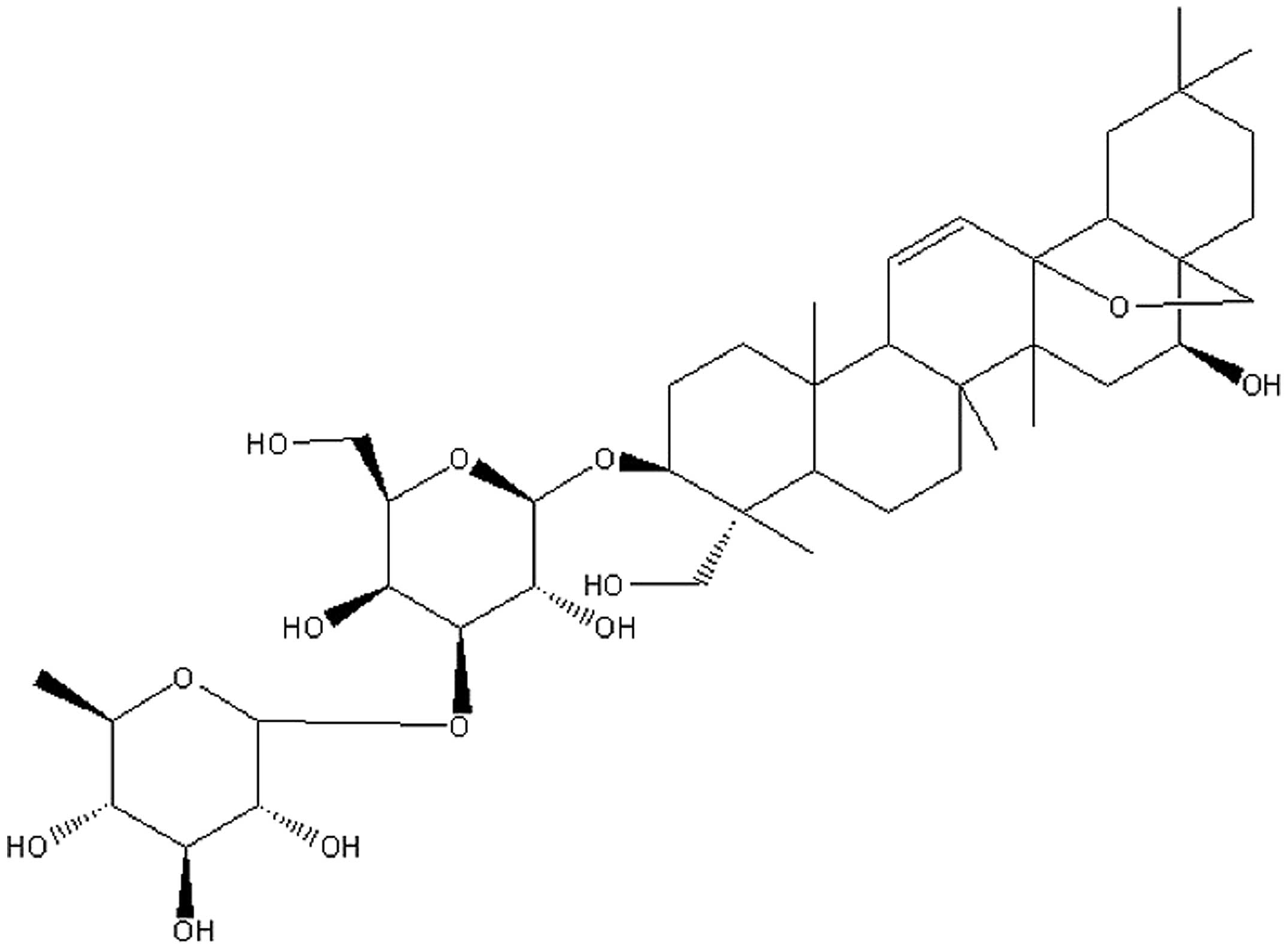
and hepatitis (7). Saikosaponin A
(SsA) is a major bioactive triterpenoid saponin isolated from RB
and has been identified as
(3β,4α,16β)-13,28-epoxy-16,23-dihydroxyolean-11-en-3-yl-6-deoxy-3-O-β-D-glucopyranosyl-β-D-galactopyranoside
(Fig. 1) (8). Previous studies have demonstrated that
SsA exhibits anti-inflammatory activity in vitro and in
vivo (9,10). However, little is known concerning
the protective effects of SsA against sepsis.
In the present study, the inhibitory effects of SsA
on the important proinflammatory cytokines, TNF-α and IL-6 in the
intestinal tissues of septic rats were investigated. Furthermore,
nucleotide-binding oligomerization domain 2 (NOD2) mRNA expression
levels and the activation of the NF-κB were examined in order to
explore the mechanism underlying the effects of SsA on the
inflammatory response.
Materials and methods
Animals
Sixty male Wistar rats weighing 180–200 g were
provided by the Laboratory Animal Center of Henan Province
[Zhengzhou, China; Certificate No. SYXK (Yu2011-0001)]. During the
experiments, all rats were kept in wire-bottomed cages at 25±2°C,
given tap water and standard pellet diet and exposed to a 12-h
light/dark cycle at 50–60% humidity. Animal use was in accordance
with the Guide for the Care and Use of Laboratory Animals of the
National Institutes of Health (4th edition, 2008). The study was
approved by the Ethics Committees of Zhengzhou University
(Zhengzhou, China).
Experimental protocol
The 60 rats were assigned equally to six groups:
Sham surgery, cecal ligation and puncture (CLP), CLP plus SsA (1.0
mg/kg), CLP plus SsA (2.5 mg/kg), CLP plus SsA (5.0 mg/kg) and sham
surgery plus SsA (2.5 mg/kg). Immediately following CLP surgery,
rats were intraperitoneally (i.p.) treated with SsA (purity >
98%; Shanghai Institute of Pharmaceutical Industry, Shanghai,
China) at the specified dose or phosphate-buffered saline (PBS; 5
ml). In each group, 2 surviving rats were sacrificed under ether
anesthesia at 1, 2, 4, 6 or 8 h after surgery respectively. The
ileal tissues were collected and stored in liquid nitrogen for
later use.
CLP procedure
CLP was performed according to the procedure
described previously (11). The rats
were anesthetized i.p. with 2.5% pentobarbital sodium (40 mg/kg;
Sigma-Aldrich, St. Louis, MO, USA). Following a 3-cm midline
incision, the cecum was exposed and ligated with a 3-0 silk suture
below the ileocecal valve. The cecum was then punctured between the
ligation and the tip of the cecum with a 20-gauge needle. After
extruding a small amount of feces from the punctured site, the
cecum was replaced into the peritoneum and the incision was closed
using a sterile 6-0 silk suture. Rats in the sham group underwent
the same laparotomy without the CLP.
Enzyme-linked immunosorbent assay
(ELISA)
The ileal tissues were thawed, washed in PBS,
blotted on filter paper, and weighed. Thereafter, homogenization
was performed in ice-cold homogenate buffer, containing 10 mM HEPES
(pH 7.9), 10 mM KCl, 2 mM MgCl2, 0.1 mM EDTA, 1.0 mM
dithiothreitol and 0.5 mM phenylmethanesulfonyl fluoride. The
homogenates were centrifuged at 3,000 × g for 15 min at 4°C. The
supernatants were collected and were stored at −80°C until assayed.
TNF-α and IL-6 levels in the tissue homogenates were determined by
ELISA using commercial kits (TNF-α ELISA kit; Diaclone, Besançon,
France; IL-6 Rat ELISA kit, cat. no. KRC0061; BioSource Europe SA,
Nivelles, Belgium). The concentrations of TNF-α and IL-6 were
expressed in units of pg of cytokine per mg total protein.
Reverse transcription-quantitative
polymerase chain reaction (RT-qPCR)
Tissues from the ileum were homogenized and total
RNA was isolated using TRIzol reagent according to the
manufacturer's instructions (Invitrogen Corp., Carlsbad, CA, USA).
The RNA was converted into cDNA using M-MLV Reverse Transcriptase
(Promega Corporation, Madison, WI, USA) and oligo(dT) primers. qPCR
analysis with SYBR Green was performed using the Rotor-Gene™ 3000
real-time DNA analysis system (Corbett Research, Sydney,
Australia). Amplifications were performed in triplicate using a
standard shuttle PCR protocol (30 sec at 94°C, 30 sec at 55°C and
30 sec at 72°C) for 40 cycles. Primer sequences were as follows
(all 5′ to 3′): NOD2 forward, ATC CCT CGG TTA CTA TGT TG; reverse,
GCT TCC TGA ATA CTC CTC CT; β-actin forward, CCC ATC TAT GAG GGT
TAC GC; reverse, TTA ATG TCA CGC ACG ATT TC. Primers were designed
with Primer Premier 6.0 software (Premier Biosoft, Palo Alto, CA,
USA). The cDNA results for NOD2 were normalized to β-actin measured
on the same plate.
Western blot analysis
Following the determination of protein
concentrations using a bicinchoninic acid (BCA) kit
(Sigma-Aldrich), proteins were separated by SDS-PAGE on a 10%
Tris-glycine gel, transferred to polyvinylidene membranes, and
incubated with phospho-NF-κB p65 rabbit polyclonal antibody
(1:1,000 dilution; Cell Signaling Technology, Danvers, MA, USA) at
4°C overnight, then washed twice with Tris-buffered saline and
Tween 20 (TBST), followed by horseradish peroxidase conjugated
secondary antibodies (Santa Cruz Biotechnology, Inc., Dallas, TX,
USA) at 1:1,000 dilution for 1 h at room temperature. After
exensive washing with TBST, protein bands were visualized by the
enhanced chemiluminescence reagent (Amersham Pharmacia Biotech,
Tokyo, Japan). The relative expression ratio was determined with
the density of the band of the protein of interest to that of a
β-actin reference band using the software UN-SCAN-IT gel version
6.1 (Silk Scientific, Inc., Orem, UT, USA). Experiments were
repeated at least three times.
Statistical analysis
Statistical analyses were performed using SPSS
software, version 15.0 (SPSS Inc., Chicago, IL, USA). Measurements
were expressed as the mean ± standard deviation (SD). Differences
among multiple groups were examined by one-way analysis of variance
(ANOVA). P<0.05 was considered to indicate a statistically
significant result.
Results
Proinflammatory cytokine
concentrations
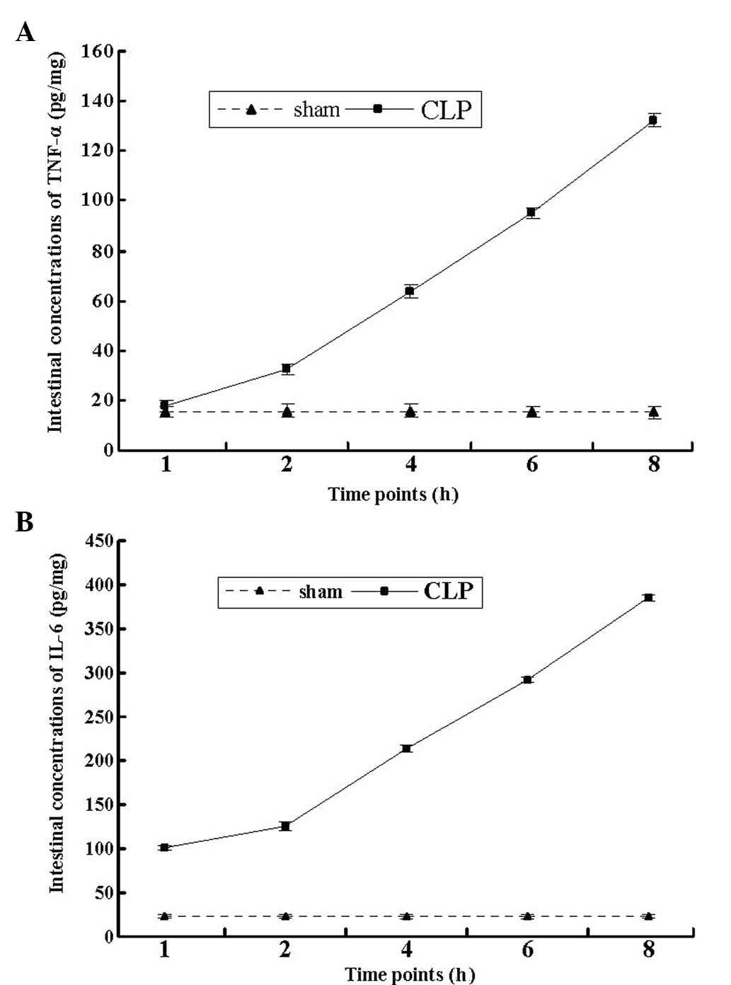
Following CLP surgery, the concentrations of TNF-α
and IL-6 in the intestines of the rats increased in a
time-dependent manner, and the maximum levels were recorded at 8 h
post-surgery (P<0.01; Fig. 2).
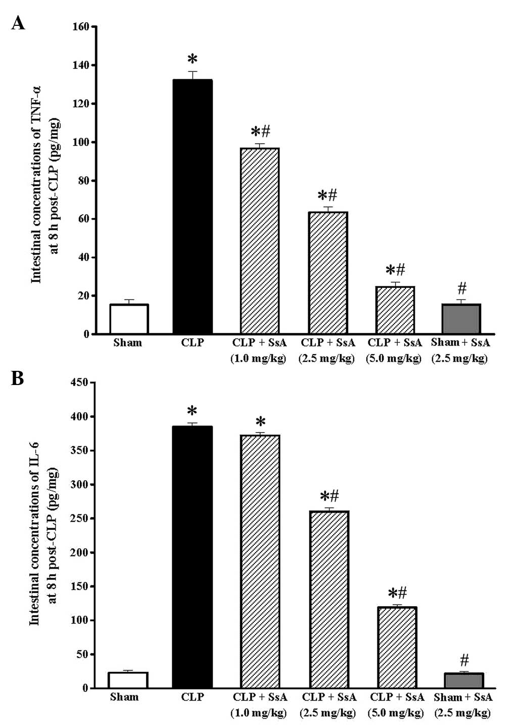
The effect of SsA on the CLP-induced increase in cytokine levels
was also studied at this time point. At doses of ≥1.0 mg/kg, SsA
significantly repressed the elevation of TNF-α (P<0.05). The
inhibitory effects on TNF-α were accompanied by a statistically
significant inhibition of IL-6 elevation at doses of ≥2.5 mg/kg
following CLP (P<0.05). Additionally, SsA did not affect the
levels of proinflammatory cytokines in the rats that did not
undergo CLP treatment (P>0.05; Fig.
3).
NOD2 mRNA expression
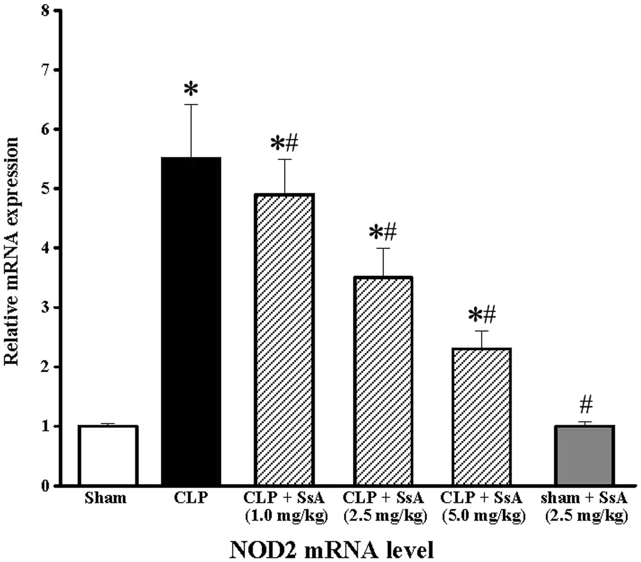
As shown in Fig. 4,
in the CLP group, NOD2 mRNA expression in the intestines was
significantly increased at 8 h post-surgery compared with that in
the sham surgery group (P<0.05), and SsA markedly suppressed the
upregulation of NOD2 mRNA expression in a dose-dependent manner
(P<0.05). In addition, the expression of NOD2 mRNA in the sham
surgery group was found to be similar to that of the sham surgery
plus SsA group.
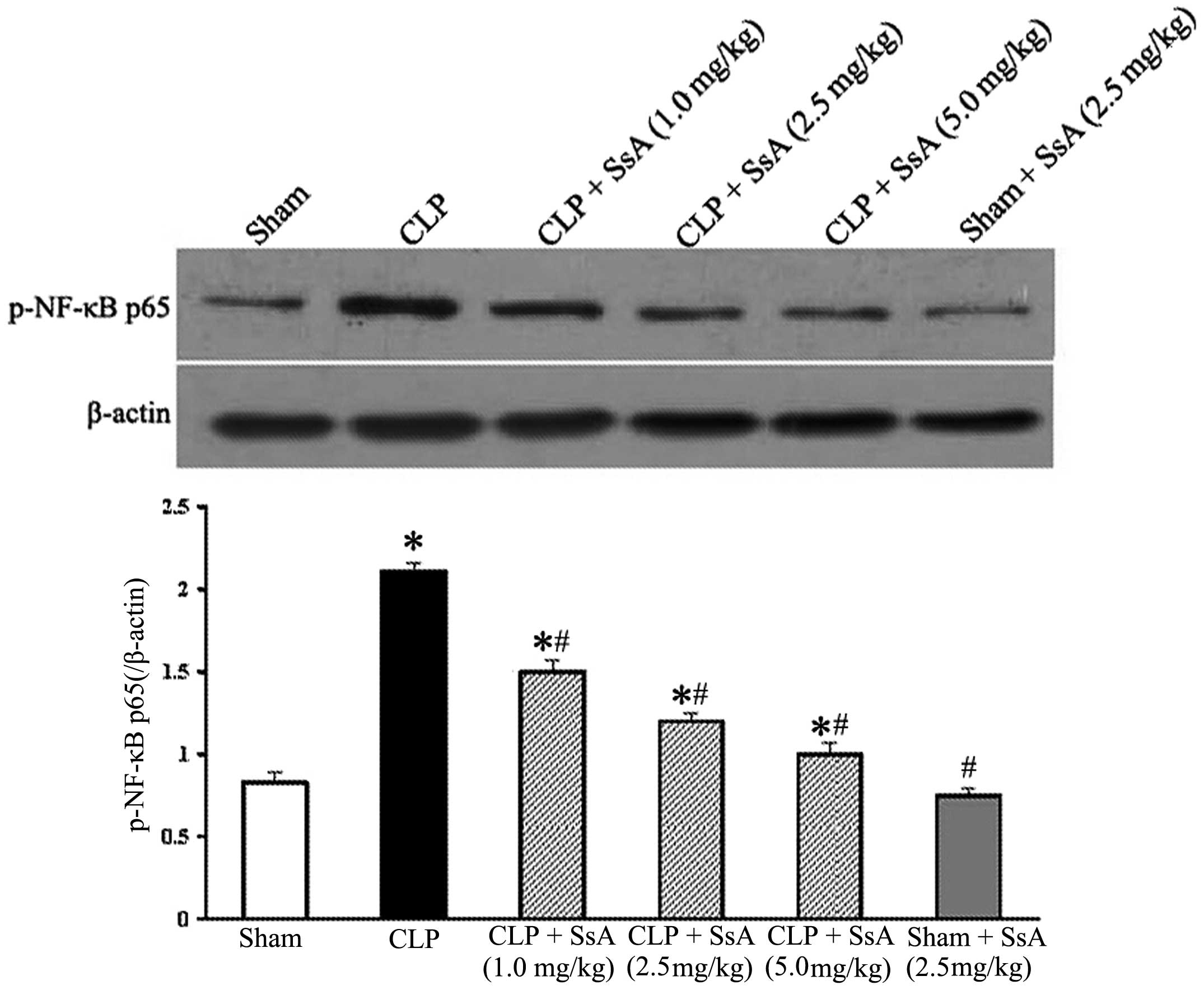
Expression of phospho-NF-κB p65
To investigate the effect of SsA on the activation
of NF-κB in the ileum, the phosphorylation of the NF-κB p65 subunit
was examined by western blotting 8 h after CLP or sham surgery. As
demonstrated in Fig. 5, the p65
level increased significantly at 8 h after CLP, while SsA treatment
markedly inhibited the activation of p65 in a dose-dependent manner
(P<0.05). Moreover, the p65 level in the sham surgery plus SsA
group was similar to that of the sham surgery group. These results
suggest that SsA blocked the elevation of NF-κB activation induced
by CLP.
Discussion
Sepsis is a complex clinical syndrome comprising a
systemic inflammatory response to invasive infection, which can
cause cell injury and progress to multi-organ dysfunction (1,12). It is
believed that the intestine is not only a major ‘victim’ that is
passively damaged, but also a driving force due to the systemic
release of inflammatory cytokines affecting the function and
integrity of other remote organs during the sepsis process
(13). To mimic clinical
polymicrobial sepsis derived from the intestinal tract, a CLP model
was established in the present study and the concentrations of
proinflammatory cytokines in the rat intestine were measured. The
results showed that the intestinal levels of TNF-α and IL-6
markedly increased following CLP in a time-dependent manner.
Several authors have reported that SsA is able to
reduce the secretion of proinflammatory mediators, such as TNF-α,
IL-1β, IL-6 and prostaglandin E2 in a number of cell types
(10,14–16).
Furthermore, SsA has been shown to suppress the contents of TNF-α,
IL-1β and IL-6, and increase the IL-10 level in rats with
CCl4-induced liver inflammation and fibrogenesis
(17,18). SsA has also been found to decrease
the serum TNF-α level in a murine model of allergic rhinitis
(15). In agreement with these
previous studies, the results of the present study demonstrated
that SsA at different doses inhibited the increases in the levels
of TNF-α and IL-6 in the ileal tissues of septic rats.
NF-κB is an inducible nuclear transcription factor,
which plays a key role in regulating the transcription of several
genes, including those encoding proinflammatory cytokines such as
TNF-α, IL-1β and IL-6 involved in severe sepsis and septic shock
(1,19). It is now well established that the
persistent activation of NF-κB is associated with a higher
mortality rate in septic patients (20). Clinical evidence has demonstrated
that the suppression of NF-κB activity may exert an beneficial
effect on sepsis (21,22). NOD2 is known to be an important
innate cytosolic receptor involved in protective immunity against
infectious agents (23). NOD2 in the
host cells senses the peptidoglycan component of gram-positive and
-negative bacteria, transmits signals to receptor-interacting
protein 2, and then triggers a NF-κB-mediated proinflammatory and
antibacterial response (24,25), which leads to a positive feedback
loop during the infection process (26). Polymorphisms in the gene encoding
NOD2 in humans have been associated with early mortality in septic
patients as they affect the ability of NOD2 to recognize bacteria
and activate NF-κB (27). From the
studies described above, a potential role of the NOD2/NF-κB pathway
in sepsis is suggested. In the present study, it was shown that the
activation of NF-κB was greatly enhanced following CLP and that SsA
suppressed NF-κB activation. Moreover, a noteworthy observation is
that NOD2 expression was markedly upregulated in the intestines of
rats following CLP and SsA inhibited its expression. Collectively,
the present findings indicate that SsA may exert a protective role
against sepsis, possibly via the downregulation of the expression
of NOD2 mRNA, which is necessary for NF-κB activation and TNF-α and
IL-6 expression.
There are certain shortcomings in the present study.
First, although NOD2 is necessary for initiating the
proinflammatory function via NF-κB activation, it is not possible
to rule out the possibility that SsA inhibits the activation of
NF-κB via other signaling pathways. Further studies are required to
explore the function of the NOD2-mediated NF-κB pathway
independently. Secondly, data in the literature indicates that the
delicate balance between proinflammatory and anti-inflammatory
mediators determines the severity of infection (28,29). In
the present study, cytokines with notably anti-inflammatory
properties, such as IL-10 and IL-13, which reduce inflammation by
suppressing NF-κB activation (30,31) were
not investigated. Further studies to demonstrate the effect of SsA
on anti-inflammatory cytokine production and the potential
mechanism involved are required.
In conclusion, the present study reveals the
protective effect of SsA during the CLP-induced septic process,
which may be achieved, at least in part, through the inhibition of
the NOD2-mediated NF-κB signaling pathway. Thus, the potent
anti-inflammatory actions of SsA indicate that it may be useful as
a new therapeutic agent for sepsis.
Acknowledgements
This study was supported by a grant from the Science
Foundation of Henan Province (no. 2013A310660) and the Research
Foundation for Talent Recruitment of Zhengzhou University.
References
|
1
|
Hotchkiss RS and Karl IE: The
pathophysiology and treatment of sepsis. N Engl J Med. 348:138–150.
2003. View Article : Google Scholar : PubMed/NCBI
|
|
2
|
Martin GS, Mannino DM, Eaton S and Moss M:
The epidemiology of sepsis in the United States from 1979 through
2000. N Engl J Med. 348:1546–1554. 2003. View Article : Google Scholar : PubMed/NCBI
|
|
3
|
Angus DC, Linde-Zwirble WT, Lidicker J,
Clermont G, Carcillo J and Pinsky MR: Epidemiology of severe sepsis
in the United States: Analysis of incidence, outcome, and
associated costs of care. Crit Care Med. 29:1303–1310. 2001.
View Article : Google Scholar : PubMed/NCBI
|
|
4
|
Parrillo JE, Parker MM, Natanson C,
Suffredini AF, Danner RL, Cunnion RE and Ognibene FP: Septic shock
in humans. Advances in the understanding of pathogenesis,
cardiovascular dysfunction, and therapy. Ann Intern Med.
113:227–242. 1990. View Article : Google Scholar : PubMed/NCBI
|
|
5
|
Pinsky MR, Vincent JL, Deviere J, Alegre
M, Kahn RJ and Dupont E: Serum cytokine levels in human septic
shock. Relation to multiple-system organ failure and mortality.
Chest. 103:565–575. 1993. View Article : Google Scholar : PubMed/NCBI
|
|
6
|
Blackwell TS and Christman JW: Sepsis and
cytokines: Current status. Br J Anaesth. 77:110–117. 1996.
View Article : Google Scholar : PubMed/NCBI
|
|
7
|
Ashour ML and Wink M: Genus Bupleurum: A
review of its phytochemistry, pharmacology and modes of action. J
Pharm Pharmacol. 63:305–321. 2011. View Article : Google Scholar : PubMed/NCBI
|
|
8
|
Park KH, Park J, Koh D and Lim Y: Effect
of saikosaponin-A, a triterpenoid glycoside, isolated from
Bupleurum falcatum on experimental allergic asthma. Phytother Res.
16:359–363. 2002. View
Article : Google Scholar : PubMed/NCBI
|
|
9
|
Lu CN, Yuan ZG, Zhang XL, Yan R, Zhao YQ,
Liao M and Chen JX: Saikosaponin a and its epimer saikosaponin d
exhibit anti-inflammatory activity by suppressing activation of
NF-κB signaling pathway. Int Immunopharmacol. 14:121–126. 2012.
View Article : Google Scholar : PubMed/NCBI
|
|
10
|
Kim SO, Park JY, Jeon SY, Yang CH and Kim
MR: Saikosaponin a, an active compound of Radix Bupleuri,
attenuates inflammation in hypertrophied 3T3-L1 adipocytes via
ERK/NF-κB signaling pathways. Int J Mol Med. 35:1126–1132.
2015.PubMed/NCBI
|
|
11
|
Rittirsch D, Huber-Lang MS, Flierl MA and
Ward PA: Immunodesign of experimental sepsis by cecal ligation and
puncture. Nat Protoc. 4:31–36. 2009. View Article : Google Scholar : PubMed/NCBI
|
|
12
|
Fry DE: Sepsis, systemic inflammatory
response, and multiple organ dysfunction: The mystery continues. Am
Surg. 78:1–8. 2012.PubMed/NCBI
|
|
13
|
Yu M, Shao D, Liu J, Zhu J, Zhang Z and Xu
J: Effects of ketamine on levels of cytokines, NF-kappaB and TLRs
in rat intestine during CLP-induced sepsis. Int Immunopharmacol.
7:1076–1082. 2007. View Article : Google Scholar : PubMed/NCBI
|
|
14
|
Zhu J, Luo C, Wang P, He Q, Zhou J and
Peng H: Saikosaponin A mediates the inflammatory response by
inhibiting the MAPK and NF-κB pathways in LPS-stimulated RAW 264.7
cells. Exp Ther Med. 5:1345–1350. 2013.PubMed/NCBI
|
|
15
|
Han NR, Kim HM and Jeong HJ: Inactivation
of cystein-aspartic acid protease (caspase)-1 by saikosaponin A.
Biol Pharm Bull. 34:817–823. 2011. View Article : Google Scholar : PubMed/NCBI
|
|
16
|
Sun Y, Cai TT, Zhou XB and Xu Q:
Saikosaponin A inhibits the proliferation and activation of T cells
through cell cycle arrest and induction of apoptosis. Int
Immunopharmacol. 9:978–983. 2009. View Article : Google Scholar : PubMed/NCBI
|
|
17
|
Wu SJ, Lin YH, Chu CC, Tsai YH and Chao
JC: Curcumin or saikosaponin A improves hepatic antioxidant
capacity and protects against CCl4-induced liver injury
in rats. J Med Food. 11:224–229. 2008. View Article : Google Scholar : PubMed/NCBI
|
|
18
|
Wu SJ, Tam KW, Tsai YH, Chang CC and Chao
JC: Curcumin and saikosaponin A inhibit chemical-induced liver
inflammation and fibrosis in rats. Am J Chin Med. 38:99–111. 2010.
View Article : Google Scholar : PubMed/NCBI
|
|
19
|
Hayden MS and Ghosh S: Shared principles
in NF-kappaB signaling. Cell. 132:344–362. 2008. View Article : Google Scholar : PubMed/NCBI
|
|
20
|
Arnalich F, Garcia-Palomero E, López J,
Jiménez M, Madero R, Renart J, Vázquez JJ and Montiel C: Predictive
value of nuclear factor kappaB activity and plasma cytokine levels
in patients with sepsis. Infect Immun. 68:1942–1945. 2000.
View Article : Google Scholar : PubMed/NCBI
|
|
21
|
Feng X, Ren B, Xie W, Huang Z, Liu J, Guan
R, Duan M and Xu J: Influence of hydroxyethyl starch 130/0.4 in
pulmonary neutrophil recruitment and acute lung injury during
polymicrobial sepsis in rats. Acta Anaesthesiol Scand.
50:1081–1088. 2006. View Article : Google Scholar : PubMed/NCBI
|
|
22
|
Feng X, Yan W, Liu X, Duan M, Zhang X and
Xu J: Effects of hydroxyethyl starch 130/0.4 on pulmonary capillary
leakage and cytokines production and NF-kappaB activation in
CLP-induced sepsis in rats. J Surg Res. 135:129–136. 2006.
View Article : Google Scholar : PubMed/NCBI
|
|
23
|
Frutuoso MS, Hori JI, Pereira MS, Junior
DS, Sônego F, Kobayashi KS, Flavell RA, Cunha FQ and Zamboni DS:
The pattern recognition receptors Nod1 and Nod2 account for
neutrophil recruitment to the lungs of mice infected with
Legionella pneumophila. Microbes Infect. 12:819–827. 2010.
View Article : Google Scholar : PubMed/NCBI
|
|
24
|
Hasegawa M, Fujimoto Y, Lucas PC, Nakano
H, Fukase K, Núñez G and Inohara N: A critical role of RICK/RIP2
polyubiquitination in Nod-induced NF-kappaB activation. EMBO J.
27:373–383. 2008. View Article : Google Scholar : PubMed/NCBI
|
|
25
|
Magalhaes JG, Lee J, Geddes K, Rubino S,
Philpott DJ and Girardin SE: Essential role of Rip2 in the
modulation of innate and adaptive immunity triggered by Nod1 and
Nod2 ligands. Eur J Immunol. 41:1445–1455. 2011. View Article : Google Scholar : PubMed/NCBI
|
|
26
|
Hu C, Sun L, Hu Y, Lu D, Wang H and Tang
S: Functional characterization of the NF-kappaB binding site in the
human NOD2 promoter. Cell Mol Immunol. 7:288–295. 2010. View Article : Google Scholar : PubMed/NCBI
|
|
27
|
Brenmoehl J, Herfarth H, Glück T, Audebert
F, Barlage S, Schmitz G, Froehlich D, Schreiber S, Hampe J,
Schölmerich J, et al: Genetic variants in the NOD2/CARD15 gene are
associated with early mortality in sepsis patients. Intensive Care
Med. 33:1541–1548. 2007. View Article : Google Scholar : PubMed/NCBI
|
|
28
|
Girardin E, Grau GE, Dayer JM,
Roux-Lombard P and Lambert PH: Tumor necrosis factor and
interleukin-1 in the serum of children with severe infectious
purpura. N Engl J Med. 319:397–400. 1988. View Article : Google Scholar : PubMed/NCBI
|
|
29
|
Damas P, Ledoux D, Nys M, Vrindts Y, De
Groote D, Franchimont P and Lamy M: Cytokine serum level during
severe sepsis in human IL-6 as a marker of severity. Ann Surg.
215:356–362. 1992. View Article : Google Scholar : PubMed/NCBI
|
|
30
|
Wang P, Wu P, Siegel MI, Egan RW and
Billah MM: Interleukin (IL)-10 inhibits nuclear factor kappaB (NF
kappaB) activation in human monocytes. IL-10 and IL-4 suppress
cytokine synthesis by different mechanisms. J Biol Chem.
270:9558–9563. 1995. View Article : Google Scholar : PubMed/NCBI
|
|
31
|
Lentsch AB, Shanley TP, Sarma V and Ward
PA: In vivo suppression of NF-kappaB and preservation of I kappaB
alpha by interleukin-10 and interleukin-13. J Clin Invest.
100:2443–2448. 1997. View Article : Google Scholar : PubMed/NCBI
|