|
1
|
Kim CM, Shin MK, Lee KS and Ahn DK: The
Encyclopedia of Oriental Herbal Medicine. Jung Dam Publishing Co.;
Seoul: 1998, pp. 3362–3372. 1998
|
|
2
|
Li JX, Kadota S, Li HY, et al: Effects of
Cimicifugae rhizoma on serum calcium and phosphate levels in low
calcium dietary rats and on bone mineral density in ovariectomized
rats. Phytomedicine. 3:379–385. 1997. View Article : Google Scholar : PubMed/NCBI
|
|
3
|
Sakurai N and Nagai M: Chemical
constituents of original plants of Cimicifugae rhizoma in Chinese
medicine. Yakugaku Zasshi. 116:850–865. 1996.(In Japanese).
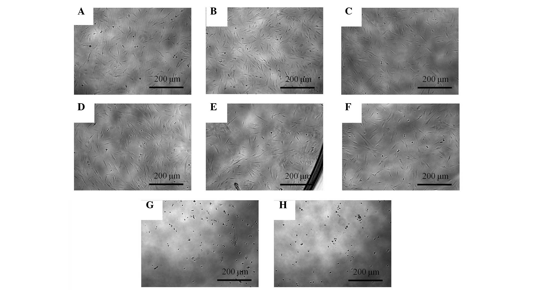
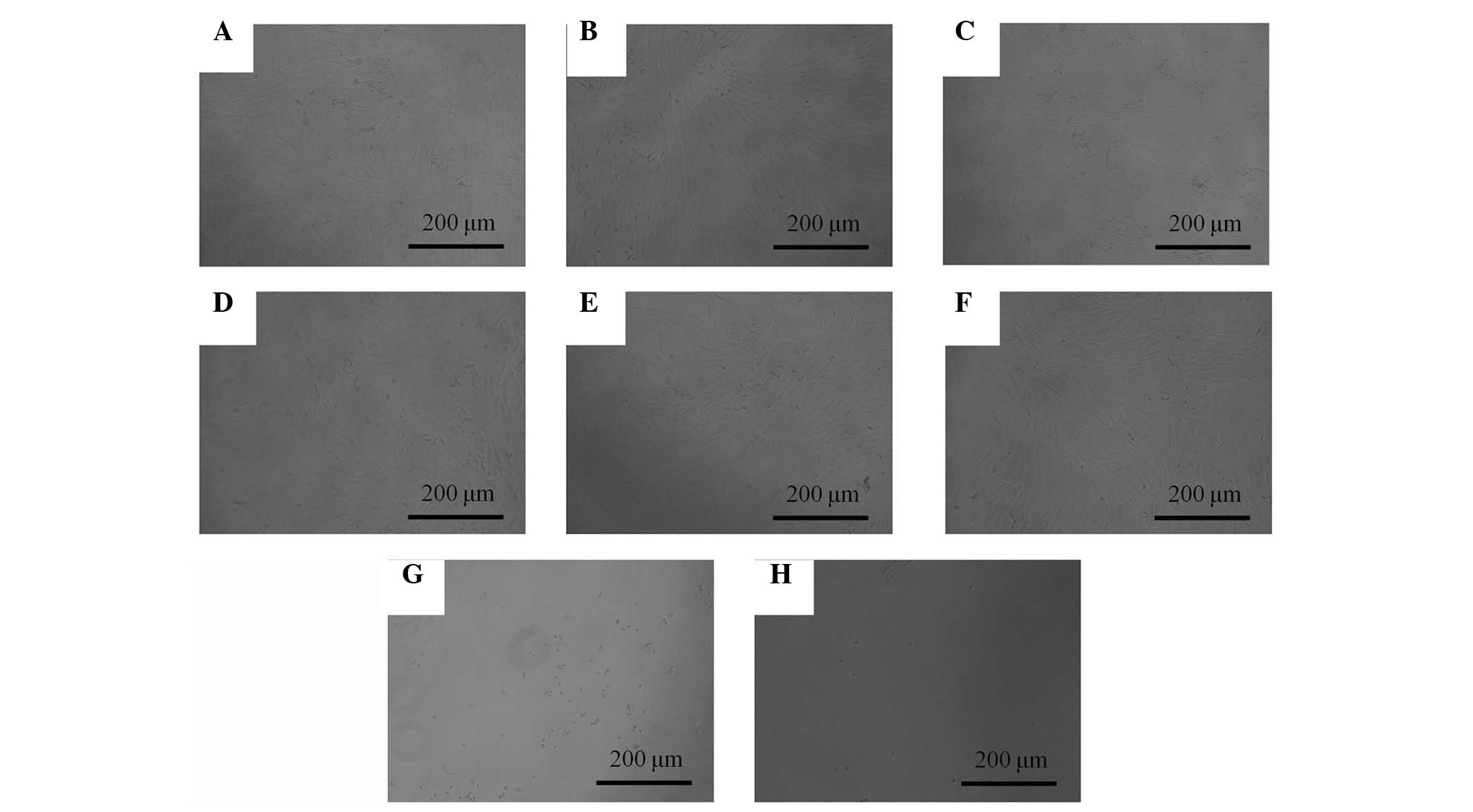
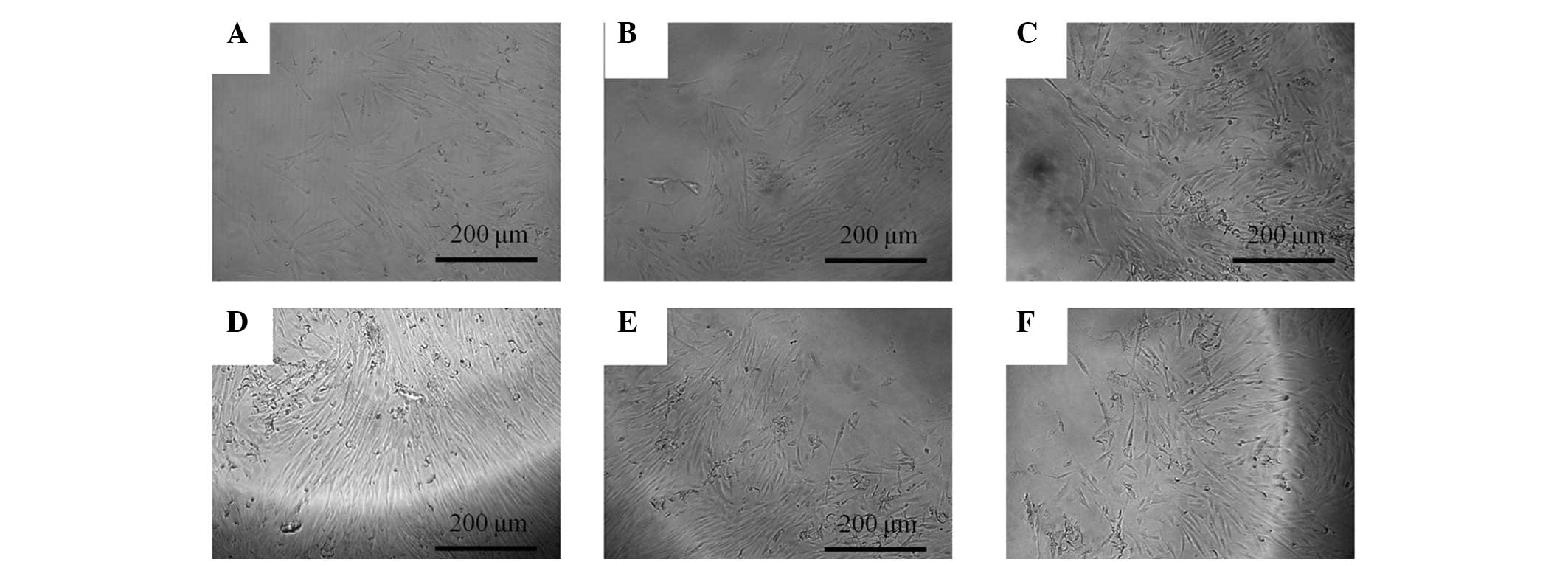
PubMed/NCBI
|
|
4
|
Liao JF, Jan YM, Huang SY, Wang HH, Yu LL
and Chen CF: Evaluation with receptor binding assay on the water
extracts of ten CNS-active Chinese herbal drugs. Proc Natl Sci
Counc Repub China B. 19:151–158. 1995.PubMed/NCBI
|
|
5
|
Kim SJ and Kim MS: Inhibitory effects of
cimicifugae rhizoma extracts on histamine, bradykinin and COX-2
mediated inflammatory actions. Phytother Res. 14:596–600. 2000.
View Article : Google Scholar : PubMed/NCBI
|
|
6
|
Kwon IH, Kim NK, Chiang HC, Lim HG and Kim
JM: Antipyretic effect of Rhizoma Cimicifugae in a rat model of
LPS-induced fever. Daehan Hanui Hakhoeji. 23:32–44. 2002.(In
Korean).
|
|
7
|
Kim DK, Kim T, Pi SH, et al: Effects of
several natural medicines on alkaline phosphatase synthesis in
MC3T3-E1 cells. J Korean Acad Periodontol. 29:751–764. 1999.
View Article : Google Scholar
|
|
8
|
Wong RW, Hägg U, Samaranayake L, Yuen MK,
Seneviratne CJ and Kao R: Antimicrobial activity of Chinese
medicine herbs against common bacteria in oral biofilm. A pilot
study. Int J Oral Maxillofac Surg. 39:599–605. 2010. View Article : Google Scholar : PubMed/NCBI
|
|
9
|
Hidaka S, Nishimura H, Nakajima K and Liu
SY: Effects of a rhubarb (Rhei rhizoma) solution and its fractions
on the formation of calcium phosphate precipitates. J Periodontal
Res. 31:408–413. 1996. View Article : Google Scholar : PubMed/NCBI
|
|
10
|
Tian Z, Pan R, Chang Q, Si J, Xiao P and
Wu E: Cimicifuga foetida extract inhibits proliferation of
hepatocellular cells via induction of cell cycle arrest and
apoptosis. J Ethnopharmacol. 114:227–233. 2007. View Article : Google Scholar : PubMed/NCBI
|
|
11
|
He CC, Dai YQ, Hui RR, et al: NMR-based
metabonomic approach on the toxicological effects of a Cimicifuga
triterpenoid. J Appl Toxicol. 32:88–97. 2012. View Article : Google Scholar : PubMed/NCBI
|
|
12
|
Lu L, Chen JC, Li Y, et al: Studies on the
constituents of Cimicifuga foetida collected in Guizhou Province
and their cytotoxic activities. Chem Pharm Bull (Tokyo).
60:571–577. 2012. View Article : Google Scholar : PubMed/NCBI
|
|
13
|
Mun L, Jun MS, Kim YM, et al:
7,8-didehydrocimigenol from Cimicifugae rhizoma inhibits
TNF-α-induced VCAM-1 but not ICAM-1expression through upregulation
of PPAR-γ in human endothelial cells. Food Chem Toxicol.
49:166–172. 2011. View Article : Google Scholar : PubMed/NCBI
|
|
14
|
Li JX and Yu ZY: Cimicifugae Rhizoma: from
origins, bioactive constituents to clinical outcomes. Curr Med
Chem. 13:2927–2951. 2006. View Article : Google Scholar : PubMed/NCBI
|
|
15
|
Li JX, Liu J, He CC, et al: Triterpenoids
from Cimicifugae rhizoma, a novel class of inhibitors on bone
resorption and ovariectomy-induced bone loss. Maturitas. 58:59–69.
2007. View Article : Google Scholar : PubMed/NCBI
|
|
16
|
Cao L, Sun H, Li Z and Pan RL: Comparison
of activities of various species of Rhizoma Cimicifugae and their
honey processed products. Zhong Yao Cai. 30:1561–1563. 2007.(In
Chinese). PubMed/NCBI
|
|
17
|
Li X, Lin J, Gao Y, Han W and Chen D:
Antioxidant activity and mechanism of Rhizoma Cimicifugae. Chem
Cent J. 6:1402012. View Article : Google Scholar : PubMed/NCBI
|
|
18
|
Kim SH, Lee SE, Oh H, et al: The
radioprotective effects of Bu-Zhong-Yi-Qi-Tang: a prescription of
traditional Chinese medicine. Am J Chin Med. 30:127–137. 2002.
View Article : Google Scholar : PubMed/NCBI
|
|
19
|
Nishida S: Effect of Hochu-ekki-to on
asymptomatic MRSA bacteriuria. J Infect Chemother. 9:58–61. 2003.
View Article : Google Scholar : PubMed/NCBI
|
|
20
|
Pan L, Huang YW, Ye YR and Wang YQ: A
model for screening anti-viral agents based on yeast killer system.
Wei Sheng Wu Xue Bao. 47:517–521. 2007.(In Chinese). PubMed/NCBI
|
|
21
|
Sekiya I, Larson BL, Smith JR, Pochampally
R, Cui JG and Prockop DJ: Expansion of human adult stem cells from
bone marrow stroma: conditions that maximize the yields of early
progenitors and evaluate their quality. Stem Cells. 20:530–541.
2002. View Article : Google Scholar : PubMed/NCBI
|
|
22
|
Kuznetsov SA, Friedenstein AJ and Robey
PG: Factors required for bone marrow stromal fibroblast colony
formation in vitro. Br J Haematol. 97:561–570. 1997. View Article : Google Scholar : PubMed/NCBI
|
|
23
|
Rodriguez AM, Elabd C, Amri EZ, Ailhaud G
and Dani C: The human adipose tissue is a source of multipotent
stem cells. Biochimie. 87:125–128. 2005. View Article : Google Scholar : PubMed/NCBI
|
|
24
|
Ballini A, De Frenza G, Cantore S, Papa F,
Grano M, Mastrangelo F, Tetè S and Grassi FR: In vitro stem cell
cultures from human dental pulp and periodontal ligament: new
prospects in dentistry. Int J Immunopathol Pharmacol. 20:9–16.
2007.PubMed/NCBI
|
|
25
|
Nagatomo K, Komaki M, Sekiya I, Sakaguchi
Y, Noguchi K, Oda S, Muneta T and Ishikawa I: Stem cell properties
of human periodontal ligament cells. J Periodontal Res. 41:303–310.
2006. View Article : Google Scholar : PubMed/NCBI
|
|
26
|
Zhang Q, Shi S, Liu Y, Uyanne J, Shi Y,
Shi S and Le AD: Mesenchymal stem cells derived from human gingiva
are capable of immunomodulatory functions and ameliorate
inflammation-related tissue destruction in experimental colitis. J
Immunol. 183:7787–7798. 2009. View Article : Google Scholar : PubMed/NCBI
|