Introduction
Rheumatoid arthritis (RA) is a clinically common
systemic autoimmune disease. In patients with RA, extensive
irreversible bone and joint destruction appears in the first 2
years of the disease and later develops into ankylosis and
deformity and leads to joint dysfunction, affecting the joint
appearance and activity of the patients. Some patients become
bed-bound, losing their ability to work and requiring care from
others, which has serious consequences on their life and that of
their family members (1–3). Early diagnosis of RA and timely
treatment can effectively reduce bone and joint destruction,
deformation and loss of function, and improve prognosis (4).
Rheumatoid factor (RF) is the only serological index
used by the American Rheumatism Association for the diagnostic
classification of RA and is currently used as an auxiliary in the
diagnosis of RA (5). RF is a highly
sensitive and accurate diagnostic marker for RA, but it has a low
specificity; as a result, the diagnosis usually relies on other
serological tests and symptom presentation (6). Vascular cell adhesion molecule-1
(VCAM-1) is generated by cells, can be found on the cell surface
and mediates the contact and binding between cells or between the
cells and stroma. As an adhesion molecule, VCAM-1 is involved in
several vital physiological and pathological processes of the
organism, including the viscosity of white blood cells and vascular
cells during inflammatory processes, immune cell recognition, lymph
node homing, tumor invasion and metastasis and intracellular signal
transduction. VCAM-1 can interact with and influence interleukin
(IL)-4, IL-8, tumor necrosis factor (TNF)-α, TNF-β and other
inflammatory cytokines (7–9). Therefore, VCAM-1 affects, to some
extent, inflammatory reactions, which may be associated with RA
occurrence and development. Reports on the changes in VCAM-1 levels
in RA and the association between the changes and the disease state
are rare (10). In the present
study, the changes in the serum VCAM-1 level and the effect of
treatment on the VCAM-1 level were analyzed in patients with RA. In
addition, Pearson correlation analysis was employed in order to
analyze the correlation between the serum VCAM-1 and RF levels in
patients with RA and provide a clinical basis for predicting the
occurrence, development and outcome of RA.
Subjects and methods
Baseline characteristics
One hundred and twenty patients with RA who had been
admitted to the Huaihe Hospital of Henan University (Kaifeng,
China) between January and December 2013 were enrolled in the study
as the observation group, and 30 healthy individuals in the
corresponding period comprised the control group. The included
patients were diagnosed with RA based on the RA classification and
diagnostic criteria defined by the American Rheumatism Association
in 1987 (11,12). The present study was conducted in
accordance with the Declaration of Helsinki and was approved by the
Ethics Committee of Huaihe Hospital of Henan University. Written
informed consent was obtained from all participants. With regard to
the severity of RA, 21 cases were classified as grade I, 44 cases
as grade II, 52 cases as grade III and 3 cases as grade IV. The
observation group consisted of 62 men and 58 women aged 28–85
years, with the mean age being 42.58±8.59 years. The patients
weighed 38–82 kg (mean, 57.24±9.85 kg) and the mean arterial
pressure was 85.98±7.24 mmHg. The control group comprised 15 men
and 15 women, aged 30–78 years (mean, 43.65±8.87 years). The
subjects weighed 40–81 kg (mean, 55.98±9.27 kg), and the mean
arterial pressure was 84.27±7.57 mmHg. No statistically significant
differences were observed in the baseline characteristics between
the two groups (P>0.05).
Treatment methods
According to the patients' conditions, nonsteroidal
anti-inflammatory drugs, such as aspirin (Bayer AG, Leverkusen,
Germany), were used for treatment. The patients also received
anti-rheumatoid drugs, and certain patients with severe syndromes
additionally underwent glucocorticoid treatment. The dose for
symptom control was 35 mg/day and the maintenance dose was 10
mg/day. With regard to the patients with a limited joint movement
range, obvious structural destruction and unbearable pain,
corresponding joint replacement surgery or synovectomy was
conducted. All patients received dietary guidance and support. When
necessary, patients were given joint immobilization and prescribed
bed-rest. The patients with mood disorders underwent psychological
nursing intervention.
Detection methods
Fasting venous blood (3 ml) was extracted from all
patients 1 day after admission, and the blood samples were
centrifuged at 4,000 × g for 5 min and then stored at −20°C until
use. Serum VCAM-1 and RF levels were detected using ELISA, and the
kits were provided by Shanghai Ruiqi Biological Technology Co.,
Ltd. (Shanghai, China). The microplate reader was provided by
Kunkeng Biological Technology Co., Ltd. (Shanghai, China). All
experiments were performed according to the manufacturers'
instructions. Following treatment, the indexes in the observation
group were checked every 12 h from the start of treatment to 1 week
after treatment and then rechecked once a week for 6 months.
Statistical analysis
All data were analyzed using SPSS 13.0 software
(SPSS, Inc., Chicago, IL, USA). The t-test was used for the
comparison between the two groups, while analysis of variance was
used for the comparison among various measurement data. The q-test
was used for further pairwise comparison, and the Pearson
correlation coefficient was used for correlation analysis.
P<0.05 was considered to indicate a statistically significant
difference.
Results
Comparison of serum VCAM-1 and RF
levels between the two groups
Prior to treatment, the serum VCAM-1 and RF levels
were significantly higher in the observation group than those in
the control group (P<0.01) (Table
I).
 | Table I.Comparison of the pretreatment serum
VCAM-1 and RF levels between the two groups. |
Table I.
Comparison of the pretreatment serum
VCAM-1 and RF levels between the two groups.
| Groups | Cases | VCAM-1 (µg/l) | RF (U/ml) |
|---|
| Observation | 120 | 1125.58±125.89 | 32.15±8.45 |
| Control | 30 | 421.25±74.25 | 13.26±4.17 |
| t-test |
| 12.47 | 8.95 |
| P-value |
| <0.01 | <0.01 |
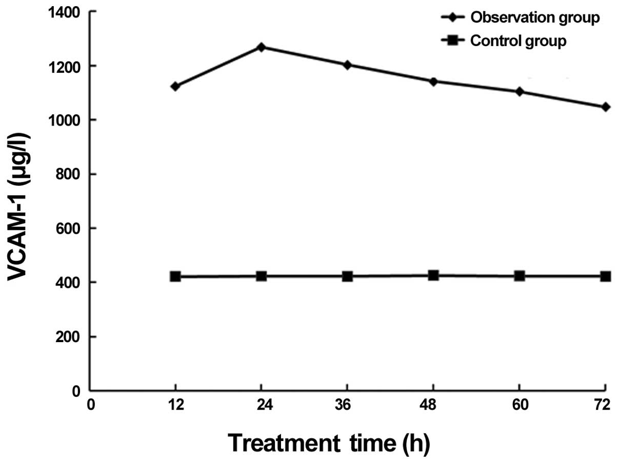
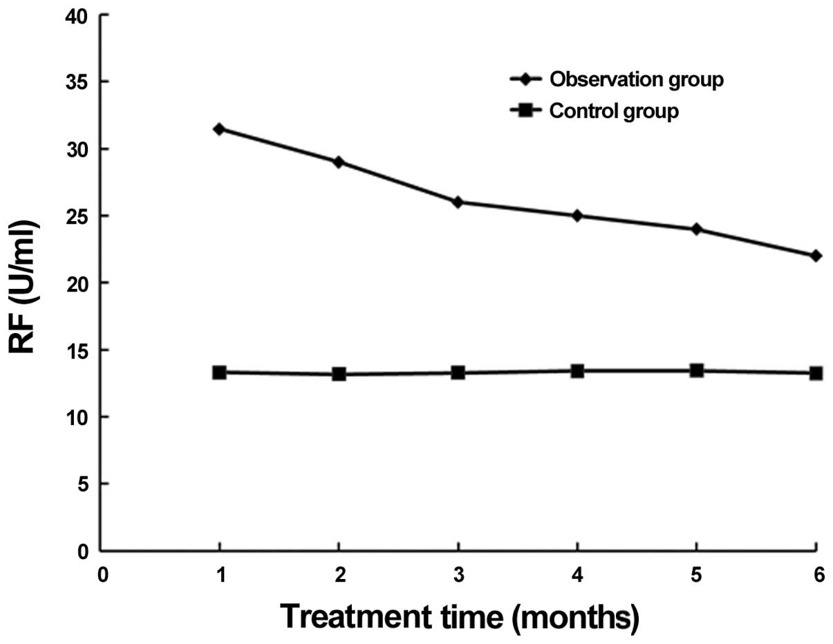
Comparison of serum VCAM-1 and RF
levels at different treatment time-points in the observation
group
The comparison of the serum VCAM-1 and RF levels at
different treatment time-points in the observation group revealed
statistically significant differences; compared with the levels of
VCAM-1 and RF prior to treatment in the observation group
(P<0.05), the levels 1, 3 and 6 months after treatment were
significantly lower, and a gradual decrease was noted over the
course of treatment (P<0.05). Twenty-four hours after treatment,
the serum VCAM-1 levels of the patients peaked, while the serum RF
levels peaked 36 h after treatment (Table II, Figs.
1–4).
| Table II.Comparison of VCAM-1 and serum RF
levels in the observation group at different treatment
time-points. |
Table II.
Comparison of VCAM-1 and serum RF
levels in the observation group at different treatment
time-points.
| Time-points | Cases | VCAM-1 (µg/l) | RF (U/ml) |
|---|
| Before treatment | 120 | 1125.58±125.89 | 32.15±8.45 |
| 1 week after
treatment | 120 | 1096.58±105.69 | 29.58±8.24 |
| 1 month after
treatment | 120 |
982.57±98.65a |
25.55±9.14a |
| 3 months after
treatment | 120 |
741.52±79.48b |
22.15±9.22b |
| 6 months after
treatment | 120 |
581.47±65.72c |
20.28±7.62c |
| t-test |
| 11.42 | 5.68 |
| P-value |
| <0.01 | <0.05 |
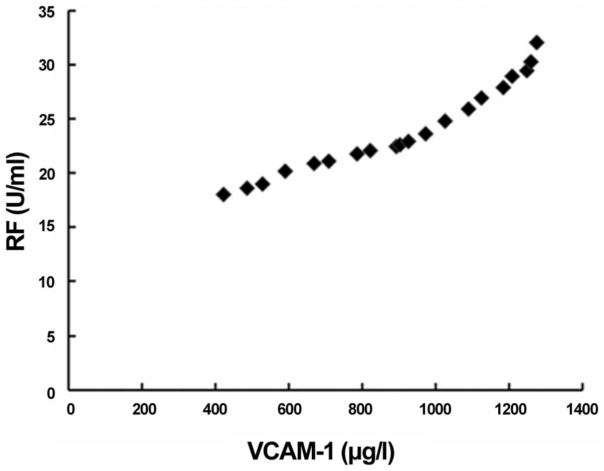
Correlation between serum VCAM-1 and
RF levels in patients with RA
The Pearson correlation analysis indicated that the
increase in the serum VCAM-1 level occurred concurrently with an
increase in the serum RF level in patients with RA, suggesting the
presence of a significantly positive correlation between the two
(r=0.852, P<0.01) (Fig. 5).
Discussion
RA is a chronic inflammatory disease that affects
several symmetrical peripheral joints, leading to clinical
manifestations of pain and swelling in the affected joints,
pathological changes and gradual dysfunction of the joints, as well
as recurrent and unhealed symptoms (13,14). The
pathological changes are characteristic of erosive synovitis, which
destroys the joint and affects several extra-articular systems. RA,
therefore, has a strongly negative influence on the quality of life
of the patients (15,16). The cause of RA still remains unknown,
as several factors can cause the autoimmune reactions of the
organs, leading to pathological changes.
The principle pathogenic factors of RA are heredity
and infections. Studies have shown that the descendants of patients
with RA may have increased RF levels, even without exhibiting any
RA symptoms, while infections caused by Mycoplasma and
Streptococcus can also result in disorders of the autoimmune
system, and thus induce RA (17,18). The
diagnosis of RA is made based on the clinical symptoms of the
patients, X-ray results and RF detection results, and the serum RF
level is the only serological indicator of RA (19,20).
VCAM-1 is an essential cell adhesion molecule and its expression
can be induced by inflammatory cytokines, such as IL-6 and TNF,
which participate in the differentiation and development of
lymphocytes and are associated with the pathological processes of
several diseases, including systemic inflammation, tumor invasion
and metastasis, autoimmune diseases and parasitic infections
(21,22). As a chronic inflammatory disease, it
is possible that RA is associated with VCAM-1; however, reports on
the changes in the levels of VCAM-1 and its association with the
development of RA are rare (10).
Determining the function and mechanism of VCAM-1 in patients with
RA could provide a strong clinical basis for the treatment of RA
and the evaluation of the curative effects of the treatment.
The results of the present study showed that the
serum VCAM-1 level was increased in the patients with RA, and that
this increase continued for a short period of time during
conventional treatment. With the prolongment of the treatment,
certain clinical symptoms were relieved and the serum RF level
decreased. The decline in the serum RF level was accompanied by a
gradual decline in the serum VCAM-1 level, which suggests that the
serum VCAM-1 level may be associated with the disease condition and
serum RF level of the patients.
Pearson correlation analysis was employed in order
to further study the correlation between the serum VCAM-1 and RF
levels of the patients with RA. The results indicated that, in
patients with RA, an increase in the serum VCAM-1 levels was
accompanied by an increase in the serum RF levels, thus revealing a
significantly positive correlation between the two. The serum
VCAM-1 level can reflect the changes in the disease condition and,
to some degree, the effect of the RA treatment. The present
findings were consistent with the hypothesis that, as a chronic
inflammatory disease, RA is associated with VCAM-1. The specific
mechanism may be associated with the interaction of VCAM-1, IL and
TNF, which adjusts levels of inflammatory cytokines and indirectly
affects inflammatory reactions. High levels of VCAM-1 can markedly
promote the release of inflammatory cytokines. VCAM-1 is involved
in the formation of lymphocytes and the adjustment of the immune
system, which also affects the immune system's function. High
levels of serum VCAM-1 may be associated with the disorder of the
immune system (23). RA is an
autoimmune and inflammatory disease, so patients with RA may
exhibit abnormal serum VCAM-1 levels. When the symptoms subside and
the inflammatory reactions are reduced, the function of the immune
system gradually returns to normal, and thus the serum VCAM-1 level
progressively decreases.
The present study had certain limitations; the
sample size was small and the observation time was short.
Furthermore, the serum VCAM-1 levels in patients with RA are
affected by several factors, and so a long-term study with a large
sample size is required for the determination of the correlation
between the VCAM-1 level in patients with RA and the curative
effects of anti-RA treatment.
In conclusion, the serum VCAM-1 level in patients
with RA first increases and then decreases, as the condition is
relieved. The serum VCAM-1 levels of patients with RA may be
associated with the autoimmune and inflammatory reactions of RA,
and therefore reflect, to some extent, the disease condition and
curative effects of treatment. The VCAM-1 levels could,
consequently, assist the prediction of the effects of anti-RA
treatment.
References
|
1
|
Paccou J, Boudot C, Renard C, Liabeuf S,
Kamel S, Fardellone P, Massy Z, Brazier M and Mentaverri R: Total
calcium-sensing receptor expression in circulating monocytes is
increased in rheumatoid arthritis patients with severe coronary
artery calcification. Arthritis Res Ther. 16:4122014. View Article : Google Scholar : PubMed/NCBI
|
|
2
|
Oiwa H, Mihara K, Kan T, Tanaka M, Shindo
H, Kumagai K and Sugiyama E: Grade 3 lymphomatoid granulomatosis in
a patient receiving methotrexate therapy for rheumatoid arthritis.
Intern Med. 53:1873–1875. 2014. View Article : Google Scholar : PubMed/NCBI
|
|
3
|
Inoue M, Yanaihara N and Okamoto A:
Salmonella ovarian abscess in a patient with rheumatoid arthritis
(RA): A case report with literature review. Clin Exp Obstet
Gynecol. 41:465–467. 2014.PubMed/NCBI
|
|
4
|
Witkowski JM: Mechanisms of the immune
system in ageing and some age-associated diseases. Postepy Biochem.
60:233–239. 2014.PubMed/NCBI
|
|
5
|
Li G, Shi F, Liu J and Li Y: The effect of
CTLA-4 A49G polymorphism on rheumatoid arthritis risk: A
meta-analysis. Diagn Pathol. 9:1572014. View Article : Google Scholar : PubMed/NCBI
|
|
6
|
Sharma JN: Basic and clinical aspects of
bradykinin receptor antagonists. Prog Drug Res. 69:1–14.
2014.PubMed/NCBI
|
|
7
|
Yaman A, Karabag F, Demir S and Koken T:
Changes in serum asymmetric dimethylarginine and endothelial
markers levels with varying periods of hemodialysis. Ther Apher
Dial. 18:361–367. 2014. View Article : Google Scholar : PubMed/NCBI
|
|
8
|
Mlinar LB, Chung EJ, Wonder EA and Tirrell
M: Active targeting of early and mid-stage atherosclerotic plaques
using self-assembled peptide amphiphile micelles. Biomaterials.
35:8678–8686. 2014. View Article : Google Scholar : PubMed/NCBI
|
|
9
|
Zheng Y, Yang W, Aldape K, He J and Lu Z:
Epidermal growth factor (EGF)-enhanced vascular cell adhesion
molecule-1 (VCAM-1) expression promotes macrophage and glioblastoma
cell interaction and tumor cell invasion. J Biol Chem.
288:31488–31495. 2013. View Article : Google Scholar : PubMed/NCBI
|
|
10
|
Navarro-Hernández RE, Oregon-Romero E,
Vázquez-Del Mercado M, Rangel-Villalobos H, Palafox-Sánchez CA and
Muñoz-Valle JF: Expression of ICAM1 and VCAM1 serum levels in
rheumatoid arthritis clinical activity. Association with genetic
polymorphisms. Dis Markers. 26:119–126. 2009. View Article : Google Scholar : PubMed/NCBI
|
|
11
|
Alarcón GS, Blackburn WD Jr, Calvo A and
Castañeda O: Evaluation of the American Rheumatism Association
preliminary criteria for remission in rheumatoid arthritis: A
prospective study. J Rheumatol. 14:93–96. 1987.PubMed/NCBI
|
|
12
|
Ariza R, Van Walsem A, Canal C, Roldán C,
Betegón L, Oyagüez I and Janssen K: Cost-minimization analysis of
subcutaneous abatacept in the treatment of rheumatoid arthritis in
Spain. Farm Hosp. 38:257–265. 2014.PubMed/NCBI
|
|
13
|
Kobelt G: Treating to target with
etanercept in rheumatoid arthritis: Cost-effectiveness of dose
reductions when remission is achieved. Value Health. 17:537–544.
2014. View Article : Google Scholar : PubMed/NCBI
|
|
14
|
Yokogawa N, Kaneko T, Nagai Y, Nunokawa T,
Sawaki T, Shiroto K, Shimada K and Sugii S: Validation of RAPID3
using a Japanese version of Multidimensional Health Assessment
Questionnaire with Japanese rheumatoid arthritis patients:
Characteristics of RAPID3 compared to DAS28 and CDAI. Mod
Rheumatol. 25:264–269. 2015. View Article : Google Scholar : PubMed/NCBI
|
|
15
|
Nazarinia M, Jalli R, Kamali Sarvestani E,
Farahangiz S and Ataollahi M: Asymptomatic atlantoaxial subluxation
in rheumatoid arthritis. Acta Med Iran. 52:462–466. 2014.PubMed/NCBI
|
|
16
|
Ponchel F, Burska AN and Vital EM:
Pharmacogenomics in rheumatoid arthritis: How close are we to the
clinic? Pharmacogenomics. 15:1275–1279. 2014. View Article : Google Scholar : PubMed/NCBI
|
|
17
|
Morgan MJ, Gamez G, Menke C, Hernandez A,
Thorburn J, Gidan F, Staskiewicz L, Morgan S, Cummings C and
Maycotte P: Regulation of autophagy and chloroquine sensitivity by
oncogenic RAS in vitro is context-dependent. Autophagy.
10:1814–1826. 2014. View Article : Google Scholar : PubMed/NCBI
|
|
18
|
Cheng T, Wang M, Chen Z, Eisenberg RA,
Zhang Y, Zou Y, Deng Y, Wang M and Zhou L: Tartrate-resistant acid
phosphatase 5b is a potential biomarker for rheumatoid arthritis: A
pilot study in Han Chinese. Chin Med J (Engl). 127:2894–2899.
2014.PubMed/NCBI
|
|
19
|
Li S, Yu Y, Yue Y, Zhang Z and Su K:
Microbial infection and rheumatoid arthritis. J Clin Cell Immunol.
4:1742013.PubMed/NCBI
|
|
20
|
Hafström I, Engvall IL, Rönnelid J, Boonen
A, van der Heijde D and Svensson B: BARFOT study group: Rheumatoid
factor and anti-CCP do not predict progressive joint damage in
patients with early rheumatoid arthritis treated with prednisolone:
A randomised study. BMJ Open. 4:e0052462014. View Article : Google Scholar : PubMed/NCBI
|
|
21
|
Tan TW, Chou YE, Yang WH, Hsu CJ, Fong YC
and Tang CH: Naringin suppress chondrosarcoma migration through
inhibition vascular adhesion molecule-1 expression by modulating
miR-126. Int Immunopharmacol. 22:107–114. 2014. View Article : Google Scholar : PubMed/NCBI
|
|
22
|
Mei H, Campbell JM, Paddock CM,
Lertkiatmongkol P, Mosesson MW, Albrecht R and Newman PJ:
Regulation of endothelial cell barrier function by antibody-driven
affinity modulation of platelet endothelial cell adhesion
molecule-1 (PECAM-1). J Biol Chem. 289:20836–20844. 2014.
View Article : Google Scholar : PubMed/NCBI
|
|
23
|
Persidsky Y, Steffan AM, Gendrault JL,
Royer C, Beyer C, Muchmore E, Kim A and Aubertin AM: Morphological
changes in lymph nodes and expression of VCAM1 and cytokines at the
late stages of SIV-induced disease in rhesus monkeys. Res Virol.
146:185–200. 1995. View Article : Google Scholar : PubMed/NCBI
|