Introduction
Adenocarcinomas comprise 10–20% of all primary
malignant neoplasms of the nasal cavity and paranasal sinuses
(1). Occupational exposure to dust
from hard wood or leather has been considered as one of the major
causes of adenocarcinomas since the 1960s (2,3).
Therefore, adenocarcinomas of the nasal cavity and paranasal
sinuses are generally considered to be occupational diseases,
although spontaneous cases do occur (4,5). The
majority of adenocarcinoma cases originating from the nasal cavity
or sinuses (85%) are located in the ethmoid sinus and upper part of
the nasal cavity (6). Approximately
10% of cases arise in the maxillary sinus, and these are not
usually associated with wood dust exposure (7). The median age of onset is between 50
and 60 years, and a higher incidence of cases are observed in males
subjects than in females (8). The
present study reports the case of a maxillary sinus adenocarcinoma
that was misdiagnosed as a frog sparganum infection, and discusses
the differential diagnosis between the two diseases.
Case report
A 52-year-old male patient was admitted to a local
hospital 20 months previously with complaints of left eye swelling
and epiphora, without light fear or pain. The swelling gradually
extended to the face and neck, without fever or pain, and remained
for ~1 year. Written informed patient consent was obtained from the
patients family. The patient was initially treated with several
courses of dacryocyst irrigation, anti-inflammatory treatment and
traditional Chinese medicine for presumed dacryocystitis in two
local hospitals. With the deterioration of symptoms, the onset of
left-side eyelid swelling and the difficulty in opening the eyes,
two endoscopy biopsies were performed, which revealed eosinophil
infiltration without any other abnormalities. The patient was
referred to the Collaborative Innovation Center for Diagnosis and
Treatment of Infectious Diseases at the First Affiliated Hospital
of Zhejiang University (Hangzhou, China), 20 months subsequent to
the first presentation at the local hospital, due to severe left
facial swelling. The general heath of the patient was satisfactory
and a gross physical examination revealed no evident abnormalities.
The patient had a history of consuming undercooked frogs and
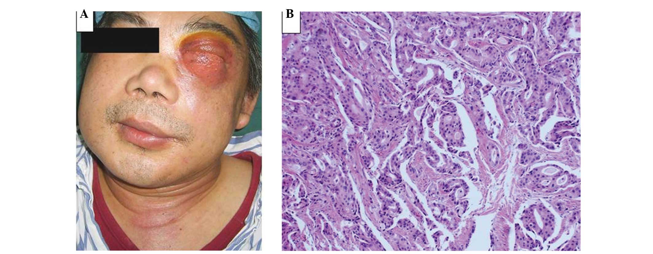
snakes. The patient exhibited diffuse soft-tissue swelling with a
firm surface on the left side of his face. The swelling extended to
the left eye, which was associated with difficulty in eyelid
opening and purulent secretions on the edge (Fig. 1A). Computed tomography (CT) and
magnetic resonance imaging (MRI) examinations of the head revealed
multiple lesions disseminating in the left orbit, maxillary sinus
and ethmoid sinus, as well as diffuse soft-tissue swelling of the
left side of the face and neck. An ELISA for Sparganum
mansoni was performed and the results were negative, using a
Sparganum mansoni antibody detection kit (Shenzhen Combined
Biotech, Co., Ltd.). However, a sparganosis infection was suspected
based on the patient's history of consuming frogs and snakes and
the detection of eosinophil infiltration in the biopsy. However,
anti-sparganosis treatment with praziquantel and anti-inflammatory
treatment, consisting of 0.5 g azithromycin, 0.4 g amikacin and 0.5
g levogloxacin for 1 week, produced no discernible improvement. The
patient underwent a left orbital mass resection and biopsy, during
which white, fish-like tissue and bone destruction was identified
in the deep left inner canthus. Pathological examination of the
biopsy specimens collected from the mass and bone revealed the
presence of an adenocarcinoma (Fig.
1B) with bone metastases. Distant bone metastases were also
observed in an emission CT scan of the skeleton. Therefore, a final
diagnosis of a carcinoma in the left maxillary sinus with bone
metastasis was established. The patient was treated with
chemotherapy and palliative care. The chemotherapy regimen
consisted of 75 mg/m2 cisplatin on day 1 and 1,250
mg/m2 on days 1 and 8, every 21 days, which was repeated
four times. However, the patient succumbed to multiple organ
failure six months following treatment.
Discussion
Primary malignant tumors of the maxillary sinuses
are rarely observed in clinical practice. The presence of large air
spaces within the sinus allows the asymptomatic expansion of sinus
tumors. At the early stages, these tumors are often asymptomatic or
mimicking inflammatory diseases, leading to a delay in diagnosis or
misdiagnosis (9). Thus, a number of
patients present with advanced-stage disease and local invasion to
the surrounding tissues, including the orbit, dura, brain,
pterygomaxillary fossa and the cribriform plate (10). In the present study, the patient
first complained of left eye swelling, which was initially
misdiagnosed as dacryocystitis and a sparganosis infection.
Sparganosis is a rare infectious disease that is
caused by the plerocercoid larva of tapeworms belonging to the
Spirometra species. The disease is most prevalent in Eastern
Asia. Humans may acquire the infection by eating undercooked meat,
particularly meat from snakes or frogs (11). The early stages of the disease are
often asymptomatic; however, the sparganum typically causes a
painful inflammatory reaction in the tissues surrounding the
subcutaneous site with growth (12).
The clinical manifestations are diverse, and the majority of
infections present with masses in the soft tissues and sinus tract,
including the sinonasal passage and eyes (13). A sparganum is typically accompanied
by granulomatous inflammation and eosinophilic infiltration in the
subcutaneous tissues, which is often identified with microscopic
examination of the excised tissue. In addition, an anti-sparganum
ELISA test can be indicative (14).
In general, a preoperative diagnosis should be established based on
the identification of exposure history and a painful, migratory,
subcutaneous nodule. However, the final diagnosis should be
confirmed by surgical removal of the worms or identification of the
parasite in a tissue specimen.
In the case of the present study, the patient lived
in a sparganum endemic area, where five cases of a sparganum
infection were reported between 2000 and 2010 (15). The current patient presented with a
progressive mass in the sinonasal passage, had an exposure history
to undercooked frog, and eosinophilic infiltration was observed in
the subcutaneous tissue. Although the anti-sparganum ELISA test was
negative, a diagnosis of a sparganosis infection was initially
proposed. However, treatment with anti-sparganosis drugs resulted
in no improvements. To confirm a definite diagnosis, a resection of
the left orbital mass was performed and histological examination
was conducted. The results confirmed the diagnosis of an
adenocarcinoma with bone metastases. Notably, a sparganosis
parasite was not identified during the surgery or the pathological
analysis of the specimens.
To the best of our knowledge, the present study was
the first to report the case of a maxillary sinus carcinoma
misdiagnosed as sparganosis. In the present case, two endoscopic
biopsies failed to provide any evidence for malignancy, and the
patient received several treatments for dacryocystitis and
sparganosis infection. The duration between the initial visit to
the local hospital and the definite diagnosis was almost two years,
during which period multiple invasive examinations and treatments
that caused physical injuries and an economical burden to the
patient were performed. Previous studies have reported that the
diagnosis of sinus malignancies is difficult due to the air-filled
nature and deep position of the structures involved (9,16). The
outcomes of the present case strongly indicated that the avoidance
of misdiagnosis at an early stage is crucial for the effective
treatment of this type of cancer. The following clinical
manifestations are strong indications for a carcinoma of the
maxillary sinus: Patients aged >40 years with a slowly
progressing unilateral nasal obstruction associated with epistaxis,
pain below the eye, hyposmia, facial numbness, facial swelling,
skin sensory loss or difficulty in opening the mouth. Since the
differentiation between a carcinoma and sparganosis is based on
histological examinations, an adequate tissue biopsy is a necessary
requirement. Maxillary sinus exploratory surgery should be
considered to eliminate the possibility of the disease. In
addition, CT scanning is helpful to delineate the extent of the
cancer, although differentiating between tumors and edematous
mucosa can be difficult with CT scanning. However, MRI may be
applied to aid the differential diagnosis.
In conclusion, the consideration of a carcinoma is
important since carcinomas and sparganosis in the maxillary sinus
are extremely rare diseases. Due to the anatomical nature of the
maxillary sinus, misdiagnosis and delayed diagnosis are common.
Therefore, a careful biopsy and pathological examination are
required for early diagnosis and treatment.
Acknowledgements
The study was supported by a grant from the National
Natural Science Foundation of China (no. 81200267).
References
|
1
|
Barnes L: Intestinal-type adenocarcinoma
of the nasal cavity and paranasal sinuses. Am J Surg Pathol.
10:192–202. 1986. View Article : Google Scholar : PubMed/NCBI
|
|
2
|
Acheson ED, Cowdell RH and Rang E:
Adenocarcinoma of the nasal cavity and sinuses in England and
Wales. Br J Ind Med. 29:21–30. 1972.PubMed/NCBI
|
|
3
|
Hayes RB, Gerin M, Raatgever JW and de
Bruyn A: Wood-related occupations, wood dust exposure, and
sinonasal cancer. Am J Epidemiol. 124:569–577. 1986.PubMed/NCBI
|
|
4
|
Choussy O, Ferron C, Védrine PO, Toussaint
B, Liétin B, Marandas P, Babin E, De Raucourt D, Reyt E, Comsmidis
A, et al: GETTEC Study Group: Adenocarcinoma of Ethmoid: A GETTEC
retrospective multicenter study of 418 cases. Laryngoscope.
118:437–443. 2008. View Article : Google Scholar : PubMed/NCBI
|
|
5
|
Luce D, Leclerc A, Bégin D, Demers PA,
Gérin M, Orlowski E, Kogevinas M, Belli S, Bugel I, Bolm-Audorff U,
et al: Sinonasal cancer and occupational exposures: a pooled
analysis of 12 case-control studies. Cancer Causes Control.
13:147–157. 2002. View Article : Google Scholar : PubMed/NCBI
|
|
6
|
Klintenberg C, Olofsson J, Hellquist H and
Sökjer H: Adenocarcinoma of the ethmoid sinuses. A review of 28
cases with special reference to wood dust exposure. Cancer.
54:482–488. 1984. View Article : Google Scholar : PubMed/NCBI
|
|
7
|
Llorente JL, Pérez-Escuredo J,
Alvarez-Marcos C, Suárez C and Hermsen M: Genetic and clinical
aspects of wood dust related intestinal-type sinonasal
adenocarcinoma: A review. Eur Arch Otorhinolaryngol. 266:1–7. 2009.
View Article : Google Scholar : PubMed/NCBI
|
|
8
|
Kleinsasser O and Schroeder HG:
Adenocarcinomas of the inner nose after exposure to wood dust.
Morphological findings and relationships between histopathology and
clinical behavior in 79 cases. Arch Otorhinolaryngol. 245:1–15.
1988. View Article : Google Scholar : PubMed/NCBI
|
|
9
|
Dulguerov P, Jacobsen MS, Allal AS,
Lehmann W and Calcaterra T: Nasal and paranasal sinus carcinoma:
Are we making progress? A series of 220 patients and a systematic
review. Cancer. 92:3012–3029. 2001. View Article : Google Scholar : PubMed/NCBI
|
|
10
|
Chu Y, Liu HG and Yu ZK: Patterns and
incidence of sinonasal malignancy with orbital invasion. Chin Med J
(Engl). 125:1638–1642. 2012.PubMed/NCBI
|
|
11
|
Williams JE: Diagnostic medical
parasitology. Parasitol Today. 14:125–126. 1998. View Article : Google Scholar : PubMed/NCBI
|
|
12
|
Mueller JF: The biology of
Spirometra. J Parasitol. 60:3–14. 1974. View Article : Google Scholar : PubMed/NCBI
|
|
13
|
Kim JR and Lee JM: A case of intramuscular
sparganosis in the sartorius muscle. J Korean Med Sci. 16:378–380.
2001. View Article : Google Scholar : PubMed/NCBI
|
|
14
|
Kimura S, Kashima M, Kawa Y, Nakamura F,
Nawa Y, Takai K and Mizoguchi M: A case of subcutaneous
sparganosis: Therapeutic assessment by an indirect
immunofluorescence antibody titration using sections of the worm
body obtained from the patient. British J Dermatol. 148:369–371.
2003. View Article : Google Scholar
|
|
15
|
Li MW, Song HQ, Li C, Lin HY, Xie WT, Lin
RQ and Zhu XQ: Sparganosis in mainland China. Int J Infect Dis.
15:e154–156. 2011. View Article : Google Scholar : PubMed/NCBI
|
|
16
|
Yoshimura R, Shibuya H, Ogura I, et al:
Trimodal combination therapy for maxillary sinus carcinoma. Int J
Radiat Oncol Biol Phys. 53:656–663. 2002. View Article : Google Scholar : PubMed/NCBI
|