Introduction
Intractable epilepsy (IE) accounts for 30–40% of the
pediatric epilepsy and poses a challenge in pediatric neurology
(1–2). In recent years, most studies
investigating the resistance mechanism of IE have focused on cell
death and reconstruction, overexpression of multidrug transporter,
and changes of intracephalic drug targets (3,4).
Previous studies focused on the association between multidrug
transporter and epilepsy tolerance (5,6,15). The multidrug-resistant associated
protein (MRP) is an important multidrug transporter, comprising
nine MRPs (MRP1-MRP9), which are associated with drug transport.
Among these, MRP1 and MRP2 may be related to the drug-resistant
mechanisms of IE (7). MRP1 and MRP2
are distributed in the kidneys, liver, lungs, testes and peripheral
blood mononuclear cells. They are also expressed in the luminal
membrane on the basal side of endothelial cells or the luminal
membrane of capillary endothelial cells in the choroid plexus,
whose function is to restrict the entry of specific substances into
the cerebrospinal fluid (8,9). The aim of the present study was to
examine the expression of MRP1 and MRP2 protein in the peripheral
blood mononuclear cells of children with IE using RT-PCR and
western blot analysis. A higher relative expression of MRP1 and
MRP2 mRNA and protein was identified in children with IE compared
to the control groups. This higher expression indicates that IE may
be relevant to the drug-resistant mechanism of IE.
Materials and methods
Subjects
For this study, 100 children with epilepsy from the
outpatient or inpatient clinic of Xuzhou Children's Hospital
(Jiangsu, China) were enrolled between November 2010 and October
2013. The patients were into two groups. The IE group comprised 50
cases, all of which conformed to the criteria formulated by the
International League Against Epilepsy (10). The criteria for enrollment were:
exact clinical diagnosis, the application of two types of
appropriate and tolerable antiepileptic drugs failed to completely
avoid epileptic seizure following adequate duration of treatment
(duration-free from any epileptic seizure was considered as ≥3
times the longest interictal period prior to treatment or 12
months) and adequate doses of monotherapy or combinatorial
treatment, with other nervous system disease history being
excluded. Since the longest interictal periods in the enrolled
patients was <3 months, 12 months was defined as the course of
treatment. In the IE group, 29 cases were male and 21 were female,
between the ages of 1.2 and 12 years, with an average age of
5.2±2.1 years, and a course of disease of 1–6 years. Of the 50
cases, 23 were cases of partial seizure, 10 of general tonic-clonic
seizure, 3 of myoclonic seizure, 3 of children atypical seizure, 6
of epileptic spasm, and 5 cases of myoclonia atonic seizure.
The epilepsy controlled by anti-epileptic drugs
(AEDs)group comprised 50 patients, diagnosed with epilepsy, but who
had not experienced a seizure for at least one year following
first-line anti-epileptic drug treatment. In this group, 26 cases
were male and 24 cases were female, between 1.6 and 10 years of
age, with an average age of 4.3±2.3 years. Of the 50 cases, 19 were
cases of partial seizure, 15 of general tonic-clonic seizure, 5 of
myoclonic seizure, 3 of atonic seizure, 3 of myoclonia atonic
seizure, 3 of absence seizure and 2 cases of epileptic spasm.
The control group comprised 50 healthy children
without epilepsy who served as the control group. In this group, 30
cases were male and 20 cases were female, aged between 2 and 12
years, with an average age of 4.5±1.9 years. A comparison of
differences in age and gender among the three groups revealed no
significant difference.
The study was approved by the Ethis Committee of
Xuzhou Children's Hospital, Xuzhou, China. Written patient consent
was obtained from each patient.
Analysis of the relative expression
amount of mRNA of MRP1 and MRP2 using RT-PCR
To conduct the reverse transcription reaction, total
RNA was extracted from the peripheral blood mononuclear cells using
the Total RNA Extraction Kit (Takara, Dalian, China) to synthesize
cDNA. The total volume of the PCR reaction was 25 µl, including
12.5 µl of premix type mixed solution, 1 µl of forward primer, 1 µl
of downstream primer, 5.0 µl of reverse transcription product, and
5.5 µl of double distilled water. The primers used were: MRP1
upstream, 5′-GAG GAA CCA TAT TAC AGG TCC GT-3′ and downstream,
5′-AGG GGA TCA TCG AAG AGG TAA AT-3′, with a product of 188 bp;
MRP2 upstream, 5′-AAT AGC ACC GAC TAT CCA GCAT-3′ and downstream,
5′-GTG AGA GTA GAT TGG GGA CCTG-3′, with a product of 456 bp;
reference of β-actin upstream, 5′-CTT AGT TGC GTT ACA CCC TTTC-3′
and downstream, 5′-GGT CAC CTT CAC CGT TCC AGT-3′, product of 526
bp. The PCR reaction conditions for MRP1 were: 94°C for 3 min,
followed by 33 cycles of 94°C for 30 sec, 55°C for 30 sec, 72°C for
1 min, and, 72°C for 5 min. The PCR reaction conditions for MRP2
were: 95°C for 2 min, followed by 35 cycles of 95°C for 30 sec,
56°C for 30 sec, 72°C for 1 min and, 72°C for 5 min. The relative
expression amount of MRP1 and MRP2 was expressed with the
corresponding amplified products and gray level ratio of
β-actin.
Analysis of the relative amount of
MRP1 and MRP2 protein expression using western blot analysis
The amount of the total protein extracted from the
single nuclear membrane was measured using the bicinchoninic acid
assay (Beyotime, Beijing, China). The amount of protein in the
samples was balanced and run on sodium dodecyl
sulfate-polyacrylamide gel electrophoresis (SDS-PAGE; Beyotime).
The proteins were then transferred to PVDF membranes (Sigma, St.
Louis, MO, USA) using the wet transfer method for 3 h, after which
the membranes were incubated with diluted primary antibodies (MRP1,
Cat. no. sc-365635, mouse monoclonal, 1:50; Santa Cruz
Biotechnology, Inc., Santa Cruz, CA, USA; MRP2, Cat. no. ab3373,
mouse monoclonal, 1:100, Abcam, Cambridge, MA, USA), and incubated
overnight at 4°C. The following day, the membranes were rewarmed
for 30 min, and washed with buffer solution (Beyotime) for 5 min ×
3 times. The membranes were then incubated with diluted secondary
antibody (goat anti-mouse antibody 1:1,000; LI-COR Biosciences,
Lincoln, NE, USA) and incubated at room temperature for 2 h. The
blots were developed after chemical staining and the gel image
processing system was used to analyze the molecular weight and net
optical density value of the target band. The optical density ratio
of the target band/β-actin band yielded the relative expression
amount of the target protein band.
Statistical analysis
The results were presented as mean ± SD. The SPSS
16.0 statistical software was applied to compare the expression of
MRP2 genes and proteins in all the groups. The ANOVA test was
applied to make comparisons between the groups, and the LSD test
was applied to make comparisons between any two groups, P<0.05
was considered to indicate statistically significant results.
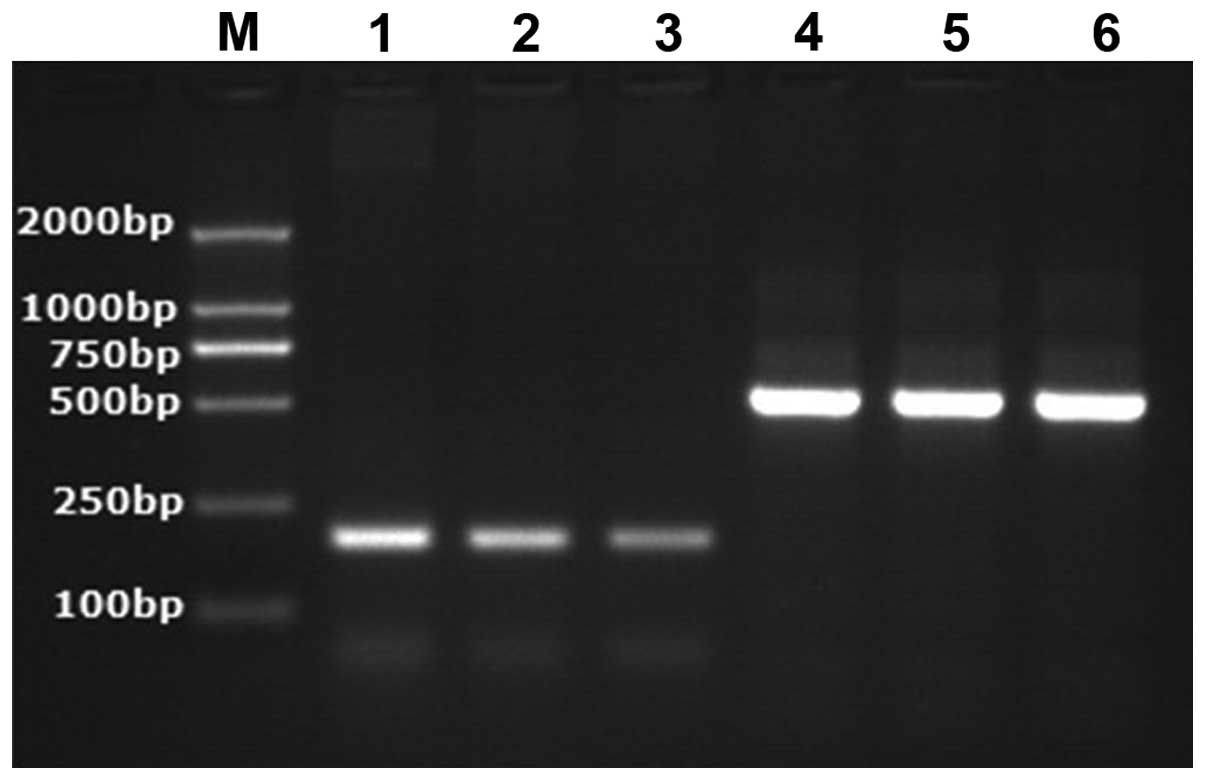
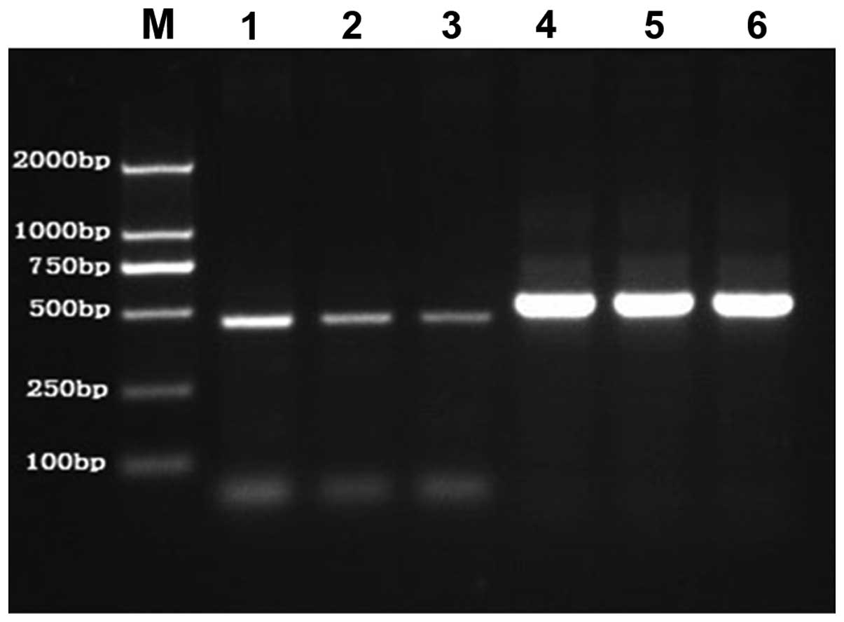
Results
Expression of mRNA of MRP1 and
MRP2
The relative expression amount of mRNA of MRP1 and
MRP2 in the IE group increased compared to the AEDs group and the
group comprising healthy children without epilepsy. The difference
was statistically significant (P<0.001). However, the relative
expression of mRNA of MRP1 and MRP2 in the AEDs group compared to
the group of healthy children without epilepsy was not
statistically significant (P>0.05) (Table I and Figs.
1 and 2).
 | Table I.Expression of mRNA of MRP1 and MRP2 in
peripheral blood mononuclear cells of children with intractable
epilepsy (mean ± SD). |
Table I.
Expression of mRNA of MRP1 and MRP2 in
peripheral blood mononuclear cells of children with intractable
epilepsy (mean ± SD).
| Group | Cases | MRP1 | MRP2 |
|---|
| Group of healthy
children without epilepsy | 50 | 0.665±0.031 | 0.654±0.029 |
| Group of children
with epilepsy controlled by anti-epileptic drugs | 50 |
0.682±0.030a |
0.675±0.021a |
| Group of intractable
epilepsy | 50 |
0.795±0.042b |
0.804±0.023b |
| F-value |
| 121.364 | 292.194 |
| P-value |
|
<0.001 |
<0.001 |
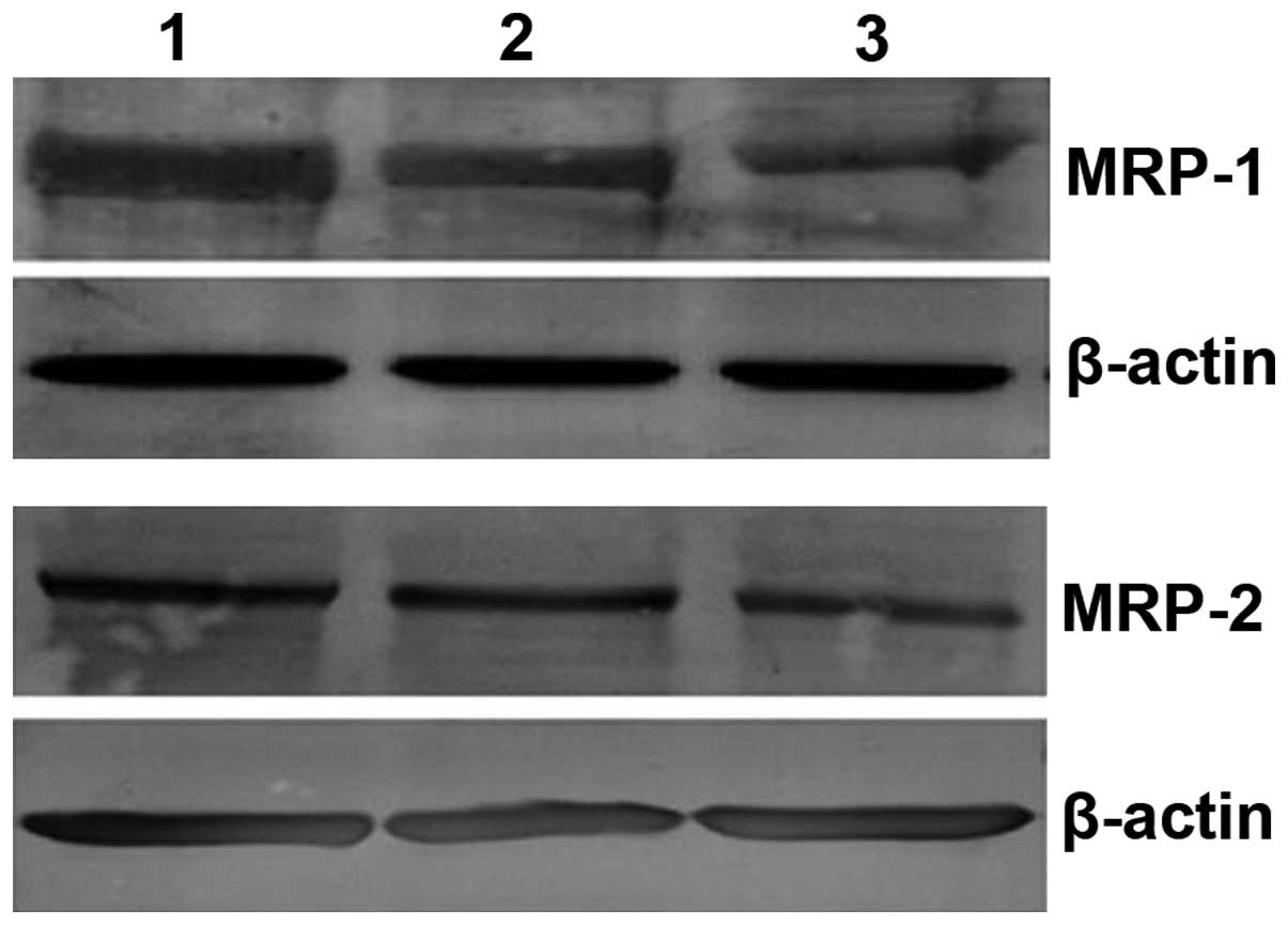
Relative expression amount of protein
of MRP1 and MRP2
The relative expression of proteins MRP1 and MRP2 in
the IE group increased compared to AEDs group and the group of
healthy children without epilepsy. The difference was statistically
significant (P<0.001). However, a comparison of the relative
expression amount of proteins MRP1 and MRP2 in the AEDs group with
that in the group of healthy children without epilepsy indicated no
statistically significant difference (P>0.05) (Table II and Fig. 3).
| Table II.Expression of MRP1 and MRP2 in
peripheral blood mononuclear cells of children with intractable
epilepsy (mean ± SD). |
Table II.
Expression of MRP1 and MRP2 in
peripheral blood mononuclear cells of children with intractable
epilepsy (mean ± SD).
| Group | Cases | MRP1 | MRP2 |
|---|
| Group of healthy
children without epilepsy | 50 | 1.093±0.023 | 1.045±0.018 |
| Group of children
with epilepsy controlled by anti-epileptic drugs | 50 |
1.131±0.042a |
1.086±0.027a |
| Group of intractable
epilepsy | 50 |
2.027±0.034b |
1.902±0.021b |
| F-value |
| 20.391 | 24.197 |
| P-value |
| <0.001 | <0.001 |
Discussion
The mechanism of resistance for intractable epilepsy
remains to be elucidated. Findings of recent studies have shown
that overexpression of the multidrug transporter may be one of the
factors for this resistance (11–13). MRP
is an important multidrug transporter that is involved in adjusting
the density of antiepileptic drugs within and beyond the cells of
epileptic individuals (14,15).
Although significant progress has been revealed in
MRP structure and function studies, the majority of these mainly
focus on tumors and blood diseases (16,17).
Investigations into the expression of MRP are relatively few and
limited to animal epileptic models or in vitro adult brain
tissue samples (18–20), which restricts the progress of
studies due to difficulty in sampling brain tissues and repeating,
as well as small sample size. However, collection from peripheral
blood is simple and convenient, and may support dynamic
observation. In addition to brain tissues, the expression of MRP
also exists in the respiratory tract, digestive tract, urinary
tract, and peripheral blood (8,21).
Antiepileptic drugs initially enter the bloodstream and then the
brain tissues through blood brain barrier. The concentration of
drugs with pharmacological effects are similar to the free
concentration in blood. Thus, MRP may be induced to overexpress
simultaneously inside the blood and brain.
The expression of mRNA of MRP increased
significantly in the peripheral blood of recurrent acute leukemic
patients and MDR-TB patients (9,22) which
indicated that a high expression of MRP in peripheral blood was
closely associated with the recurrence of leukemia and multidrug
resistance of tuberculosis. In addition, it has been identified
that the expression of MDR genes and MRP1 mRNA in the peripheral
blood of IE adult patients was significantly higher than that of
the group controlled by anti-epileptic drugs and the healthy
control group, which indicated that MDR1 and MRP1 may be associated
with the tolerance of epilepsy (23,24).
However, the number of studies focusing on MRP expression in
peripheral blood of epileptic children, especially IE children, are
limited. Thus, in the present study, we concentrated on examining
the expression of the MRP gene and protein in peripheral blood
mononuclear cells of IE children, to discuss the role of MRP in the
pathogenesis of IE children. The result showed that MRP1 and MRP2
was expressed in the peripheral blood mononuclear cells of, not
only IE children, but also the AEDs children and the healthy
children without epilepsy (25,26).
Compared to the AEDs and the children without epilepsy, a higher
mRNA and protein expression of MRP1 and MRP2 for IE children was
observed, and the difference was statistically significant. By
contrast, no difference was identified between the AEDs group and
the healthy control group (27). The
results indicate that MRP1 and MRP2 was distributed extensively in
the peripheral blood of the different groups. Of note, single drug
induction did not cause the increase of MRP1 and MRP2 in peripheral
blood, thus, MRP1 and MRP2 may be involved in the resistance
process of the IE group. This result was consistent with the
findings of Lan et al following an investigation of the MRP1
in the peripheral blood of adult IE patients (28).
Repeated abnormal discharges of the neurons in the
brain and epileptic seizures are considered the major induction
factor of a high MRP expression. For example, studies on rat in the
status epilepticus have shown that the expression of MRP1 and MRP2
in neurons in the hippocampus and surrounding cortex (29), vascular endothelial cells as well as
astrocyte increased significantly. The long-term intervention
treatment of certain anti-epileptic drugs, such as oxcarbazepine,
may also induce the expression of MRP1 in rat (30). However, investigations into tuberous
sclerosis identified that the expression of MRPs in certain
patients already existed prior to the treatment of anti-epileptic
drugs (31).
The results of the present study have shown that the
application of anti-epileptic drugs failed to increase MRP1 and
MRP2 in the peripheral blood of the AEDs. This finding indicates
that besides the effects of repeated epileptic seizure and
anti-epileptic drugs, elevation of MRP may also be the result of
multiple factors and mechanisms, such as the types and acting time
of the influential factors, including genetics and immunity.
Additionally, the polymorphism and haplotype of MRP genes may
affect the reactions of epileptics towards anti-epileptic drugs,
thereby resulting in IE. Therefore, the exact mechanism of MRP in
IE tolerance requires intensive and extensive investigations.
References
|
1
|
Sugano H and Arai H: Epilepsy surgery for
pediatric epilepsy: Optimal timing of surgical intervention. Neurol
Med Chir (Tokyo). 55:399–406. 2015. View Article : Google Scholar : PubMed/NCBI
|
|
2
|
Epilepsy: Benefits and risks of
reoperation after failed surgery for intractable epilepsy. Nat Rev
Neurol. 11:2472015. View Article : Google Scholar : PubMed/NCBI
|
|
3
|
Sebe JY and Baraban SC: The promise of an
interneuron-based cell therapy for epilepsy. Dev Neurobiol.
71:107–117. 2011. View Article : Google Scholar : PubMed/NCBI
|
|
4
|
Jose M and Thomas SV: Role of multidrug
transporters in neurotherapeutics. Ann Indian Acad Neurol.
12:89–98. 2009. View Article : Google Scholar : PubMed/NCBI
|
|
5
|
Xu J, Deng Y and Gao B: Blood-CSF barrier
related translocator and intractable epilepsy. J Appl Clin Pediatr.
27:1900–1902. 2012.
|
|
6
|
Luna-Tortós C, Fedrowitz M and Löscher W:
Evaluation of transport of common antiepileptic drugs by human
multidrug resistance-associated proteins (MRP1, 2 and 5) that are
overexpressed in pharmacoresistant epilepsy. Neuropharmacology.
58:1019–1032. 2010. View Article : Google Scholar : PubMed/NCBI
|
|
7
|
Marquez B and Van Bambeke F: ABC multidrug
transporters: target for modulation of drug pharmacokinetics and
drug-drug interactions. Curr Drug Targets. 12:600–620. 2011.
View Article : Google Scholar : PubMed/NCBI
|
|
8
|
Schinkel AH and Jonker JW: Mammalian drug
efflux transporters of the ATP binding cassette (ABC) family: An
overview. Adv Drug Deliv Rev. 55:3–29. 2003. View Article : Google Scholar : PubMed/NCBI
|
|
9
|
Keppler D: Multidrug resistance proteins
(MRPs, ABCCs): Importance for pathophysiology and drug therapy.
Handbook Exp Pharmacol. 201:299–323. 2011. View Article : Google Scholar
|
|
10
|
Kwan P, Arzimanoglou A, Berg AT, Brodie
MJ, Allen Hauser W, Mathern G, Moshé SL, Perucca E, Wiebe S and
French J: Definition of drug resistant epilepsy: Consensus proposal
by the ad hoc Task Force of the ILAE Commission on Therapeutic
Strategies. Epilepsia. 51:1069–1077. 2010. View Article : Google Scholar : PubMed/NCBI
|
|
11
|
Rong H, Jin L, Wei W, Wang X and Xi Z:
Alpha-synuclein is a potential biomarker in the serum and CSF of
patients with intractable epilepsy. Seizure. 27:6–9. 2015.
View Article : Google Scholar : PubMed/NCBI
|
|
12
|
Grote A, Witt JA, Surges R, von Lehe M,
Pieper M, Elger CE, Helmstaedter C, Ormond DR, Schramm J and Delev
D: A second chance-reoperation in patients with failed surgery for
intractable epilepsy: long-term outcome, neuropsychology and
complications. J Neurol Neurosurg Psychiatry. pii:jnnp-2015–310322.
2015.
|
|
13
|
Fernandez L, Gedela S, Tamber M and Sogawa
Y: Vagus nerve stimulation in children less than 3 years with
medically intractable epilepsy. Epilepsy Res. 112:37–42. 2015.
View Article : Google Scholar : PubMed/NCBI
|
|
14
|
Yi JH, Cho YJ, Kim WJ, Lee MG and Lee JH:
Genetic variations of ABCC2 gene associated with adverse drug
reactions to valproic acid in Korean epileptic patients. Genomics
Inform. 11:254–262. 2013. View Article : Google Scholar : PubMed/NCBI
|
|
15
|
Chen YH, Wang CC, Xiao X, Wei L and Xu G:
Multidrug resistance-associated protein 1 decreases the
concentrations of antiepileptic drugs in cortical extracellular
fluid in amygdale kindling rats. Acta Pharmacol Sin. 34:473–479.
2013. View Article : Google Scholar : PubMed/NCBI
|
|
16
|
Xu Y, Wang L, Zheng X, Liu G, Wang Y, Lai
X and Li J: Positive expression of p53, c-erbB2 and MRP proteins is
correlated with survival rates of NSCLC patients. Mol Clin Oncol.
1:487–492. 2013.PubMed/NCBI
|
|
17
|
Kovalev AA, Tsvetaeva DA and Grudinskaja
TV: Role of ABC-cassette transporters (MDR1, MRP1, BCRP) in the
development of primary and acquired multiple drug resistance in
patients with early and metastatic breast cancer. Exp Oncol.
35:287–290. 2013.PubMed/NCBI
|
|
18
|
Nasilowska-Adamska B, Solarska I,
Paluszewska M, Malinowska I, Jedrzejczak WW and Warzocha K:
FLT3-ITD and MLL-PTD influence the expression of MDR-1, MRP-1, and
BCRP mRNA but not LRP mRNA assessed with RQ-PCR method in adult
acute myeloid leukemia. Ann Hematol. 93:577–593. 2014. View Article : Google Scholar : PubMed/NCBI
|
|
19
|
Calatozzolo C, Pollo B, Botturi A,
Dinapoli L, Carosi M, Salmaggi A and Maschio M: Multidrug
resistance proteins expression in glioma patients with epilepsy. J
Neurooncol. 110:129–135. 2012. View Article : Google Scholar : PubMed/NCBI
|
|
20
|
Yao D, Liu L, Jin S, Li J and Liu XD:
Overexpression of multidrug resistance-associated protein 2 in the
brain of pentylenetetrazole-kindled rats. Neuroscience.
227:283–292. 2012. View Article : Google Scholar : PubMed/NCBI
|
|
21
|
Zhang Y, Dai Y, Rui L, et al: Effect of
Oxcarbazepine on multidrug resistance associated protein 1
expression in hippocampus of kainic acid-induced seizure in rats
during development period rats. Chin J Appl Clin. 28:1170–1171.
2013.
|
|
22
|
Zhan L, Ping W and Jing Z: The expression
of multi-drug resistance transmembrane transporter in the
peripheral blood of patients with multi-drug resistant
tuberculosis. Zhonghua Jie He He Hu Xi Za Zhi. 34:520–522. 2011.(In
Chinese). PubMed/NCBI
|
|
23
|
Song Y, Bing L and Zhong L: Expression of
multi-drug resistance-associated protein genein acute leukemia.
Acta Acad Med Qingdao Univ. 37:41–43. 2001.
|
|
24
|
Ban JJ, Jung KH, Chu K, Lee ST, Jeon D,
Park KI, Moon HJ, Kim H, Kim S, Lee SK, et al: Profiles of
multidrug resistance protein-1 in the peripheral blood mononuclear
cells of patients with refractory epilepsy. PLoS One. 7:e369852012.
View Article : Google Scholar : PubMed/NCBI
|
|
25
|
Murakami N, Morioka T, Hashiguchi K,
Suzuki SO, Shigeto H, Sakata A and Sasaki T: Clinical and
histological characteristics of ictal onset zone in cases of
intractable epilepsy associated with dysembryoplastic
neuroepithelial tumor. Brain Nerve. 67:525–532. 2015.(In Japanese).
PubMed/NCBI
|
|
26
|
Mateo-Carrasco H, Serrano-Castro PJ,
Molina-Cuadrado E, Goodwin M, Nguyen TV and Kotecha PN: Role of
high-dose levetiracetam as add-on therapy for intractable epilepsy:
Case report and brief review of the literature. Int J Clin Pharm.
37:559–562. 2015. View Article : Google Scholar : PubMed/NCBI
|
|
27
|
Meador KJ, Kapur R, Loring DW, Kanner AM
and Morrell MJ: RNS® System Pivotal Trial Investigators: Quality of
life and mood in patients with medically intractable epilepsy
treated with targeted responsive neurostimulation. Epilepsy Behav.
45:242–247. 2015. View Article : Google Scholar : PubMed/NCBI
|
|
28
|
Lan T, Teng M, Song J, et al: The
expression of multidrug resistance protein-1 in peripheral blood of
patients with intractable epilepsy. Zhonghua Jie He He Hu Xi Za
Zhi. 39:403–404. 2006.(In Chinese).
|
|
29
|
van Vliet EA, Redeker S, Aronica E,
Edelbroek PM and Gorter JA: Expression of multidrug transporters
MRP1, MRP2, and BCRP shortly after status epilepticus, during the
latent period, and in chronic epileptic rats. Epilepsia.
46:1569–1580. 2005. View Article : Google Scholar : PubMed/NCBI
|
|
30
|
Lazarowski A, Lubieniecki F, Camarero S,
Pomata H, Bartuluchi M, Sevlever G and Taratuto AL: Multidrug
resistance proteins in tuberous sclerosis and refractory epilepsy.
Pediatr Neurol. 30:102–106. 2004. View Article : Google Scholar : PubMed/NCBI
|
|
31
|
Qu J, Zhou BT, Yin JY, Xu XJ, Zhao YC, Lei
GH, Tang Q, Zhou HH and Liu ZQ: ABCC2 polymorphisms and haplotype
are associated with drug resistance in Chinese epileptic patients.
CNS Neurosci Ther. 18:647–651. 2012. View Article : Google Scholar : PubMed/NCBI
|