Introduction
Nonalcoholic fatty liver disease (NAFLD) is
characterized by an accumulation of lipids within hepatocytes, and
is considered the hepatic manifestation of the metabolic syndrome
(1). NAFLD develops progressively
from simple steatosis to nonalcoholic steatohepatitis (NASH),
fibrosis and cirrhosis (2,3). Previous studies have suggested a
‘two-hit’ theory for the pathogenesis of NAFLD, including insulin
resistance, oxidative stress, lipid peroxidation and mitochondrial
dysfunction (4,5), with insulin resistance considered the
major factor.
Proliferator-activated receptor (PPAR)-γ is a member
of the nuclear hormone receptor family and as such is activated by
its ligand (6). Previous studies
have demonstrated that this transcription factor is associated with
insulin resistance, lipid metabolism and the regulation of
inflammation in fatty liver disease (7–9). The
insulin receptor (IR) is a well-studied receptor tyrosine kinase,
which is associated with the regulation of peripheral glucose
metabolism (10,11); therefore, a defect in IR may directly
affect the role of insulin in the body, leading to insulin
resistance and abnormal glucose tolerance (12). Previous studies have demonstrated
that PPAR-γ and IR are closely associated with insulin resistance
(13,14), and PPAR-γ agonists, such as
thiazolidinediones (TZDs), are able to increase insulin sensitivity
in adipose, liver and skeletal muscle tissue (15). However, TZDs possess numerous side
effects, including significant weight gain and peripheral edema
(16,17); therefore, it is necessary that novel
agents that target PPAR-γ with reduced adverse effects be
developed.
Previous studies have revealed the therapeutic
effects of integrative medicine in treating patients with metabolic
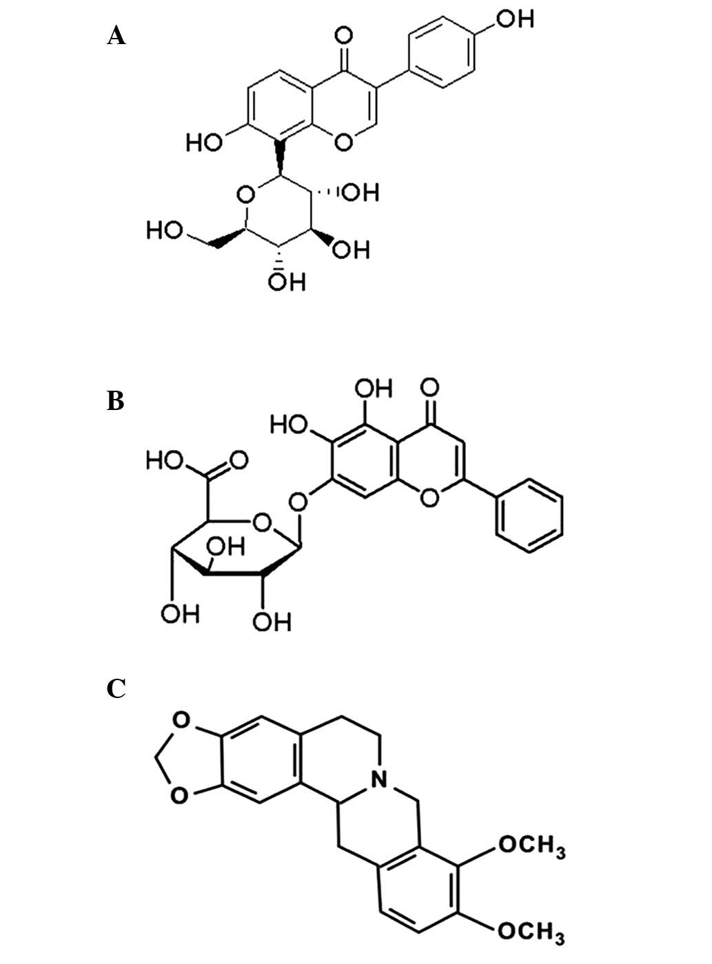
syndrome (18,19). Puerarin, baicalin and berberine
(Fig. 1A–C), are three major
constituents of the Chinese herbs: Puerariae Radix [roots of
Pueraria lobata (Willd.) Ohwi, PR], Scutellariae
Radix (roots of Scutellaria baicalensis Georgi SR) and
Coptidis Rhizoma (rhizomes of Coptis chinensis
Franch, CR), which have demonstrated therapeutic effects on rats
models of NAFLD (20–22). It is believed that a combination of
agents may effectively reduce side effects and improve adaptive
resistance. In the present study, an orthogonal experimental design
was used to investigate the various effects of puerarin, baicalin
and berberine on rats with high-fat diet-induced NAFLD. The present
study aimed to elucidate the complex interactions of this
multicomponent disease, and further clarify the potential for
natural combination pharmacological therapies for patients with
NAFLD.
Materials and methods
Experimental animals and modeling
A total of 96 adult male Sprague-Dawley rats
weighing 150–180 g were obtained from the Animal Breeding Center of
the Beijing Vital River Laboratories Company (Beijing, China). The
rats were individually housed at 22±2°C with a relative humidity of
50±10% and a 12 h light/dark cycle. After 7 days of adaptive
feeding, the rats were randomly divided into control (n=8), model
(n=8), rosiglitazone treatment (n=8), and nine orthogonal
experiment groups (n=8×9). The control group was fed a normal
laboratory diet with free drinking water, whereas the model group,
rosiglitazone treatment group (R) and orthogonal experiment groups
(A-I) were fed a normal diet supplemented with 2% cholesterol and
10% lard, for 8 weeks. This study was approved by the Review Board
of the Beijing University of Chinese Medicine (Beijing, China). All
animal studies were conductedc in accordance with the regulations
and guidelines for the use and care of experimental animals of the
Beijing University of Chinese Medicine (Beijing, China).
Therapeutic agents and reagents. Puerarin, baicalin,
and berberine (Fig. 1) were
purchased from Nanjing Zelang Medical Technology Co., Ltd.
(Jiangsu, China). Rosiglitazone was purchased from Chengdu Hengrui
Biotech Co., Ltd (Chengdu, China). Colorimetric kits for testing
triglyceride (TG), total cholesterol (TC), high-density lipoprotein
(HDL), low-density lipoprotein (LDL), alanine aminotransferase
(ALT) and aspartate aminotransferase (AST) were purchased from
BioSino Biotechnology and Science, Inc. (Beijing, China). ELISA
kits for tumor necrosis factor (TNF)-α and interleukin (IL)-6 were
purchased from Shanghai Yanji Biotechnology Co., Ltd. (Shanghai,
China). Rabbit anti-PPARγ (ab27649, 1:2,000), mouse anti-IR (cat.
no. ab69508, 1:5,000) and goat anti-GAPDH (ab9483, 1:20,000)
antibodies were purchased from Abcam (Cambridge, UK). Additional
reagents were obtained from Sigma-Aldrich (St. Louis, MO, USA).
Orthogonal experimental design and intervention.
Based on single-factor experimental results, three independent
parameters: Puerarin, baicalin and berberine doses (mg), were
confirmed as the major influencing factors (23). A L9(34) orthogonal experimental design was
conducted to evaluate the effects of the independent variables on
the treatment of rats with NAFLD. The levels and ranges of the
individual variables are outlined in Table I. The experiment design is presented
in Table II, along with the
experimental data. All experiments were performed in duplicate.
 | Table I.Factors and levels of orthogonal
experimental design. |
Table I.
Factors and levels of orthogonal
experimental design.
| Levels | Puerarin (mg) | Baicalin (mg) | Berberine (mg) |
|---|
| 1 | 200 | 100 | 100 |
| 2 | 100 | 50 | 50 |
| 3 | 50 | 25 | 25 |
| Table II.Orthogonal experimental design. |
Table II.
Orthogonal experimental design.
| Test | Puerarin | Baicalin | Berberine |
|---|
| A | 1 | 1 | 1 |
| B | 1 | 2 | 2 |
| C | 1 | 3 | 3 |
| D | 2 | 1 | 2 |
| E | 2 | 2 | 3 |
| F | 2 | 3 | 1 |
| G | 3 | 1 | 3 |
| H | 3 | 2 | 1 |
| I | 3 | 3 | 2 |
Measurement of serum biochemical parameters. Blood
samples from the rats were obtained via abdominal aortic puncture
and centrifuged at 644 × g for 10 min, in order to isolate the
serum. Serum ALT, AST, TC, TG, LDL, and HDL concentrations were
determined using a Vitros 250 automatic biochemical analyzer
(Johnson & Johnson, Rochester, NY, USA).
Histological examination. All rats survived to the
final analysis and rats were sacrificed by an intraperitoneal
injection of 10% chloral hydrate at the 8th week. In
accordance with the frozen section technique, one fresh section of
liver tissue from each rat was maintained in liquid nitrogen for
4–10 sec prior to staining with Oil Red O (Sigma-Aldrich). An
additional liver tissue section was fixed by immersion in 10%
buffered formalin for paraffin embedding, prior to staining with
hematoxylin and eosin (Beijing CellChip Biotechnology Co. Ltd.,
Beijing, China). The samples were examined using an Olympus CK40
inverted microscope (Olympus, Tokyo, Japan).
Detection of TNF-α and IL-6 in the serum and liver
tissue. The concentrations of TNF-α and IL-6 in the serum and liver
tissue of the rats were analyzed using commercially available ELISA
kits, according to the manufacturer's instructions. Briefly, the
tissue was homogenized in an ice bath with ultrasonic irradiation.
After 30 min, the homogenate was centrifuged for 15 min at 15,000 ×
g at a temperature of 4°C. The supernatant was extracted in order
to determine the concentration of protein. All samples were diluted
to 1:10 and the absorbance was read at 450 nm using a microplate
reader (Multiskan MK3; Thermo Fisher Scientific Inc., Rockford, IL,
USA). Samples and standards were performed in triplicate.
Detection of the mRNA expression levels of PPAR-γ
and IR by reverse transcription-quantitative polymerase chain
reaction (RT-qPCR). Total RNA was isolated from the liver samples
using TRIzol® reagent (Invitrogen Life Technologies, Carlsbad, CA,
USA) and subsequently reverse-transcribed at 42°C for 60 min using
the oligodT primers and Super Script RT (Invitrogen Life
Technologies). The primer sequences were as follows: PPAR-γ,
forward 5′-TACCACGGTTGATTTCTC-3′, reverse 5′-GCTCTACTTTGATCGCAC
T-3′; IR, forward 5′-CTAAGGCAGATGACATCGTT-3′, reverse
5′-GCTCCTCATCACCATATCGC-3′; GAPDH, forward
5′-TGGAGTCTACTGGCGTCTT-3′, reverse 5′-TGTCATATTTCTCGTGGTTCA-3′. All
primers were synthesized by Shanghai Yingjun Biotechnology Company
(Shanghai, China). Following cDNA synthesis, 3 µl cDNA was used in
a 50-µl quantitative RT-qPCR reaction including 1X SYBR® Green Mix
(Toyobo, Osaka, Japan) and 7.5 pmol of each specific primer. qPCR
was conducted using a 7900HT Fast Real-Time PCR system (Applied
Biosystems, CA, USA) with the following cycle conditions: 50°C for
2 min, 95°C for 10 min and then 40 cycles of 95°C for 5 min, 94°C
for 15 sec, 60°C for 30 sec, 72°C for 30 sec and a final extension
of 72°C for 5 min. The PCR products with the highest content were
diluted by 1:1, 1:10, 1:100, 1:1,000, 1:10,000. PPAR-γ/IR mRNA
expression levels were calculated using the ΔΔCt method
(24) using Sequence Detection
system 2.1 software (Eppendorf Co., Ltd., Hamburg, Germany). The
relative PPAR-γ/IR mRNA expression levels were normalized to the
expression of GAPDH in the samples. PCR amplifications were
simultaneously performed in duplicate to avoid systematic
errors.
Western blot analysis for the detection of PPAR-γ
and IR protein expression. Proteins in the liver tissue homogenates
were extracted using ice-cold tissue lysis buffer. Protein
concentrations were determined using a bicinchoninic acid (BCA)
protein assay kit (Promega, Madison, WI, USA). The liver tissue
samples were homogenized in radioimmunoprecipitation assay lysis
buffer (BioTeke, Co. Ltd., Beijing, China) containing a
phenylmethylsulfonyl fluoride protease inhibitor (BioTeke, Co.
Ltd.), and the total protein concentrations were subsequently
determined using a BCA protein assay. Proteins (~50 µg) were loaded
onto 5–10% SDS-PAGE gels and transferred to a polyvinylidene
difluoride membrane (Bio-Rad Laboratories Inc., Hercules, CA, USA)
for 70 min at 100 V. The membranes were then incubated in blocking
buffer for 2 h prior to the addition of primary antibodies,
including PPAR-γ and IR, which were then incubated at 4°C
overnight. The membranes were immunoblotted with primary antibodies
that recognized PPARγ (dilution, 1:2,000), IR (dilution, 1:5,000)
and GAPDH (dilution, 1:5,000). The secondary antibodies [goat
anti-rabbit immunoglobulin G (IgG; cat.no. 554020) and goat anti
mouse IgG (cat.no. 551011) secondary antibodies; BD Biosciences,
Franklin Lakes, NJ, USA)] were diluted to 1:1,000 and incubated at
room temperature for 2 h. Proteins were detected via enhanced
chemiluminescence, and the intensity of protein bands was
quantified using ImageJ version 1.37 software (National Institutes
of Health, Bethesda, MD, USA).
Statistical analysis
All data were analyzed using SPSS software version
17.0 (SPSS, Inc., Chicago, IL, USA). Comparisons between the groups
were analyzed for statistical significance using one-way analysis
of variance and Wilcoxon's signed-rank test. P<0.05 was
considered to indicate a statistically significant difference.
Results
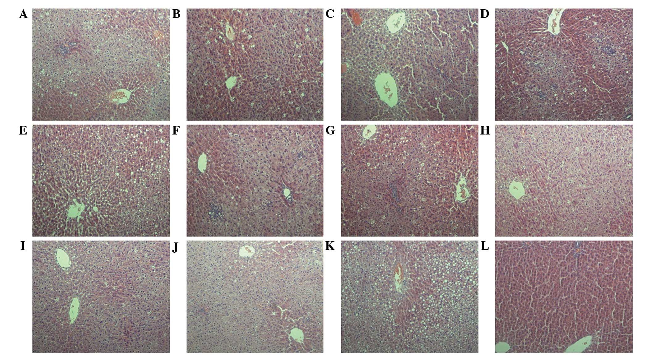
Histological changes in the liver. Typical
steatosis, balloon degeneration in hepatocytes and evident
infiltration by inflammatory cells in the intercellular substance,
were observed in the model group liver tissue samples (Fig. 2K), as compared with that of the
control group (Fig.2L). However,
these changes in pathology were alleviated in the orthogonal
experiment and rosiglitazone treatment groups (Figs. 2A-I and J, respectively).
Furthermore, Oil Red O staining was used to detect the quantities
of lipids in the hepatocytes, the results of which demonstrated
that hepatocyte lipidosis was significantly increased in the model
group (Fig. 3K); however, the
deposition of lipid droplets in the hepatocytes was markedly
reduced in the orthogonal experiment groups and rosiglitazone
treatment group (Figs. 3A-I and J,
respectively).
Effects of puerarin, baicalin and
berberine on the biochemical parameters
Plasma HDL, LDL, TG and TC levels
Compared with the control group, the serum HDL
levels in the model group were significantly decreased (P<0.01);
whereas the levels of LDL, TG, and TC were significantly increased
(P<0.01). However, no significant differences in the HDL levels
of rats were determined between the orthogonal experiment groups
(P>0.05, Fig. 4C), and the model
group. The levels of TG in groups A, D, F, G and R were
significantly decreased (P<0.05, Fig.
4A), as were the TC levels in groups A-C and E (P<0.05,
Fig. 4B), and the levels of LDL in
groups A-D and R (P<0.05, Fig.
4D), as compared with the model group.
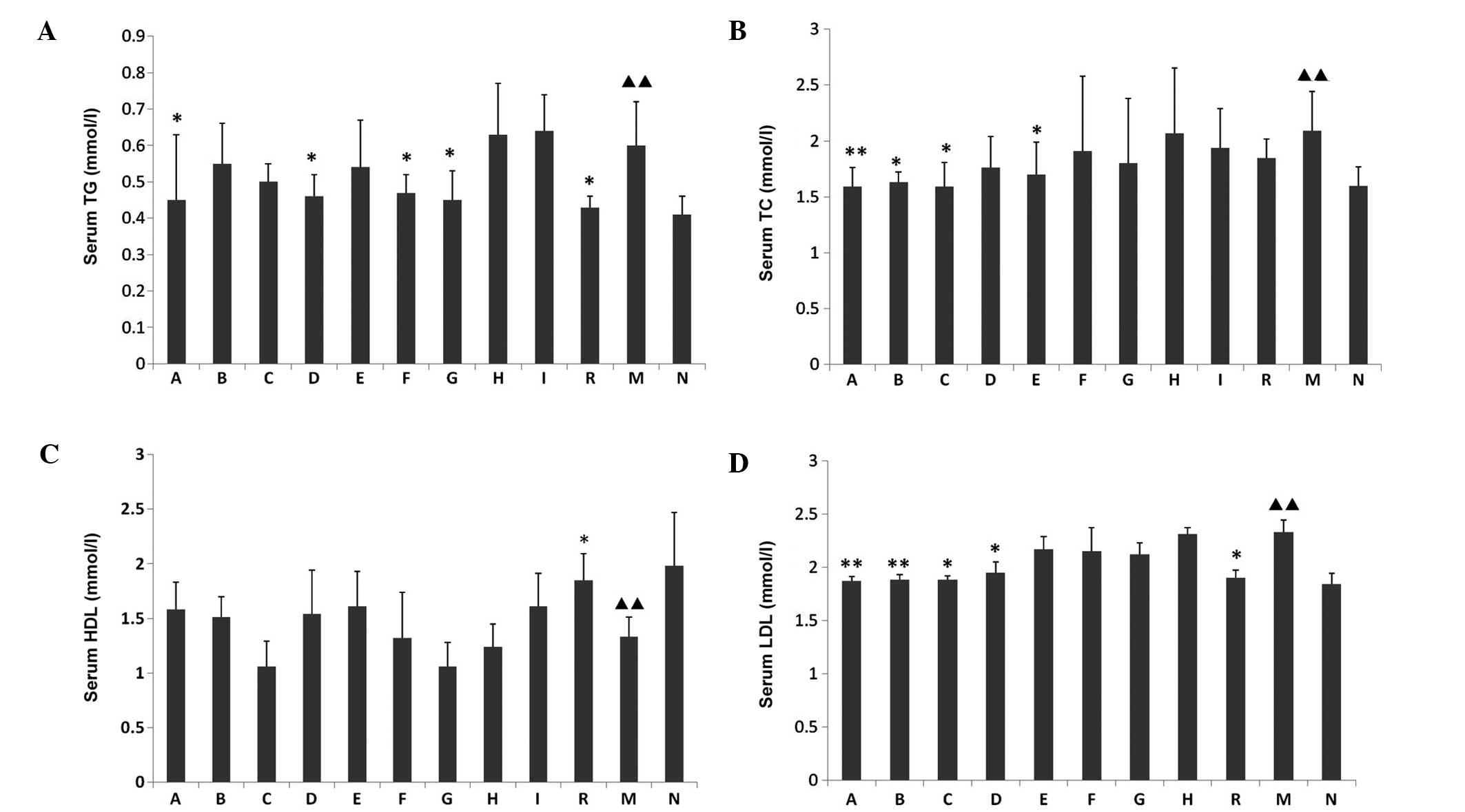 | Figure 4.Effects of puerarin, baicalin, and
berberine on the serum levels of (A) TG, (B) TC, (C) HDL and (D)
LDL in all experimental groups. Data are presented as the mean ±
standard deviation. ▲▲P<0.01, as compared with the
normal control group;*P<0.05 and **P<0.01, as compared with
the model group. TG, triglycerides; TC, total cholesterol; HDL,
high-density lipoproteins; LDL, low-density lipoproteins; A-I,
orthogonal experiment groups A-I; R, rosiglitazone treatment group;
M, model group; N, control group. |
Plasma ALT and AST levels
The serum levels of ALT and AST were significantly
increased (P<0.01) in the model group, as compared with the
control group. Furthermore, in groups F, G, H, I and R the serum
levels of AST were significantly decreased (P<0.01, Fig. 5A), as were the levels of ALT in
groups A, B, D, E, I and R (P<0.01, Fig. 5B), as compared with the model
group.
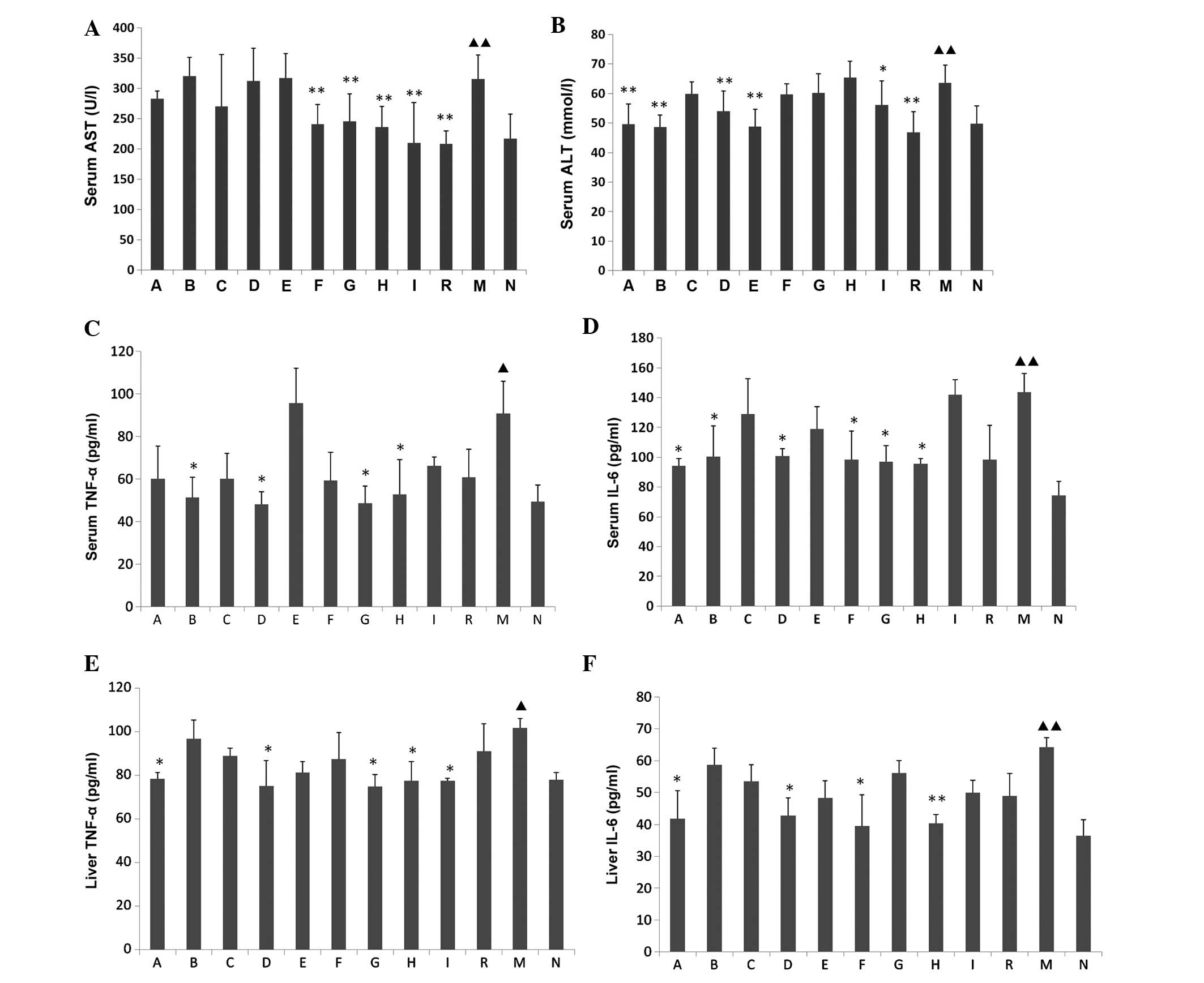 | Figure 5.Effects of puerarin, baicalin, and
berberine on (A) AST, (B) ALT, (C and E) TNF-α and (D and F) IL-6
levels in all experimental groups. Data are presented as the mean ±
standard deviation. ▲P<0.05 and
▲▲P<0.01, vs. the control group; *P<0.05 and
**P<0.01, vs. the model group. AST, aspartate aminotransferase;
ALT, alanine aminotransferase; TNF-α, tumor necrosis factor-α;
IL-6, interleukin-6; A-I, orthogonal experiment groups A-I; R,
rosiglitazone treatment group; M, model group; N, control
group. |
Effects of puerarin, baicalin, and berberine on
the levels of TNF-α and IL-6
Compared with the control group, the levels of TNF-α
and IL-6 in the serum and liver samples of the model group were
significantly increased (P<0.05); whereas, compared with the
model group, the serum levels of TNF-α in groups B, D, G and H were
significantly decreased (P<0.05, Fig.
5C), and the serum levels of IL-6 were significantly decreased
in groups A, B, D and F-H (P<0.05, Fig. 5D). Furthermore, in groups A, D, G, H
and I, the levels of TNF-α in the liver were significantly
decreased (P<0.05, Fig. 5E), and
IL-6 levels in the liver were decreased in groups A, D, F and H
(P<0.05, Fig. 5F), as compared
with the model group.
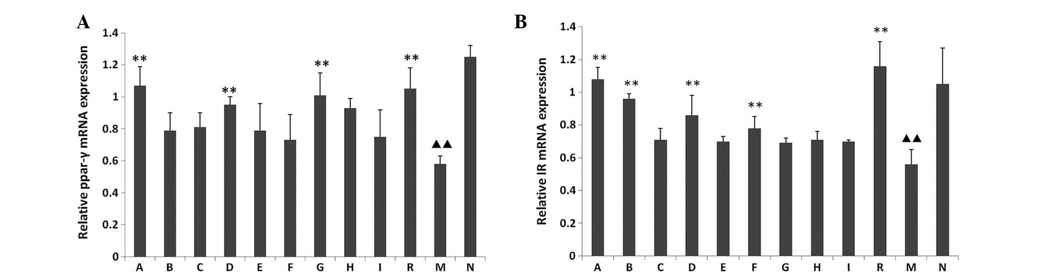
mRNA expression levels of PPAR-γ and IR
In the model group, the mRNA expression levels of
PPAR-γ and IR were significantly decreased (P<0.01), as compared
with the control group. Furthermore, in groups A, D, G and R the
mRNA expression levels of PPAR-γ were significantly increased
(P<0.01, Fig. 6A), as were the IR
mRNA expression levels in groups A, B, D, F and R (P<0.01,
Fig. 6B), as compared with the model
group. However, no significant changes were determined between the
rosiglitazone treatment group and the orthogonal experiment groups
(P>0.05).
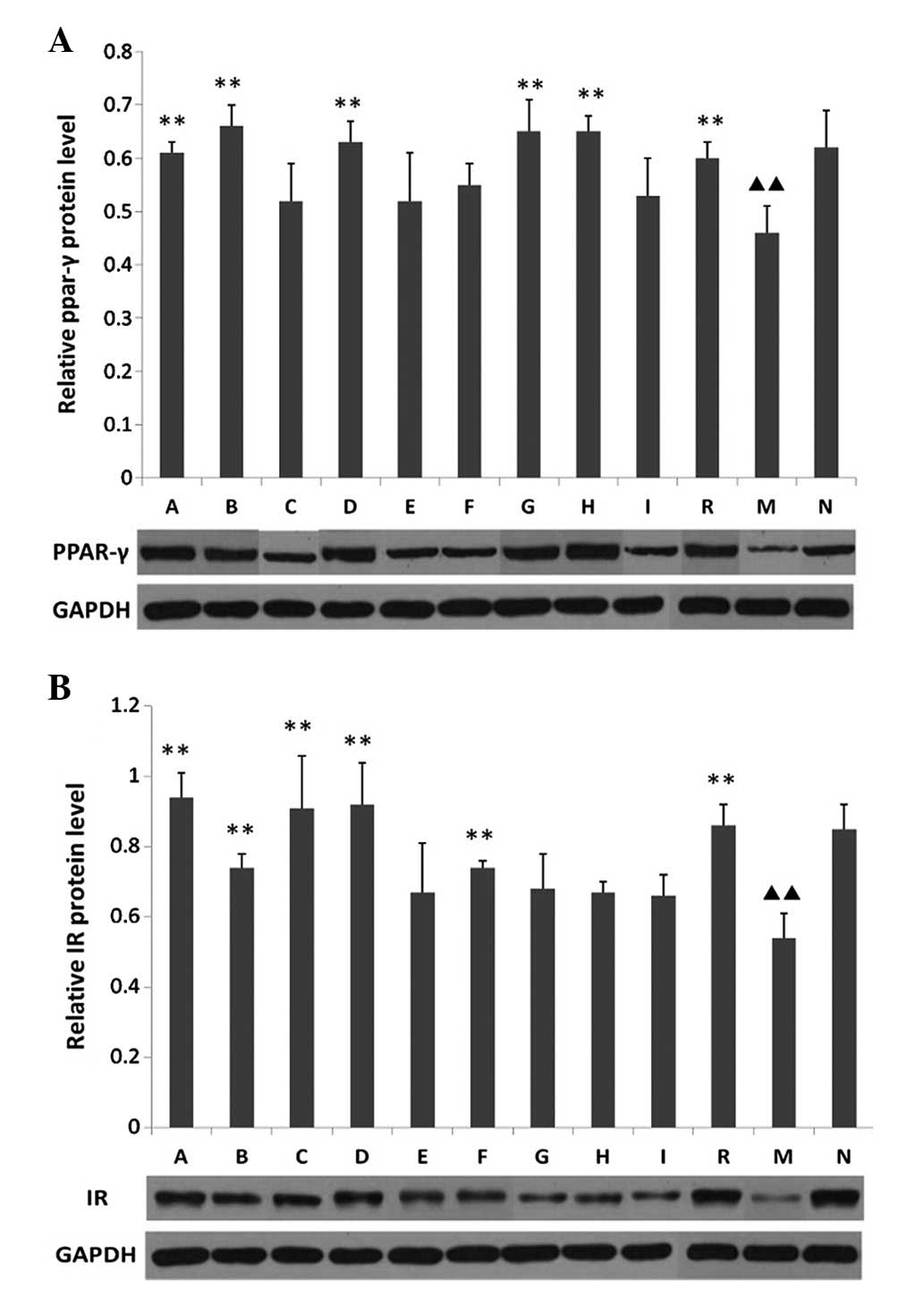
PPAR-γ and IR protein expression levels, as
determined by western blotting
Compared with the control group, the protein
expression levels of PPAR-γ and IR in the model group were
significantly decreased (P<0.01). Furthermore, the PPAR-γ
protein expression levels in groups A, B, D, G, H and R were
significantly increased, as compared with the model group
(P<0.01, Fig. 7A); and in groups
A-D, F and R the protein expression levels of IR were significantly
increased (P<0.01, Fig. 7B).
However, no differences were determined between the rosiglitazone
treatment and orthogonal experiment groups (P>0.05).
Discussion
NAFLD, which has a clinical spectrum ranging from
simple fatty liver disease to NASH and cirrhosis (25), is currently the most common type of
liver disease (26). Although the
etiology of NAFLD has yet to be fully elucidated, the ‘two-hit’
theory has been accepted as the most popular mechanism (27). The ‘first hit’ is associated with
hepatic TG accumulation and insulin resistance, whereas the ‘second
hit’ involves oxidative stress and the induction of inflammatory
cytokines (28). PPAR-γ is a
sequence-specific and ligand-dependent nuclear transcription
factor. With its four isoforms: γ1, γ2, γ3 and γ4, PPAR-γ is
associated with the control of lipid storage and the
differentiation of adipocytes and macrophages (29). TZDs are high affinity synthetic
PPAR-γ ligands that can stimulate adipocyte differentiation and
improve insulin resistance via PPAR-γ activation (30). One TZD in particular, rosiglitazone,
has been established as a widely used therapeutic agent in the
treatment of insulin resistance. Rosiglitazone has previously been
demonstrated to improve sensitivity to insulin as well as
transaminases and liver histology (31). In the present study, rosiglitazone
was used as a positive pharmacological control, in order to compare
the effects of puerarin, baicalin, and berberine on rats with
high-fat diet-induced NAFLD. IR was initially identified as a
homodimer, with extrinsic disulfide bonds that generate the
functional receptor. Each monomer of IR is composed of one α and
one β subunit bridged by an intrinsic disulfide bond (32,33). The
lack of IR may directly affect the distribution of insulin in the
body, causing insulin resistance and abnormal glucose tolerance.
Previous studies have demonstrated that PPAR-γ and IR may be
important for the ‘first hit’ stage of NAFLD, since they are
associated with insulin resistance (34,35).
Increased PPAR-γ also has an anti-inflammatory role as it inhibits
the production of various inflammatory cytokines including TNF-α,
IL-6, IL-1, matrix metalloproteinase-9 and transforming growth
factor-β (36). Therefore, in the
present study TNF-α and IL-6 were selected as representative
inflammatory markers involved in the ‘second hit’ stage.
Since there is still no clear curative treatment for
NAFLD, patients may use anti-diabetic, lipid-lowering or
anti-hypertensive agents to control NAFLD co-morbidities. Chinese
herbs have been traditionally used for treating liver disease
worldwide (37), and there are
numerous herbal products that are believed to benefit patients with
NAFLD, with acceptable levels of safety. For example, previous
studies have demonstrated that puerarin, baicalin and berberine,
which are three major constituents of the Chinese herbs:
Puerariae Radix, Scutellariae Radix and Coptidis
Rhizoma, exhibit therapeutic effects on rats with NAFLD
(20–22). However, there have been few reports
regarding the compatibility of these constituents. The orthogonal
experiment is an efficient and economical test, which has the
advantage of simultaneously balancing samples and reducing test
times, thus ensuring that each test has a strong representation
(38). In order to identify improved
treatment options for puerarin, baicalin and berberine combination
therapy through a small number of experiments that determine the
best therapeutic effect, the orthogonal experiment was applied to
the overall design, comprehensive comparison and statistical
analysis of the present study.
In the present study, rats with high-fat
diet-induced NAFLD were employed to evaluate the efficacies of
puerarin, baicalin and berberine against NAFLD. The high-fat diet
model has been widely used in previous NAFLD studies (39,40). The
histological results of the present study observed NAFLD features
in the model rats, indicating that an NAFLD model was successfully
generated. Furthermore, as expected, the rosiglitazone treatment
and orthogonal experiment groups significantly attenuated hepatic
steatosis, inflammation and fibrosis in the rats.
The pharmaceutical compositions examined in the
present study demonstrated that a combination of puerarin, baicalin
and berberine may exert promising lipid-lowering, hepatoprotective
and anti-inflammatory activities. The various orthogonal experiment
groups had numerous effects on biochemical parameters and
pro-inflammatory cytokines. The present study demonstrated that
serum levels of TC, LDL and ALT were significantly reduced in the
puerarin-dominated groups, whereas serum levels of TNF-α and IL-6
were improved in the baicalin- and berberine-dominated groups.
Therefore, the results of the present study suggested that puerarin
may preferentially affect lipid metabolism, whereas baicalin and
berberine may impact the inflammatory response. Furthermore,
PPAR-γ/IR protein and mRNA expression levels were also reduced in
the model group, thus suggesting that insulin resistance had
developed in response to decreased PPAR-γ and IR in the NAFLD model
rats. Notably, the rosiglitazone treatment group and certain
orthogonal experiment groups improved insulin resistance; however,
no obvious trends in the various combinations of puerarin, baicalin
and berberine were determined. In addition, no differences were
demonstrated between the rosiglitazone treatment and orthogonal
experiment groups. Therefore, we hypothesize that a combination of
puerarin, baicalin and berberine may be used in the early
pathological stage (‘first hit’) of NAFLD to improve insulin
resistance via PPAR-γ and IR upregulation. Since puerarin treatment
is associated with the regulation of lipid metabolism, puerarin may
be used in the early stages of simple fatty liver disease; whereas
baicalin and berberine may be used in NASH, as they may alleviate
the inflammatory response. However, it was not possible to clarify
the effects on liver cirrhosis in the present study due to a lack
of detection of liver fibrosis.
In conclusion, the combination of puerarin, baicalin
and berberine induced favorable therapeutic effects on rats with
high-fat diet-induced NAFLD. Furthermore, variably positive effects
were demonstrated in the various stages of NAFLD following
treatment with numerous proportions of monomer compositions. This
suggests that a therapeutic combination of puerarin, baicalin and
berberine may regulate lipid metabolism by upregulating the
expression of hepatic PPAR-γ and IR, leading to improvements in
insulin resistance, which may be useful in the prevention and
treatment of NAFLD, especially simple fatty liver disease and NASH.
Further studies focusing on the effects of combination treatment
with puerarin, baicalin and berberine on liver fibrosis are
required.
Acknowledgements
The present study was supported by the Youth Fund of
National Natural Science Foundation of China (grant no. 81503407),
Self-selected subject of Beijing University of Chinese Medicine
(grant no. 2015-JYB-JSMS125), and the Wang Bao-enLiver Fibrosis
Research Fund (grant no. 2013-xjs).
References
|
1
|
Marchesini G and Marzocchi R: Metabolic
syndrome and NASH. Clin Liver Dis. 11105–117. (ix)2007. View Article : Google Scholar : PubMed/NCBI
|
|
2
|
Fabbrini E, Sullivan S and Klein S:
Obesity and nonalcoholic fatty liver disease: Biochemical,
metabolic and clinical implications. Hepatology. 51:679–689. 2010.
View Article : Google Scholar : PubMed/NCBI
|
|
3
|
Song HY, Zhang L, Pan JL, Yang LL and Ji
G: Bioactivity of five components of Chinese herbal formula
Jiangzhi granules against hepatocellular steatosis. J Integr Med.
11:262–268. 2013. View Article : Google Scholar : PubMed/NCBI
|
|
4
|
Stein LL, Dong MH and Loomba R: Insulin
sensitizers in nonalcoholic fatty liver disease and
steatohepatitis: Current status. Adv Ther. 26:893–907. 2009.
View Article : Google Scholar : PubMed/NCBI
|
|
5
|
Sun L and Lü SZ: Association between
non-alcoholic fatty liver disease and coronary artery disease
severity. Chin Med J (Engl). 124:867–872. 2011.PubMed/NCBI
|
|
6
|
Zhao JS, Zhu FS, Liu S, Yang CQ and Chen
XM: Pioglitazone ameliorates nonalcoholic steatohepatitis by
down-regulating hepatic nuclear factor-kappa B and
cyclooxygenases-2 expression in rats. Chin Med J (Engl).
125:2316–2321. 2012.PubMed/NCBI
|
|
7
|
Zhao CY, Jiang LL, Li L, Deng ZJ, Liang BL
and Li JM: Peroxisome proliferator activated receptor-gamma in
pathogenesis of experimental fatty liver disease. World J
Gastroenterol. 10:1329–1332. 2004.PubMed/NCBI
|
|
8
|
Nan YM, Fu N, Wu WJ, Liang BL, Wang RQ,
Zhao SX, Zhao JM and Yu J: Rosiglitazone prevents nutritional
fibrosis and steatohepatitis in mice. Scand J Gastroenterol.
44:358–365. 2009. View Article : Google Scholar : PubMed/NCBI
|
|
9
|
Lutchman G, Modi A, Kleiner DE, Promrat K,
Heller T, Ghany M, Borg B, Loomba R, Liang TJ and Premkumar A: The
effects of discontinuing pioglitazone in patients with nonalcoholic
steatohepatitis. Hepatology. 46:424–429. 2007. View Article : Google Scholar : PubMed/NCBI
|
|
10
|
Chiu SL and Cline HT: Insulin receptor
signaling in the development of neuronal structure and function.
Neural Dev. 5:72010. View Article : Google Scholar : PubMed/NCBI
|
|
11
|
Zhang Q, Xiao XH, Li M, Li WH, Yu M, Zhang
HB, Ping F, Wang ZX and Zheng J: Chromium-containing traditional
Chinese medicine, Tianmai Xiaoke Tablet improves blood glucose
through activating insulin-signaling pathway and inhibiting PTP1B
and PCK2 in diabetic rats. J Integr Med. 12:162–170. 2014.
View Article : Google Scholar : PubMed/NCBI
|
|
12
|
Samuel VT and Shulman GI: Integrating
mechanisms for insulin resistance: Common threads and missing
links. Cell. 148:852–8712012. View Article : Google Scholar
|
|
13
|
Mohammadi A, Gholamhoseinian A and Fallah
H: Zataria multiflora increases insulin sensitivity and
PPAR-γ gene expression in high fructose fed insulin resistant rats.
Iran J Basic Med Sci. 17:263–270. 2014.PubMed/NCBI
|
|
14
|
Rani S and O'Driscoll L: Analysis of
changes in phosphorylation of receptor tyrosine kinases: Antibody
arrays. Methods Mol Biol. 1233:15–23. 2015. View Article : Google Scholar : PubMed/NCBI
|
|
15
|
Promrat K, Lutchman G, Uwaifo GI, Freedman
RJ, Soza A, Heller T, Doo E, Ghany M, Premkumar A, Park Y, et al: A
pilot study of pioglitazone treatment for nonalcoholic
steatohepatitis. Hepatology. 39:188–196. 2004. View Article : Google Scholar : PubMed/NCBI
|
|
16
|
Nesto RW, Bell D, Bonow RO, Fonseca V,
Grundy SM, Horton ES, Le Winter M, Porte D, Semenkovich CF, Smith
S, et al: Thiazolidinedione use, fluid retention, and congestive
heart failure: A consensus statement from the American Heart
Association and American Diabetes Association. Diabetes Care.
27:256–263. 2004. View Article : Google Scholar : PubMed/NCBI
|
|
17
|
Parulkar AA, Pendergrass ML, Granda-Ayala
R, Lee TR and Fonseca VA: Nonhypoglycemic effects of
thiazolidinediones. Ann Intern Med. 134:61–71. 2001. View Article : Google Scholar : PubMed/NCBI
|
|
18
|
Wang WJ: Enhancing the treatment of
metabolic syndrome with integrative medicine. J Integr Med.
11:153–156. 2013. View Article : Google Scholar : PubMed/NCBI
|
|
19
|
Yao Z, Zhang L and Ji G: Efficacy of
polyphenolic ingredients of Chinese herbs in treating dyslipidemia
of metabolic syndromes. J Integr Med. 12:135–146. 2014. View Article : Google Scholar : PubMed/NCBI
|
|
20
|
Wang Y and Li J, Zhuge L, Su D, Yang M,
Tao S and Li J: Comparison between the efficacies of curcumin and
puerarin in C57BL/6 mice with steatohepatitis induced by a
methionine- and choline-deficient diet. Exp Ther Med. 7:663–668.
2014.PubMed/NCBI
|
|
21
|
Guo HX, Liu DH, Ma Y, Liu JF, Wang Y, Du
ZY, Wang X, Shen JK and Peng HL: Long-term baicalin administration
ameliorates metabolic disorders and hepatic steatosis in rats given
a high-fat diet. Acta Pharmacol Sin. 30:1505–1512. 2009. View Article : Google Scholar : PubMed/NCBI
|
|
22
|
Chang X, Yan H, Fei J, Jiang M, Zhu H, Lu
D and Gao X: Berberine reduces methylation of the MTTP promoter and
alleviates fatty liver induced by a high-fat diet in rats. J Lipid
Res. 51:2504–2515. 2010. View Article : Google Scholar : PubMed/NCBI
|
|
23
|
Cui W, Li X, Zhou S and Weng J:
Investigation on process parameters of electrospinning system
through orthogonal experimental design. J Appl Polym Sci.
103:3105–3112. 2007. View Article : Google Scholar
|
|
24
|
Khan F, Choong WL, Du Q and Jovanovi'c A:
Real-time RT-PCR Ct values for blood GAPDH correlate with measures
of vascular endothelial function in humans. Clin Transl Sci.
6:481–484. 2013. View Article : Google Scholar : PubMed/NCBI
|
|
25
|
Matteoni CA, Younossi ZM, Gramlich T,
Boparai N, Liu YC and McCullough AJ: Nonalcoholic fatty liver
disease: A spectrum of clinical and pathological severity.
Gastroenterology. 116:1413–1419. 1999. View Article : Google Scholar : PubMed/NCBI
|
|
26
|
Adams LA and Angulo P: Treatment of
non-alcoholic fatty liver disease. Postgrad Med J. 82:315–322.
2006. View Article : Google Scholar : PubMed/NCBI
|
|
27
|
Day CP and James OF: Steatohepatitis: A
tale of two ‘hits’? Gastroenterology. 114:842–845. 1998. View Article : Google Scholar : PubMed/NCBI
|
|
28
|
Postic C and Girard J: Contribution of de
novo fatty acid synthesis to hepatic steatosis and insulin
resistance: Lessons from genetically engineered mice. J Clin
Invest. 118:829–838. 2008. View
Article : Google Scholar : PubMed/NCBI
|
|
29
|
Law RE, Goetze S, Xi XP, Jackson S, Kawano
Y, Demer L, Fishbein MC, Meehan WP and Hsueh WA: Expression and
function of PPARgamma in rat and human vascular smooth muscle
cells. Circulation. 101:1311–1318. 2000. View Article : Google Scholar : PubMed/NCBI
|
|
30
|
Harrison SA: Thiazolidinedione therapy for
nonalcoholic steatohepatitis: Go, stop, or proceed with caution?
Hepatology. 51:366–369. 2010. View Article : Google Scholar : PubMed/NCBI
|
|
31
|
Nan YM, Fu N, Wu WJ, Liang BL, Wang RQ,
Zhao SX, Zhao JM and Yu J: Rosiglitazone prevents nutritional
fibrosis and steatohepatitis in mice. Scand J Gastroenterol.
44:358–365. 2009. View Article : Google Scholar : PubMed/NCBI
|
|
32
|
Cheatham B and Kahn CR: Cysteine 647 in
the insulin receptor is required for normal covalent interaction
between alpha-andbeta-subunits and signal transduction. J Biol
Chem. 267:7108–7115. 1992.PubMed/NCBI
|
|
33
|
Seino S and Bell GI: Alternative splicing
of human insulin receptor messenger RNA. Biochem Biophys Res
Commun. 159:312–316. 1989. View Article : Google Scholar : PubMed/NCBI
|
|
34
|
El-Bassossy HM, Abo-Warda SM and Fahmy A:
Chrysin and luteolin alleviate vascular complications associated
with insulin resistance mainly through PPAR-γ activation. Am J Chin
Med. 42:1153–1167. 2014. View Article : Google Scholar : PubMed/NCBI
|
|
35
|
Shamsi BH, Ma C, Naqvi S and Xiao Y:
Effects of pioglitazone mediated activation of PPAR-γ on CIDEC and
obesity related changes in mice. PLoS One. 9:e1069922014.
View Article : Google Scholar : PubMed/NCBI
|
|
36
|
Wang RC and Jiang DM: PPAR-γ agonist
pioglitazone affects rat gouty arthritis by regulating cytokines.
Genet Mol Res. 13:6577–6781. 2014. View Article : Google Scholar : PubMed/NCBI
|
|
37
|
Seeff LB, Lindsay KL, Bacon BR, Kresina TF
and Hoofnagle JH: Complementary and alternative medicine in chronic
liver disease. Hepatology. 34:595–603. 2001. View Article : Google Scholar : PubMed/NCBI
|
|
38
|
Liu RJ, Zhang YW, Wen CW and Tang J: Study
on the design and analysis methods of orthogonal experiment. Exp
Technol and Manage. 27:52–552010.
|
|
39
|
Silva RN, Bueno PG, Avó LR, Nonaka KO,
Selistre-Araújo HS and Leal AM: Effect of physical training on
liver expression of activin A and follistatin in a nonalcoholic
fatty liver disease model in rats. Braz J Med Biol Res. 47:746–752.
2014. View Article : Google Scholar : PubMed/NCBI
|
|
40
|
Kucera O and Cervinkova Z: Experimental
models of non-alcoholic fatty liver disease in rats. World J
Gastroenterol. 20:8364–8376. 2014. View Article : Google Scholar : PubMed/NCBI
|