Introduction
Gliomas, which are tumors that originate from
neuroglial cells, commonly occur in the brain and occasionally in
the spinal cord (1). Gliomas account
for 80% of primary tumors in the brain and central nervous system,
and for 30% of malignant tumors in the brain (2). Patients with low-malignancy gliomas
[World Health Organization (WHO) grade II] have good prognoses,
with a 10-year survival rate of up to 47% (3), and reportedly as long as 17 years
(4). By contrast, high-malignancy
gliomas (WHO grades III–IV) are rich in vessels and their size
increases rapidly. Hypoxic and necrotic tissues in the tumor center
can damage the surrounding tissues and destroy the blood-brain
barrier; thus, these tumors may relapse even when completely
resected. The 1- and 2-year survival rates of high-malignancy
gliomas are only 50 and 25%, respectively (5).
Cancer stem cells (CSCs) have the same
characteristics as other types of stem cells, but can also
differentiate into various types of cells in the tumor tissue
(6,7). Thus, CSCs closely affect tumor
recurrence and metastasis. Glioma CSCs are important in the
development of invasive tumor growth, insensitivity to chemotherapy
and radiotherapy, and poor prognosis (8,9). Since
glioma CSCs were successfully isolated and cultured in vitro
in 2004 (10), their characteristics
have been studied extensively. Currently known glioma CSC markers
include CD133, nestin, (sex determining region Y)-box 2 (SOX2),
ATP-binding cassette sub-family G member 2 (ABCG2) and musashi-1
(11–15), with CD133 considered to be the most
important of these markers. CD133+ glioma CSCs form cell
spheres in culture medium that contains growth factors (16), and differentiate into neurons and
glial cells following the removal of the growth factors (17–19).
However, certain CD133− glioma cells also exhibit
similar characteristics to CSCs (20). In addition, certain types of cells,
such as endothelial cells, are also CD133+ (21). Therefore, CD133, even alongside other
biomarkers, may not be a specific biomarker for glioma CSCs.
Vascular endothelial growth factor (VEGF) is highly
expressed in glioma cells (22–24), and
its expression is directly associated with the malignancy and
prognosis of gliomas (25).
Phosphoinositide-3-kinase (PI3K) is a lipid second messenger
associated with intracellular signal transduction that can catalyze
the formation of phosphoinositide-3 phosphate, which is the
phosphorylated product of the third hydroxyl of inositol phosphate
(26). PI-3,4,5-P3 is the
phosphorylated product of PI3K, which is gathered in the inner
surface of the cell membrane. Protein kinase B (Akt) combines with
PI-3,4,5-P3 and is subsequently activated. The activated
Akt then enters the cell membrane, where it is phosphorylated by
PDK1 and PDK2, and regulates a series of functions, including the
cell cycle, growth and survival (27). PI3K comprises of five subtypes,
including p55α, p55γ, p85α, p85β and p50α, all of which are
expressed in neuronal cells in the rat brain, indicating that PI3K
is important in signal transduction in the brain (28). However, the association between
PI3K/Akt gene expression and glioma remains unclear.
In the present study, specimens from glioma patients
were divided into the following two groups according to clinical
grading: Low-malignancy (WHO grade II) and high-malignancy (WHO
grades III–IV) groups (29). Stem
cells were extracted from fresh tumor tissues, and the expression
levels of CD133, nestin, SOX2, VEGF and PI3K were detected by
reverse transcription-quantitative polymerase chair reaction
(RT-qPCR), in order to identify the association of glioma CSCs with
the VEGF and PI3K signal transduction systems. To the best of our
knowledge, this is the first study investigating the expression
levels of VEGF and PI3K in glioma CSCs obtained from glioma
patients. The results will provide first-hand information for
further study of drugs that target glioma.
Materials and methods
Sample collection
Samples were collected in strict accordance with the
scientific research sample collection guidelines of the Department
of Neurosurgery the Affiliated Hospital of Beihua University
(Jilin, Jilin). Glioma samples were successfully collected from 27
patients with glioma who were undergoing resection surgery at the
Department of Neurosurgery, between 2010 and 2013. Tissue samples
were diagnosed by pathological section and classified into 12 low-
and 15 high-malignancy gliomas, according to the WHO guidelines
(29–33). The tissue samples immediately
underwent tissue digestion and cell isolation. This study was
conducted in accordance with the declaration of Helsinki and was
approved by the Ethics Committee of Jilin University (Changchun,
China). Written informed consent was obtained from all the
participants.
Isolation and purification of glioma
CSCs
Several 6-well plates were coated with 20 µg/ml
poly-ornithine (Sigma-Aldrich, Carlsbad, CA, USA) and incubated in
a cell incubator at 37°C for at least 2 h. Subsequently, the
poly-ornithine was removed by washing once with deionized water,
the plates were rinsed once with phosphate-buffered saline (PBS),
and were then incubated with a final concentration of 5 µg/ml
laminine (Sigma-Aldrich) for 1 h.
Fresh glioma tumor tissues were cut into small
sections (7 µm) and rinsed twice with Hank's balanced salt solution
(HBSS; Gibco Life Technologies, Grand Island, NY, USA) containing
20% fetal bovine serum (FBS; Thermo Fisher Scientific, Inc.,
Pittsburgh, PA, USA). The specimens were then rinsed three times
with HBSS without FBS in order to remove blood cells and then
digested with 0.25% trypsin (Gibco Life Technologies) at 37°C for
10 min. Subsequently, the digestion was terminated using 3 ml HBSS
containing 20% FBS, and the samples were centrifuged at 400 × g at
4°C for 3 min. The supernatant was then discarded, and the cells
were resuspended using the neural stem cell culture medium (Thermo
Fisher Scientific, Inc., Waltham, MA, USA). The cell suspension was
filtered through a 70-µm nylon mesh (Mumford Industries, Inc., CA,
USA) and the cells were transferred to the 6-well plates that had
been prepared previously. To further evaulate biomarker expression,
U251 human glioma cells (Sigma-Aldrich) were used as a control. The
U251 and primary neural stem cells were cultured in medium
containing 10% FBS, 40 ng/ml fibroblast growth factor 2, 20 ng/ml
epidermal growth factor (EGF) and 20 ng/ml platelet-derived growth
factor (all from Gibco Life Technologies) on the first day. On the
second day, the medium was replaced with fresh medium without FBS,
and thereafter, half the medium was changed every day, with the
addition of fresh growth factors. The cells were observed and
images were captured under 200× magnification using a microscope
(BX51-32H01; Olympus Corporation, Tokyo, Japan).
Detection of glioma CSCs using
RT-qPCR
The total RNA of glioma CSCs and U251 cells was
extracted using an RNeasy kit (Qiagen, Hilden, Germany). Next, 1 µg
total RNA was reverse transcribed into cDNA using iScript cDNA
Synthesis kit (Bio-Rad Laboratories, Inc., Hercules, CA, USA), and
PCR was performed using PCR Master Mix (Applied Biosystems, Life
Technologies, Rockville, MD, USA). The PCR protocol comprised of
initial denaturation at 95°C for 10 min, 35 cycles at 95°C for 30
sec, 54°C for 30 sec and 72°C for 30 sec, and then extension at
72°C for 10 min. The primers used in the present study are listed
in Table I. PCR products were
seperated by 1% agarose gel electrophoresis, and results were
quantified using model-driven agility software (Kepler 4.3; Eclipse
Foundation, Inc., Ottawa, ON, Canada).
 | Table I.Primers used in the present
study. |
Table I.
Primers used in the present
study.
| Gene | Forward primer
(5′→3′) | Reverse primer
(5′→3′) |
|---|
| CD133 |
acactgctggtgtgctgac |
cccaaggaccacttcacagt |
| SOX2 |
cacaactcggagatcagcaa |
ctccgggaagcgtgtactta |
| Nestin |
aggctgagaactctcgcttg |
attaggcaagggggaagaga |
| VEGF |
cacgaacgagtccctagagc |
atggtgatgcggttttcttc |
| PI3K |
attacgctagttacactgca |
tggacctggccatcgactga |
| GAPDH |
accacagtccatgccatcac |
tccaccaccctgttgctgta |
Statistical analysis
VEGF and PI3K mRNA expression levels were normalized
to GAPDH. Student's t-test was used to analyze the VEGF and PI3K
expression levels between the two glioma groups. All analyses were
conduced using SPSS 20 software (IBM SPSS, Armonk, NY, USA).
P<0.05 was considered to indicate a statistically significance
difference.
Results
Comparison of clinical conditions
All the glioma patients included in the present
study had not received any treatment prior to surgery. The patients
were divided into the low-malignancy (WHO grade II; n=12) and
high-malignancy groups (WHO grades III–IV; n=15). No statistically
significant differences in the gender, age, tumor site or
pathological type were observed between the two groups (P>0.05;
Table II).
| Table II.Comparison of clinical
characteristics of patients with glioma. |
Table II.
Comparison of clinical
characteristics of patients with glioma.
| Characteristic | Low-malignancy
group (n=12) | High-malignancy
group (n=15) |
|---|
| Mean age,
years | 42 | 45 |
| Age range,
years | 33–52 | 28–56 |
| Male: female
ratio | 1.8:1 | 1.6:1 |
| Tumor site |
|
|
| Frontal
lobe | 5 | 5 |
|
Temporal lobe | 4 | 6 |
|
Parietal lobe | 3 | 4 |
| Pathological
type |
|
|
|
Glioblastoma | 5 | 4 |
|
Oligodendroglioma | 2 | 4 |
| Diffuse
astroglioma | 5 | 7 |
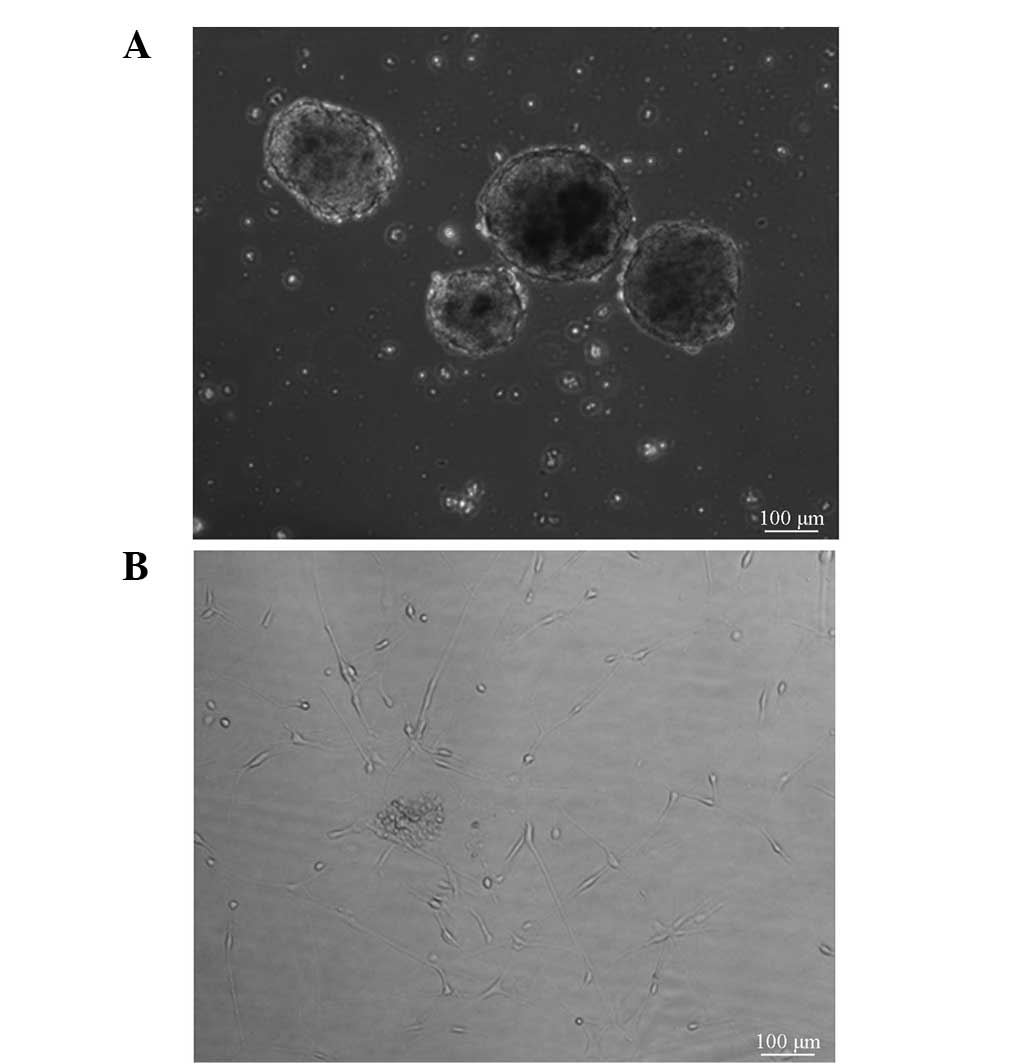
Morphology of glioma CSCs
Glioma CSCs were observed in the suspended and
adherent state (Fig. 1). These two
cell states display different shapes. The suspended cells formed
spheres, whereas the adherent cells exhibited a spindle shape.
Notably, the amount of sphere-shaped CSCs was increased in the
high-malignancy group, as compared with in the low malignancy
group. Taken together, various states of CSCs were detected in the
cell culture.
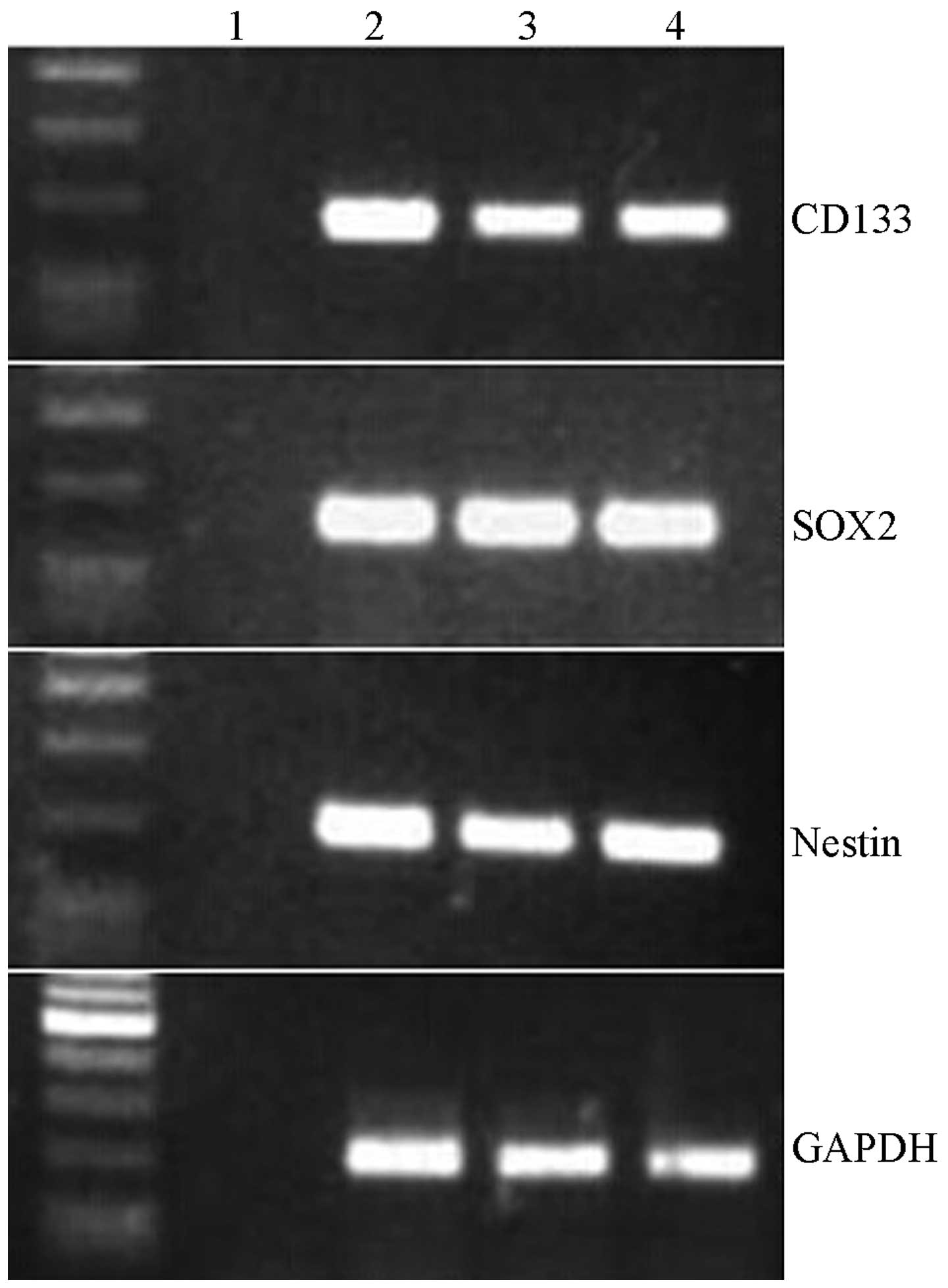
Identification of glioma stem cell
markers
To validate the expression levels of conventional
CSC biomarkers, the mRNA expression levels of CD133, SOX2 and
nestin were detected in the high- and low-malignancy glioma CSCs.
CD133 expression levels were markedly lower in the low-malignancy
group, as compared with in the high-malignancy group; however,
there was no significant difference with the U251 cells (Fig. 2). Furthermore, neither SOX2 nor
nestin expression levels differed between the glioma CSCs and U251
cells. These results suggest that the transcriptional levels of
CD133, SOX2 and nestin may not be able to efficiently evaluate the
malignant stage of glioma CSCs.
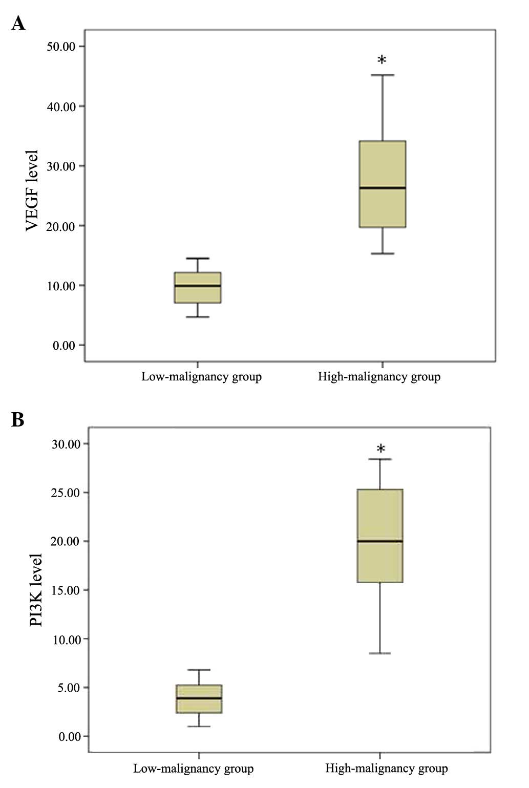
Expression levels of VEGF and
PI3K
The present study further investigated the VEGF and
PI3K mRNA expression levels between the two glioma groups. VEGF was
significantly upregulated in the high-malignancy group, as compared
with the low-malignancy group (T value=12.655, P=0.002). In
addition, the mRNA expression levels of PI3K exhibited a similar
tendency (T value=15.948, P=0.001) (Table III; Fig.
3A and B). These results suggest that the transcriptional
levels of VEGF and PI3K may significantly differ in the various
malignant stages of glioma.
| Table III.Differential expression of VEGF and
PI3K in the low- and high-malignancy glioma CSCs. |
Table III.
Differential expression of VEGF and
PI3K in the low- and high-malignancy glioma CSCs.
| A, Low-malignancy
glioma CSCs (n=12) |
|---|
| Patient | VEGF levels | PI3K levels |
|---|
| 1 | 14.5±1.0 | 4.6±0.2 |
| 2 | 10.9±0.6 | 6.4±0.1 |
| 3 | 5.0±0.2 | 2.6±0.6 |
| 4 | 4.7±0.5 | 5.8±0.7 |
| 5 | 13.2±2.0 | 6.8±0.6 |
| 6 | 9.5±1.5 | 1.7±0.1 |
| 7 | 11.7±1.2 | 1.0±0.1 |
| 8 | 7.8±0.4 | 3.6±0.2 |
| 9 | 12.6±1.7 | 2.4±0.1 |
| 10 | 10.3±1.6 | 4.7±0.3 |
| 11 | 6.9±0.3 | 2.4±0.7 |
| 12 | 7.2±0.3 | 4.2±0.8 |
|
| B, High-malignancy
glioma CSCs (n=15) |
|
| Patient | VEGF levels | PI3K levels |
| 1 | 28.4±2.5 | 15.7±0.9 |
| 2 | 18.9±1.7 | 18.5±1.2 |
| 3 | 37.9±2.6 | 25.8±2.7 |
| 4 | 15.4±1.2 | 28.4±3.0 |
| 5 | 19.5±1.3 | 8.5±0.5 |
| 6 | 32.5±2.0 | 26.8±0.4 |
| 7 | 45.2±2.4 | 9.8±0.5 |
| 8 | 26.3±2.9 | 20.0±2.1 |
| 9 | 35.8±2.4 | 15.8±1.9 |
| 10 | 24.7±1.3 | 13.8±1.4 |
| 11 | 38.9±2.1 | 23.3±1.8 |
| 12 | 19.9±1.2 | 24.8±2.1 |
| 13 | 32.4±2.3 | 28.4±1.9 |
| 14 | 15.3±0.5 | 23.0±1.2 |
| 15 | 23.6±1.6 | 17.2±2.1 |
Discussion
The incidence of glioma is 3.8/100,000 for men and
3.1/100,000 for women in developing countries, and 5.8/100,000 for
men and 4.4/100,000 for women in developed countries (34). In the United States, malignant glioma
accounts for 70% of newly-diagnosed cases of glioma, of which
glioblastoma accounts for 60–70% (35,36). The
glioma classification system of the World Health Organization (WHO)
includes four grades, based on the pathological atypia, mitotic
condition and anaplasia. The present study aimed to detect the
differences in the transcriptional expression of CD133, SOX2,
nestin, VEGF and PI3K between high-malignancy and low-malignancy
glioma CSCs. The results of the present study suggested a robust
association among tumor grade, invasion of CSCs and prognosis. In
addition, the expression levels of VEGF and PI3K in WHO grade
III–IV samples were found to be significantly higher compared with
those in grade II samples, which implies that the expression levels
of these two biomarkers may be used as criteria for the clinical
grading of gliomas. However, a larger-scale study is required to
further confirm this finding.
Mutation in the isocitrate dehydrogenase (IDH) gene
is widely detected in low-grade gliomas and is generally considered
to be an important predictor of prognosis in glioma patients
(37,38). The IDH mutation may induce abnormal
formation of 2-hydroxyglutarate, which inhibits
ketoglutarate-dependent dioxygenase, resulting in abnormal histone
and DNA methylation, and thus to poor prognosis for glioma patients
(38,39).
Methylation of the O6-alkylguanine DNA
alkyltransferase (MGMT) gene promoter is also associated with
glioma prognosis (40). Hegi et
al confirmed that MGMT gene promoter methylation was associated
with long-term survival in glioma patients who received only
chemotherapy treatment (41).
Furthermore, Brandes et al revealed that MGMT gene
demethylation increased the vascular permeability of glioblastomas
(42). MGMT promoter demethylation
plays different roles in gliomas of different grades and
pathological types. For instance, the types of gene mutations in
WHO grade IV gliomas differ from those in other grades of glioma.
Mutations of the IDH and p53 genes are rare, whereas the mutation
of EGF receptor (EGFR) is common (43). Further experiments with larger-scale
samples are required to detect the significance of mutations in the
IDH, MGMT, EGFR and p53 genes.
CSCs not only have the same characteristics as other
types of stem cells, but can also differentiate into various types
of cells in tumor tissue, and greatly affect tumor recurrence and
metastasis. Since Bonnet and Dick isolated leukemia stem cells in
1997 (44), researchers have
identified CSCs in specimens of brain, lung, colon, ovary, pancreas
and prostate cancer (45–50). Neurosphere assay is a primary in
vitro method for the culture of neural stem cells, which can
then differentiate into neurons and glial cells (51–53).
Neural stem cells are gathered to form neurospheres subsequent to
cell division, and the size of neurospheres increases along with
the increase of cell division times, so as to achieve a rise in the
number of cells. Since glioma CSCs were first isolated and
successfully cultured in vitro in 2004 (10), thorough research has been conducted
on these cells. Glioma CSCs have been found to express CD133, SOX2,
nestin and other markers. However, the role of glioma CSCs in tumor
grading has not received considerable attention.
In conclusion, the results of the present study
revealed that the expression levels of VEGF and PI3K mRNA in glioma
CSCs from patients with grade III–IV glioma were significantly
higher compared with those in glioma CSCs of patients with grade II
disease. Thus, the expression of VEGF and PI3K mRNA in glioma stem
cells may play a key role in the clinical grading of glioma and the
accurate prediction of the disease prognosis.
References
|
1
|
Mamelak AN and Jacoby DB: Targeted
delivery of antitumoral therapy to glioma and other malignancies
with synthetic chlorotoxin (TM-601). Expert Opin Drug Deliv.
4:175–186. 2007. View Article : Google Scholar : PubMed/NCBI
|
|
2
|
Goodenberger ML and Jenkins RB: Genetics
of adult glioma. Cancer Genet. 205:613–621. 2012. View Article : Google Scholar : PubMed/NCBI
|
|
3
|
Smoll NR, Gautschi OP, Schatlo B, Schaller
K and Weber DC: Relative survival of patients with supratentorial
low-grade gliomas. Neuro Oncol. 14:1062–1069. 2012. View Article : Google Scholar : PubMed/NCBI
|
|
4
|
Olson JD, Riedel E and DeAngelis LM:
Long-term outcome of low-grade oligodendroglioma and mixed glioma.
Neurology. 54:1442–1448. 2000. View Article : Google Scholar : PubMed/NCBI
|
|
5
|
Ohgaki H and Kleihues P: Population-based
studies on incidence survival rates and genetic alterations in
astrocytic and oligodendroglial glioma. J Neuropathol Exp Neurol.
64:479–489. 2005.PubMed/NCBI
|
|
6
|
Plaks V, Kong N and Werb Z: The cancer
stem cells niche, How essential is the niche in regulating stemness
of tumor cells? Cell Stem Cell. 16:225–238. 2015. View Article : Google Scholar : PubMed/NCBI
|
|
7
|
Wang L, Xue Y, Shen Y, Li W, Cheng Y, Yan
X, Shi W, Wang J, Gong Z, Yang G, et al: Claudin 6: A novel surface
marker for characterizing mouse pluripotent stem cells. Cell Res.
22:1082–1085. 2012. View Article : Google Scholar : PubMed/NCBI
|
|
8
|
Gilbertson RJ and Rich JN: Making a
tumour's bed: Glioblastoma stem cells and the vascular niche. Nat
Rev Cancer. 7:733–736. 2007. View
Article : Google Scholar : PubMed/NCBI
|
|
9
|
Chen L, Chen XR, Chen FF, Liu Y, Li P,
Zhang R, Yan K, Yi YJ, Xu ZM and Jiang XD: MicroRNA-107 inhibits
U87 glioma stem cells growth and invasion. Cell Mol Neurobiol.
33:651–657. 2013. View Article : Google Scholar : PubMed/NCBI
|
|
10
|
Galli R, Binda E, Orfanelli U, Cipelletti
B, Gritti A, De Vitis S, Fiocco R, Foroni C, Dimeco F and Vescovi
A: Isolation and characterization of tumorigenic, stem-like neural
precursors from human glioblastoma. Cancer Res. 64:7011–7021. 2004.
View Article : Google Scholar : PubMed/NCBI
|
|
11
|
Gaedicke S, Braun F, Prasad S, Machein M,
Firat E, Hettich M, Gudihal R, Zhu X, Klingner K, Schüler J, et al:
Noninvasive positron emission tomography and fluorescence imaging
of CD133+ tumor stem cells. Proc Natl Acad Sci USA. 111:E692–E701.
2014. View Article : Google Scholar : PubMed/NCBI
|
|
12
|
Lubanska D, Market-Velker BA, deCarvalho
AC and Mikkelsen T: Fidalgo daS ilva E and Porter LA: The
cyclin-like protein Spy1 regualtes growth and division
characteristics of the CD133+ population in human
glioma. Cancer Cell. 25:64–76. 2014. View Article : Google Scholar : PubMed/NCBI
|
|
13
|
Jin X, Jin X, Jung JE, Beck S and Kim H:
Cell surface nestin is a biomarker for glioma stem cells. Biochem
Biophys Res Commun. 433:496–501. 2013. View Article : Google Scholar : PubMed/NCBI
|
|
14
|
Dell'Albani P: Stem cell markers in
gliomas. Neurochem Res. 33:2407–2415. 2008. View Article : Google Scholar : PubMed/NCBI
|
|
15
|
Strojnik T, Røsland GV, Sakariassen PO,
Kavalar R and Lah T: Neural stem cell markers, nestin and musashi
proteins, in the progrssion of human glioma, Correlation of nestin
with prognosis of patient survival. Surg Neurol. 68:133–143. 2007.
View Article : Google Scholar : PubMed/NCBI
|
|
16
|
Raso A, Negri F, Gregorio A, Nozza P,
Mascelli S, De Marco P, Merello E, Milanaccio C, Ravegnani M, Cama
A, et al: Successful isolation and long-term establishment of a
cell line with stem cell-like features from an anaplastic
medulloblastoma. Neuropathol Appl Neurobiol. 34:306–315. 2008.
View Article : Google Scholar : PubMed/NCBI
|
|
17
|
Hemmati HD, Nakano I, Lazareff JA,
Masterman-Smith M, Geschwind DH, Bronner-Fraser M and Kornblum HI:
Cancerous stem cells can arise from pediatric brain tumors. Proc
Natl Acad Sci USA. 100:15178–15183. 2003. View Article : Google Scholar : PubMed/NCBI
|
|
18
|
Ignatova TN, Kukekov VG, Laywell ED,
Suslov ON, Vrionis FD and Steindler DA: Human cortical glial tumors
contain neural stem-like cells expressing astroglial and neuronal
markers in vitro. Glia. 39:193–206. 2002. View Article : Google Scholar : PubMed/NCBI
|
|
19
|
Dolgova EV, Alyamkina EA, Efremov YR,
Nikolin VP, Popova NA, Tyrinova TV, Kozel AV, Minkevich AM,
Andrushkevich OM, Zavyalov EL, et al: Identification of cancer stem
cells and a strategy for their elimination. Cancer Biol Ther.
15:1378–1394. 2014. View Article : Google Scholar : PubMed/NCBI
|
|
20
|
Beier D, Hau P, Proescholdt M, Lohmeier A,
Wischhusen J, Oefner PJ, Aigner L, Brawanski A, Bogdahn U and Beier
CP: CD133(+) and CD133(−) glioblastoma-derived cancer stem cells
show differential growth characteristics and molecular profiles.
Cancer Res. 67:4010–4015. 2007. View Article : Google Scholar : PubMed/NCBI
|
|
21
|
Bao S, Wu Q, McLendon RE, Hao Y, Shi Q,
Hjelmeland AB, Dewhirst MW, Bigner DD and Rich JN: Glioma stem
cells promote radioresistance by preferential activation of the DNA
damage response. Nature. 444:756–760. 2006. View Article : Google Scholar : PubMed/NCBI
|
|
22
|
Ma C, Li Y, Zhang X, Zhao G and Xu H:
Levels of vascular endothelial growth factor and matrix
metalloproteinase-9 proteins in patients with glioma. J Int Med
Res. 42:198–204. 2014. View Article : Google Scholar : PubMed/NCBI
|
|
23
|
Li Q, Qiao G, Ma J and Li Y:
Downregulation of VEGF expression attenuates maligant biological
behavior of C6 glioma stem cells. Int J Oncol. 44:1581–1588.
2014.PubMed/NCBI
|
|
24
|
Plate KH, Breier G, Millauer B, Ullrich A
and Risau W: Up-regulation of vascular endothelial growth factor
and its cognate receptors in a rat glioma model of tumor
angiogenesis. Cancer Res. 53:5822–5827. 1993.PubMed/NCBI
|
|
25
|
Das S and Marsden PA: Angiogenesis in
glioblastoma. N Engl J Med. 369:1561–1563. 2013. View Article : Google Scholar : PubMed/NCBI
|
|
26
|
Whitman M, Downes CP, Keeler M, Keller T
and Cantley L: Type I phosphatidylinositol kinase makes a novel
inositol phospholipid, phosphatidylinositol-3-phosphate. Nature.
332:644–646. 1988. View
Article : Google Scholar : PubMed/NCBI
|
|
27
|
Vara Fresno JA, Casado E, de Castro J,
Cejas P, Belda-Iniesta C and González-Barón M: PI3K/Akt signaling
pathway and cancer. Cancer Treat Rev. 30:193–204. 2004. View Article : Google Scholar : PubMed/NCBI
|
|
28
|
Shin BC, Suzuki M, Inukai K, Anai M, Asano
T and Takata K: Multiple isoforms of the regulatory subunit for
phosphatidylinositol 3 kinase (PI3-kinase) are expressed in neurons
in the rat brain. Biochem Biophys Res Commun. 246:313–319. 1998.
View Article : Google Scholar : PubMed/NCBI
|
|
29
|
Weller M, Weber RG and Willscher E:
Molecular classification of diffuse cerebral WHO grade II/III
gliomas using genome- and transcriptome-wide profiling improves
stratification of prognostically distinct patient groups. Acta
Neuropathol. 129:679–693. 2015. View Article : Google Scholar : PubMed/NCBI
|
|
30
|
Simonetti G, Gaviani P, Botturi A,
Innocenti A, Lamperti E and Silvani A: Clinical management of grade
III oligodendroglioma. Cancer Manag Res. 7:213–223. 2015.
View Article : Google Scholar : PubMed/NCBI
|
|
31
|
Zhang C, Bao Z, Zhang W and Jiang T:
Progress on molecular biomarkers and classification of malignant
gliomas. Front Med. 7:150–156. 2013. View Article : Google Scholar : PubMed/NCBI
|
|
32
|
Kahramancetin N and Tihan T: Aggressive
behavior and anaplasia in pleomorphic xanthoastrocytoma a plea for
a revision of the current WHO classification. CNS Oncol. 2:523–530.
2013. View Article : Google Scholar : PubMed/NCBI
|
|
33
|
Schafer ML, Maurer MH, Synowitz M,
Wustefeld J, Marnitz T, Streitparth F and Wiener E: Low- (WHO II)
and anaplastic (WHO III) gliomas, Differences in morphology and MRI
signal intensities. Eur Radiol. 23:2846–2853. 2013. View Article : Google Scholar : PubMed/NCBI
|
|
34
|
Ferlay J, Shin HR, Bray F, Forman D,
Mathers C and Parkin DM: Estimates of worldwide burden of cancer in
2008: GLOBOCAN 2008. Int J Cancer. 127:2893–2917. 2010. View Article : Google Scholar : PubMed/NCBI
|
|
35
|
Louis DN, Ohgaki H, Wiestler OD, Cavenee
WK, Burger PC, Jouvet A, Scheithauer BW and Kleihues P: The 2007
WHO classification of tumours of the central nervous system. Acta
Neurophthol. 114:97–109. 2007. View Article : Google Scholar
|
|
36
|
Ostrom QT, Gittleman H, Farah P, Ondracek
A, Chen Y, Wolinsky Y, Stroup NE, Kruchko C and Barnholtz-Sloan JS:
CBTRUS statistical report Primary brain and central nervous system
tumors diagnosed in the United States in 2006-2010. Neuro Oncol 15
Suppl. 2:ii1–ii56. 2013.
|
|
37
|
Olar A and Aldape KD: Biomarkers
classification and therapeutic decision-making for malignant
gliomas. Curr Treat Options Oncol. 13:417–436. 2012. View Article : Google Scholar : PubMed/NCBI
|
|
38
|
Ichimura K: Molecular pathogenesis of IDH
mutations in gliomas. Brain Tumor Pathol. 29:131–139. 2012.
View Article : Google Scholar : PubMed/NCBI
|
|
39
|
Birner P, Pusch S, Christov C, Mihaylova
S, Toumangelova-Uzeir K, Natchev S, Schoppmann SF, Tchorbanov A,
Streubel B, Tuettenberg J and Guentchev M: Mutant IDH1 inhibits
PI3K/Akt signaling in human glioma. Cancer. 120:2440–2447. 2014.
View Article : Google Scholar : PubMed/NCBI
|
|
40
|
Olson RA, Brastianos PK and Palma DA:
Prognostic and predictive value of epigenetic silencing of MGMT in
patients with high grade gliomas, A systematic review and
meta-analysis. J Neuroncol. 105:325–335. 2011. View Article : Google Scholar
|
|
41
|
Hegi ME, Diserens AC, Gorlia T, Hamou MF,
de Tribolet N, Weller M, Kros JM, Hainfellner JA, Mason W, Mariani
L, et al: MGMT gene silencing and benefit from temozolomide in
glioblastoma. N Eng J Med. 352:997–1003. 2005. View Article : Google Scholar
|
|
42
|
Brandes AA, Franceschi E, Tosoni A, Blatt
V, Pession A, Tallini G, Bertorelle R, Bartolini S, Calbucci F,
Andreoli A, et al: MGMT promoter methylation status can predict the
incidence and outcome of pseudoprogression after concomitant
radiochemotherapy in newly diagnosed glioblastoma patients. J Clin
Oncol. 26:2192–2197. 2008. View Article : Google Scholar : PubMed/NCBI
|
|
43
|
Ohgaki H and Kleihues P: Genetic pathways
to primary and secondary glioblastoma. Am J Pathol. 170:1445–1453.
2007. View Article : Google Scholar : PubMed/NCBI
|
|
44
|
Bonnet D and Dick JE: Human acute myeloid
leukemia is organized as a hierarchy that originates from a
primitive hematopoietic cell. Nat Med. 3:730–737. 1997. View Article : Google Scholar : PubMed/NCBI
|
|
45
|
Singh SK, Clarke ID, Terasaki M, Bonn VE,
Hawkins C, Squire J and Dirks PB: Identification of a cancer stem
cell in human brain tumors. Cancer Res. 63:5821–5828.
2003.PubMed/NCBI
|
|
46
|
Al-Hajj M, Wicha MS, Benito-Hernandez A,
Morrison SJ and Clarke MF: Prospective identification of
tumorigenic breast cancer cells. Proc Natl Acad Sci USA.
100:3983–3988. 2003. View Article : Google Scholar : PubMed/NCBI
|
|
47
|
O'Brien CA, Pollett A, Gallinger S and
Dick JE: A human colon cancer cell capable of initiating tumor
growth in immunodeficient mice. Nature. 445:106–110. 2007.
View Article : Google Scholar : PubMed/NCBI
|
|
48
|
Zhang S, Balch C, Chan MW, Lai HC, Matei
D, Schilder JM, Yan PS, Huang TH and Nephew KP: Identification and
characterization of ovarian cancer-initiating cells from primary
human tumors. Cancer Res. 68:4311–4320. 2008. View Article : Google Scholar : PubMed/NCBI
|
|
49
|
Li C, Heidt DG, Dalerba P, Burant CF,
Zhang L, Adsay V, Wicha M, Clarke MF and Simeone DM: Identification
of pancreatic cancer stem cells. Cancer Res. 67:1030–1037. 2007.
View Article : Google Scholar : PubMed/NCBI
|
|
50
|
Maitland NJ and Collins AT: Prostate
cancer stem cells: A new target for therapy. J Clin Oncol.
26:2862–2870. 2008. View Article : Google Scholar : PubMed/NCBI
|
|
51
|
Temple S: Division and differentiation of
isolated CNS blast cells in microculture. Nature. 340:471–473.
1989. View Article : Google Scholar : PubMed/NCBI
|
|
52
|
Reynolds BA, Tetzlass W and Weiss S: A
Multipotent EGF-responsive striatal embryonic progenitor cell
produces neurons and astrocytes. J Neurosci. 12:4564–4574.
1992.
|
|
53
|
Reynolds BA and Weiss A: Generation of
neurons and astrocytes from isolated cells of the adult mammalian
central nervous system. Science. 255:1707–1710. 1992. View Article : Google Scholar : PubMed/NCBI
|