|
1
|
Purcell P, Joo BW, Hu JK, Tran PV,
Calicchio ML, O'Connell DJ, Maas RL and Tabin CJ: Temporomandibular
joint formation requires two distinct hedgehog-dependent steps.
Proc Natl Acad Sci USA. 106:18297–18302. 2009. View Article : Google Scholar : PubMed/NCBI
|
|
2
|
Li X, Liu H, Gu S, Liu C, Sun C, Zheng Y
and Chen Y: Replacing Shox2 with human SHOX leads to congenital
disc degeneration of the temporomandibular joint in mice. Cell
Tissue Res. 355:345–354. 2014. View Article : Google Scholar : PubMed/NCBI
|
|
3
|
Owtad P, Park JH, Shen G, Potres Z and
Darendeliler MA: The biology of TMJ growth modification, A review.
J Dent Res. 92:315–321. 2013. View Article : Google Scholar : PubMed/NCBI
|
|
4
|
Bravetti P, Membre H, El Haddioui A,
Gérard H, Fyard JP, Mahler P and Gaudy JF: Histological study of
the human temporo-mandibular joint and its surrounding muscles.
Surg Radiol Anat. 26:371–378. 2004. View Article : Google Scholar : PubMed/NCBI
|
|
5
|
Soydan SS, Deniz K, Uckan S, Unal AD and
Tutuncu NB: Is the incidence of temporomandibular disorder
increased in polycystic ovary syndrome? Br J Oral Maxillofac Surg.
52:822–826. 2014. View Article : Google Scholar : PubMed/NCBI
|
|
6
|
Li X, Liang W, Ye H, Weng X, Liu F, Lin P
and Liu X: Overexpression of Indian hedgehog partially rescues
short stature homeobox 2-overexpression-associated congenital
dysplasia of the temporomandibular joint in mice. Mol Med Rep.
12:4157–4164. 2015.PubMed/NCBI
|
|
7
|
Li X, Liang W, Ye H, Weng X, Liu F and Liu
X: Overexpression of Shox2 leads to congenital dysplasia of the
temporomandibular joint in mice. Int J Mol Sci. 15:13135–13150.
2014. View Article : Google Scholar : PubMed/NCBI
|
|
8
|
Wang Y, Liu C, Rohr J, Liu H, He F, Yu J,
Sun C, Li L, Gu S and Chen Y: Tissue interaction is required for
glenoid fossa development during temporomandibular joint formation.
Dev Dyn. 240:2466–2473. 2011. View Article : Google Scholar : PubMed/NCBI
|
|
9
|
Gu S, Wei N, Yu L, Fei J and Chen Y:
Shox2-deficiency leads to dysplasia and ankylosis of the
temporomandibular joint in mice. Mech Dev. 125:729–742. 2008.
View Article : Google Scholar : PubMed/NCBI
|
|
10
|
Velasco Mérida JR, Rodríguez Vázquez JF,
De la Cuadra Blanco C, Campos López R, Sánchez M and Mérida Velasco
JA: Development of the mandibular condylar cartilage in human
specimens of 10-15 weeks' gestation. J Anat. 214:56–64. 2009.
View Article : Google Scholar : PubMed/NCBI
|
|
11
|
Yokohama-Tamaki T, Maeda T, Tanaka TS and
Shibata S: Functional analysis of CTRP3/cartducin in Meckel's
cartilage and developing condylar cartilage in the fetal mouse
mandible. J Anat. 218:517–533. 2011. View Article : Google Scholar : PubMed/NCBI
|
|
12
|
Wu Y, Gong Z, Li J, Meng Q, Fang W and
Long X: The pilot study of fibrin with temporomandibular joint
derived synovial stem cells in repairing TMJ disc perforation.
Biomed Res Int. 2014:4540212014. View Article : Google Scholar : PubMed/NCBI
|
|
13
|
Gu S, Wu W, Liu C, Yang L, Sun C, Ye W, Li
X, Chen J, Long F and Chen Y: BMPRIA mediated signaling is
essential for temporomandibular joint development in mice. PLoS
One. 9:e1010002014. View Article : Google Scholar : PubMed/NCBI
|
|
14
|
Vinkka-Puhakka H and Thesleff I:
Initiation of secondary cartilage in the mandible of the Syrian
hamster in the absence of muscle function. Arch Oral Biol.
38:49–54. 1993. View Article : Google Scholar : PubMed/NCBI
|
|
15
|
Kenzaki K, Tsuchikawa K and Kuwahara T: An
immunohistochemical study on the localization of type II collagen
in the developing mouse mandibular condyle. Okajimas Folia Anat
Jpn. 88:49–55. 2011. View Article : Google Scholar : PubMed/NCBI
|
|
16
|
Loughner B, Miller J, Broumand V and
Cooper B: The development of strains, forces and nociceptor
activity in retrodiscal tissues of the temporomandibular joint of
male and female goats.
|
|
17
|
Owtad P, Potres Z, Shen G, Petocz P and
Darendeliler MA: A histochemical study on condylar cartilage and
glenoid fossa during mandibular advancement. Angle Orthod.
81:270–276. 2011. View Article : Google Scholar : PubMed/NCBI
|
|
18
|
Liu C, Kaneko S and Soma K: Glenoid fossa
responses to mandibular lateral shift in growing rats. Angle
Orthod. 77:660–667. 2007. View Article : Google Scholar : PubMed/NCBI
|
|
19
|
Yamaki Y, Tsuchikawa K, Nagasawa T and
Hiroyasu K: Embryological study of the development of the rat
temporomandibular joint, Highlighting the development of the
glenoid fossa. Odontology. 93:30–34. 2005. View Article : Google Scholar : PubMed/NCBI
|
|
20
|
Li Q, Zhang M, Chen YJ, Zhou Q, Wang YJ
and Liu J: Psychological stress alters microstructure of the
mandibular condyle in rats. Physiol Behav 110-111. 129–139. 2013.
View Article : Google Scholar
|
|
21
|
Ricks ML, Farrell JT, Falk DJ, Holt DW,
Rees M, Carr J, Williams T, Nichols BA, Bridgewater LC, Reynolds
PR, et al: Osteoarthritis in temporomandibular joint of Col2a1
mutant mice. Arch Oral Biol. 58:1092–1099. 2013. View Article : Google Scholar : PubMed/NCBI
|
|
22
|
Willard VP, Arzi B and Athanasiou KA: The
attachments of the temporomandibular joint disc, A biochemical and
histological investigation. Arch Oral Biol. 57:599–606. 2012.
View Article : Google Scholar : PubMed/NCBI
|
|
23
|
Gu Z, Feng J, Shibata T, Hu J and Zhang Z:
Type II collagen and aggrecan mRNA expression by in situ
hybridization in rabbit temporomandibular joint posterior
attachment following disc displacement. Arch Oral Biol. 48:55–62.
2003. View Article : Google Scholar : PubMed/NCBI
|
|
24
|
Garnero P, Rousseau JC and Delmas PD:
Molecular basis and clinical use of biochemical markers of bone,
cartilage, and synovium in joint diseases. Arthritis Rheum.
43:953–968. 2000. View Article : Google Scholar : PubMed/NCBI
|
|
25
|
Zuber M, Zia F, Zia KM, Tabasum S, Salman
M and Sultan N: Collagen based polyurethanes-A review of recent
advances and perspective. Int J Biol Macromol. 80:366–374. 2015.
View Article : Google Scholar : PubMed/NCBI
|
|
26
|
Natiella JR, Burch L, Fries KM, Upton LG
and Edsberg LE: Analysis of the collagen I and fibronectin of
temporomandibular joint synovial fluid and discs. J Oral Maxillofac
Surg. 67:105–113. 2009. View Article : Google Scholar : PubMed/NCBI
|
|
27
|
Huang Q, Opstelten D, Samman N and Tideman
H: Experimentally induced unilateral tooth loss, Expression of type
II collagen in temporomandibular joint cartilage. J Oral Maxillofac
Surg. 61:1054–1060. 2003. View Article : Google Scholar : PubMed/NCBI
|
|
28
|
Aspberg A: The different roles of aggrecan
interaction domains. J Histochem Cytochem. 60:987–996. 2012.
View Article : Google Scholar : PubMed/NCBI
|
|
29
|
Gu Z, Jin X, Feng J, Shibata T, Hu J, Zhan
J and Hu Y: Type II collagen and aggrecan mRNA expressions in
rabbit condyle following disc displacement. J Oral Rehabil.
32:254–259. 2005. View Article : Google Scholar : PubMed/NCBI
|
|
30
|
Mori-Akiyama Y, Akiyama H, Rowitch DH and
de Crombrugghe B: Sox9 is required for determination of the
chondrogenic cell lineage in the cranial neural crest. Proc Natl
Acad Sci USA. 100:9360–9365. 2003. View Article : Google Scholar : PubMed/NCBI
|
|
31
|
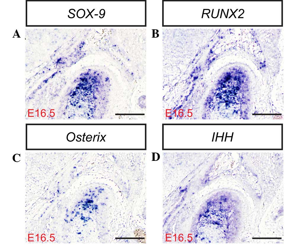
Shibata S, Suda N, Suzuki S, Fukuoka H and
Yamashita Y: An in situ hybridization study of Runx2, Osterix, and
Sox9 at the onset of condylar cartilage formation in fetal mouse
mandible. J Anat. 208:169–177. 2006. View Article : Google Scholar : PubMed/NCBI
|
|
32
|
Ochiai T, Shibukawa Y, Nagayama M, Mundy
C, Yasuda T, Okabe T, Shimono K, Kanyama M, Hasegawa H, Maeda Y, et
al: Indian hedgehog roles in post-natal TMJ development and
organization. J Dent Res. 89:349–354. 2010. View Article : Google Scholar : PubMed/NCBI
|
|
33
|
Wu MJ, Gu ZY and Sun W: Effects of
hydrostatic pressure on cytoskeleton and BMP-2, TGF-beta, SOX-9
production in rat temporomandibular synovial fibroblasts.
Osteoarthritis Cartilage. 16:41–47. 2008. View Article : Google Scholar : PubMed/NCBI
|
|
34
|
Jing J, Hinton RJ, Jing Y, Liu Y, Zhou X
and Feng JQ: Osterix couples chondrogenesis and osteogenesis in
post-natal condylar growth. J Dent Res. 93:1014–1021. 2014.
View Article : Google Scholar : PubMed/NCBI
|
|
35
|
Ishizuka Y, Shibukawa Y, Nagayama M,
Decker R, Kinumatsu T, Saito A, Pacifici M and Koyama E: TMJ
degeneration in SAMP8 mice is accompanied by deranged Ihh
signaling. J Dent Res. 93:281–287. 2014. View Article : Google Scholar : PubMed/NCBI
|
|
36
|
Shibukawa Y, Young B, Wu C, Yamada S, Long
F, Pacifici M and Koyama E: Temporomandibular joint formation and
condyle growth require Indian hedgehog signaling. Dev Dyn.
236:426–434. 2007. View Article : Google Scholar : PubMed/NCBI
|
|
37
|
Suda N, Shibata S, Yamazaki K, Kuroda T,
Senior PV, Beck F and Hammond VE: Parathyroid hormone-related
protein regulates proliferation of condylar hypertrophic
chondrocytes. J Bone Miner Res. 14:1838–1847. 1999. View Article : Google Scholar : PubMed/NCBI
|
|
38
|
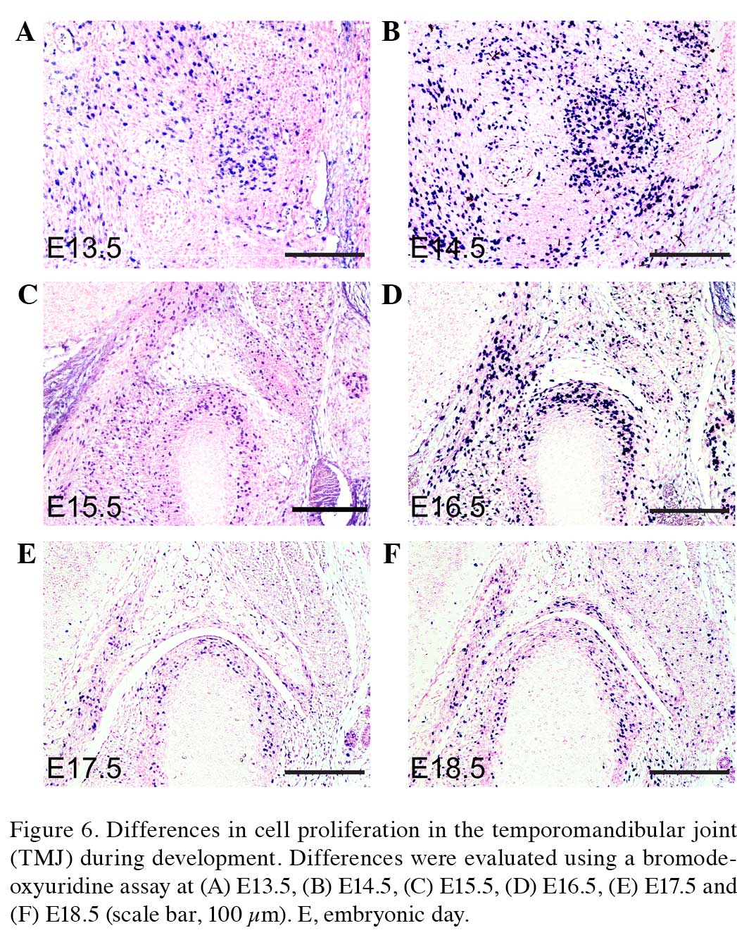
Hume WJ and Thompson J: Double labelling
of cells with tritiated thymidine and bromodeoxyuridine reveals a
circadian rhythm-dependent variation in duration of DNA synthesis
and S phase flux rates in rodent oral epithelium. Cell Tissue
Kinet. 23:313–323. 1990.PubMed/NCBI
|
|
39
|
Herring SW, Decker JD, Liu ZJ and Ma T:
Temporomandibular joint in miniature pigs, Anatomy, cell
replication, and relation to loading. Anat Rec. 266:152–166. 2002.
View Article : Google Scholar : PubMed/NCBI
|
|
40
|
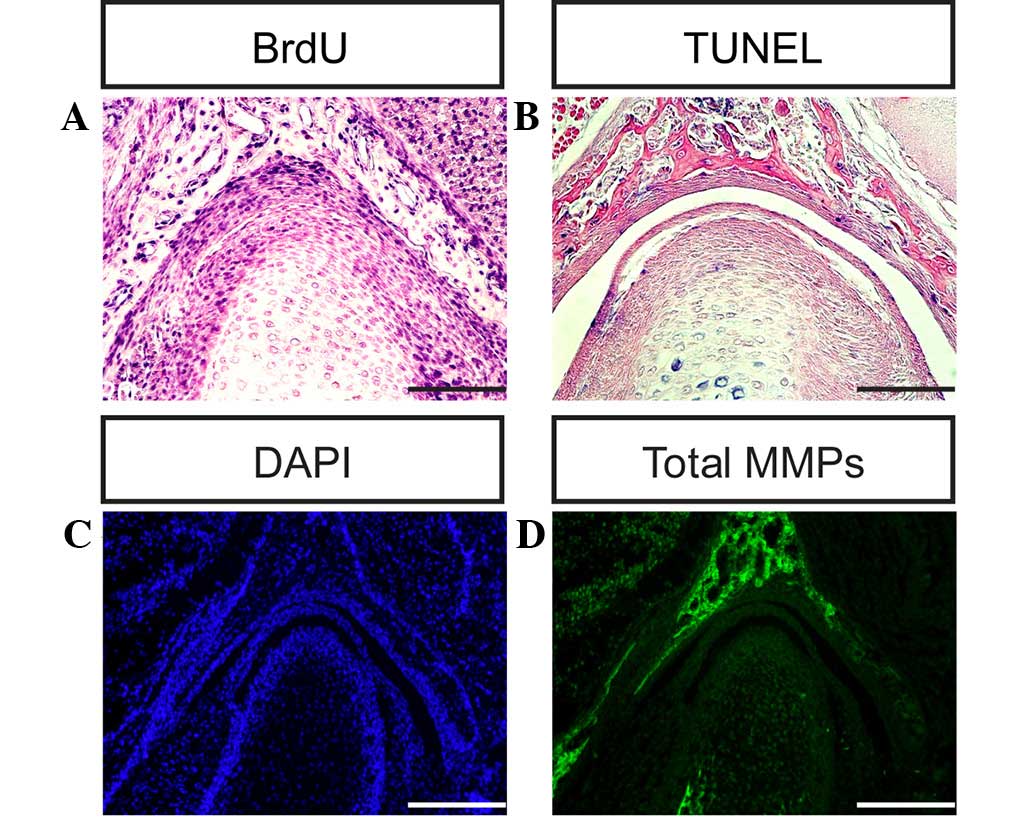
Matsuda S, Mishima K, Yoshimura Y, Hatta T
and Otani H: Apoptosis in the development of the temporomandibular
joint. Anat Embryol (Berl). 196:383–391. 1997. View Article : Google Scholar : PubMed/NCBI
|
|
41
|
Sato I, Uneno R, Miwa Y and Sunohara M:
Distribution of tenascin-C and tenascin-X, apoptotic and
proliferating cells in postnatal soft-diet rat temporomandibular
joint (TMJ). Ann Anat. 188:127–136. 2006. View Article : Google Scholar : PubMed/NCBI
|
|
42
|
Wattanachai T, Yonemitsu I, Kaneko S and
Soma K: Functional lateral shift of the mandible effects on the
expression of ECM in rat temporomandibular cartilage. Angle Orthod.
79:652–659. 2009. View Article : Google Scholar : PubMed/NCBI
|
|
43
|
Gao Y, Liu S, Huang J, Guo W, Chen J,
Zhang L, Zhao B, Peng J, Wang A, Wang Y, Xu W, Lu S, Yuan M and Guo
Q: The ECM-cell interaction of cartilage extracellular matrix on
chondrocytes. Biomed Res Int. 2014:6484592014. View Article : Google Scholar : PubMed/NCBI
|
|
44
|
Okada Y: Matrix-degrading
metalloproteinases and their roles in joint destruction. Mod
Rheumatol. 10:121–128. 2000. View Article : Google Scholar : PubMed/NCBI
|
|
45
|
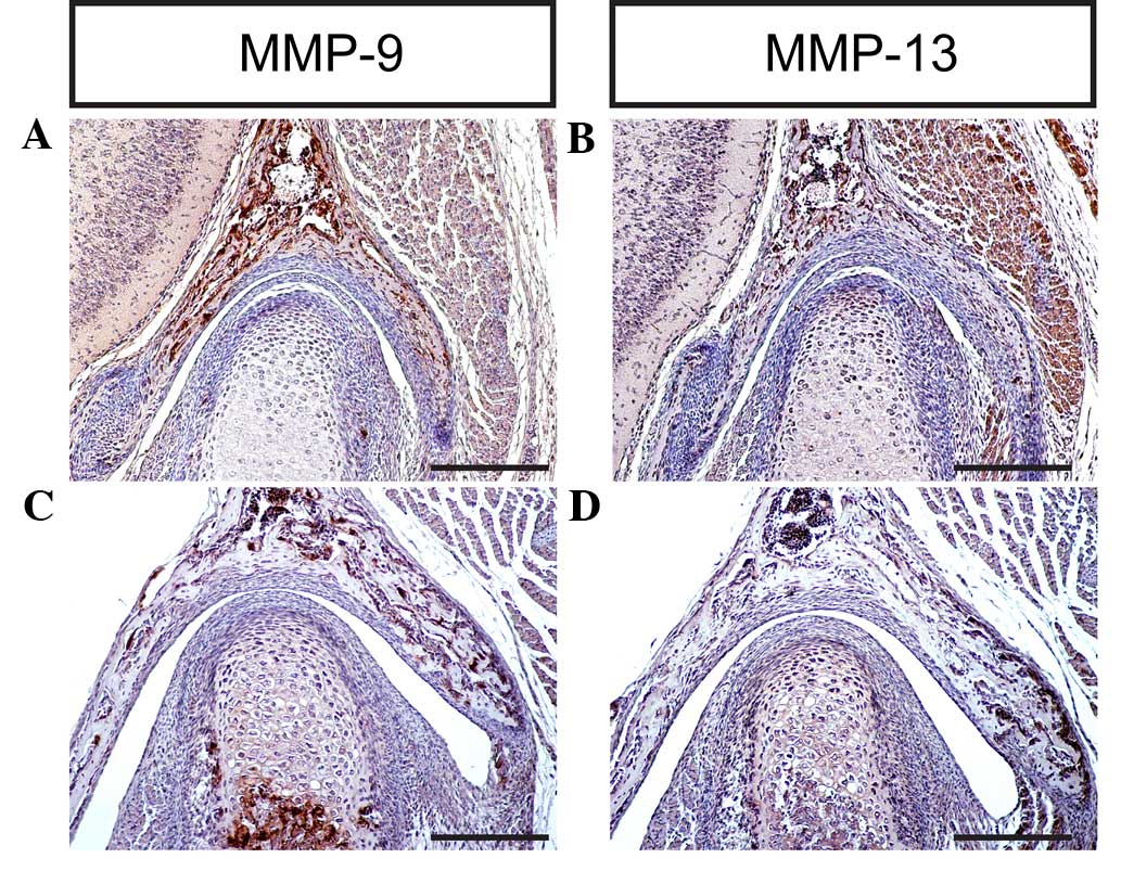
Almeida LE, Caporal K, Ambros V, Azevedo
M, Noronha L, Leonardi R and Trevilatto PC: Immunohistochemical
expression of matrix metalloprotease-2 and matrix metalloprotease-9
in the disks of patients with temporomandibular joint dysfunction.
J Oral Pathol Med. 44:75–79. 2015. View Article : Google Scholar : PubMed/NCBI
|
|
46
|
Malemud CJ: Matrix metalloproteinases: R
ole in skeletal development and growth plate disorders. Front
Biosci. 11:1702–1715. 2006. View
Article : Google Scholar : PubMed/NCBI
|
|
47
|
Burrage PS, Mix KS and Brinckerhoff CE:
Matrix metalloproteinases, Role in arthritis. Front Biosci.
11:529–543. 2006. View
Article : Google Scholar : PubMed/NCBI
|
|
48
|
Stickens D, Behonick DJ, Ortega N, Heyer
B, Hartenstein B, Yu Y, Fosang AJ, Schorpp-Kistner M, Angel P and
Werb Z: Altered endochondral bone development in matrix
metalloproteinase 13-deficient mice. Development. 131:5883–5895.
2004. View Article : Google Scholar : PubMed/NCBI
|
|
49
|
Inada M, Wang Y, Byrne MH, Rahman MU,
Miyaura C, López-Otín C and Krane SM: Critical roles for
collagenase-3 (Mmp13) in development of growth plate cartilage and
in endochondral ossification. Proc Natl Acad Sci USA.
101:17192–17197. 2004. View Article : Google Scholar : PubMed/NCBI
|
|
50
|
Oka K, Oka S, Sasaki T, Ito Y, Bringas P
Jr, Nonaka K and Chai Y: The role of TGF-beta signaling in
regulating chondrogenesis and osteogenesis during mandibular
development. Dev Biol. 303:391–404. 2007. View Article : Google Scholar : PubMed/NCBI
|