Introduction
Bone tissue engineering therapy has emerged as a
promising strategy for the repair of bone defects (1,2), and
consists of three elements: A scaffold, cells and cytokines
(3). For an ideal construct, the
scaffold should be derived from homologous materials, be
biodegradable and provide support for cell retention, and space for
cell proliferation and matrix synthesis (4,5). In
addition, the seed cells should be autologous, differentiate into
osteoblasts, and enhance bone formation and angiogenesis (6,7). The
cytokines must be readily produced, exhibit poor immunogenicity,
and be able to promote cell proliferation and differentiation into
the osteoblast lineage (8,9). Therefore, in order to meet these three
requirements simultaneously, it is important to explore novel
engineered bone materials.
β-Tricalcium phosphate (β-TCP), which is a
well-known bioactive ceramic, has been used extensively as a bone
substitute due to its similarity to the mineral composition of
human bone, excellent biocompatibility and osteoconductivity
(10–13). In addition, β-TCP exhibits a moderate
degradation rate to match the rate of osteogenesis (14,15).
Furthermore, β-TCP is able to support the attachment, proliferation
and differentiation of various seed cells, including osteoblasts,
and adipose- and bone-marrow-derived stem cells (16,17).
Adipose-derived stem cells (ADSCs), which are
recognized as a type of mesenchymal stem cell (18), have a similar morphology,
differentiation capacity, and phenotype to bone marrow stromal
cells (BMSCs) (19). Zuk et
al (20,21) initially reported the existence of a
mesenchymal stromal/stem cell isolated from adipose tissue, and
since then the osteogenic potential of ADSCs in vitro and
in vivo has been demonstrated in numerous studies (19,22,23). As
compared with BMSCs, ADSCs are easier to obtain and can be
harvested in large numbers from small volumes of adipose tissue. In
addition, they are associated with a lower donor site morbidity and
an increased rate of growth (24,25).
ADSCs have a greater potential for application due to their
proliferative capacity and ability to maintain their function.
Furthermore, the bone-forming capacity of ADSCs has been shown to
be greater than that of BMSCs (26,27).
Therefore, ADSCs are considered an excellent resource for bone
tissue engineering.
Platelet-rich plasma (PRP) is an autogenous blood
fraction with high platelet concentrations that contains >300
active molecules, which influence the tissue regeneration process
(28,29). PRP containing various growth factors
has been widely used to enhance bone formation (30,31).
Furthermore, previous studies have demonstrated that PRP combined
with various biomaterials and cell sources exerts positive effects
on bone regeneration (32–35). Therefore, PRP is considered to be
highly beneficial to bone regeneration.
The present study aimed to investigate the
properties of a novel construct for bone regeneration, which was
composed of ADSCs attached to a porous β-TCP scaffold via
biotin-avidin bridging and PRP. Its potential application as a
scaffold for repairing bone defects was evaluated by analyzing the
porosity, compressive strength, in vitro biocompatibility
and in vivo bone-forming capacity in a rabbit
mandibulofacial defect model.
Materials and methods
β-TCP preparation
The β-TCP powder was synthesized by a modified wet
chemical precipitation reaction of 98% calcium nitrate
tetra-hydrate (Ca(NO3)2•4H2O) with 99% diammonium hydrogen
phosphate ((NH4)2HPO4; both Sinopharm Chemical Reagent Co., Ltd.,
Shanghai, China) (36). Briefly,
(NH4)2HPO4 solution was prepared at room temperature and mixed with
Ca(NO3)2•4H2O powder, with agitation. The Ca:P ratio was 1:5. The
pH of the mixture solution was maintained at 6.0–8.0 by adding 25%
NH4OH. The obtained white suspension was stirred for 4 h and then
left to precipitate for 12 h at 35°C. The synthesized precipitate
was centrifuged at 3,000 × g and washed with distilled water, after
which it was dried at 90°C for 24 h. Subsequently, the dried powder
underwent calcination in an alumina crucible at 700°C for 2 h, in
order to generate β-TCP crystals.
The crystalline phase of β-TCP was investigated
using the Ultima IV X-Ray Diffraction (XRD) system (Rigaku, Tokyo,
Japan), using monochromated Cu Kα radiation (λ=1.5405 A°; 20
mA; 40 kV) in a continuous scan mode. The 2θ range was 5–80° at a
scanning speed of 8°/min. XRD data were analyzed using the JADE 6.0
software (Materials Data, Inc., Livermore, CA, USA), and the mean
crystallite size (D) was calculated from the XRD line
broadening measurement using the Scherrer equation (37): D = 0.89 λ/β cosθ, where λ is
the wavelength of the Cu Kα radiation, β is the full width
at the half maximum of the β-TCP line, and θ is the diffraction
angle.
β-TCP crystals were ground in an agate mortar, mixed
with dry KBr powder, and pressed into thin tablets using a manual
hydraulic press for 1 min at 16 MPa (FW-4A; Tianjin Tuopu
Instrument Co., Ltd., Tianjin, China). Infrared spectra were
obtained using Fourier transform infrared spectroscopy (FTIR;
EQUINOX 55; Bruker, Ettlingen, Germany) in the 400–4,000 cm-1
wavenumber range.
Porous scaffold fabrication and
characterization
Porous β-TCP was prepared by mixing β-TCP powder
with ammonium chloride (NH4Cl) pore-forming agent. Briefly,
following drying for 24 h in a dryer, the NH4Cl powder was ground
in an agate mortar and screened by progressively sieving at 200,
150, 100 and 60 µm. Filter materials with an average diameter of
150–200 µm and 60–100 µm were selected as the macroporous and
microporous pore-forming agents in NH4Cl, respectively. β-TCP
powder (1 g), 0.6 g macroporous pore-forming agent and 0.4 g
microporous agent were transferred into a circular mould and
subsequently processed by 1 min compression moulding (20 Mpa), in
order to form a discoid green body (diameter, 10 mm; height, 4 mm).
Upon sintering the green body using the temperature programming
method, a porous β-TCP scaffold was obtained. In the temperature
programming, firstly, the temperature was increased from 20 to
400°C, for 20 min at each 20°C interval, respectively. Then, the
temperature was kept at 400°C for 1 h. Subsequently, the
temperature was increased from 400 to 1,100°C, for 30 min at each
100°C interval, respectively. The temperature was kept at 1,100°C
for 4 h, then decreased to 900°C. The temperature was kept at 900°C
for 2 h, then allowed to decrease to room temperature naturally.
The surface morphology and microstructure of the porous β-TCP
scaffold were analyzed using a light microscope (CKX41; Olympus
Corporation, Tokyo, Japan) and scanning electron microscope (SEM;
Nova NanoSEM 600; FEI Company, Hillsboro, OR, USA).
The porosity of the β-TCP scaffold was measured
using Archimedes' Principle, as described in a previous study
(38). Briefly, a specific gravity
bottle filled with ethanol was weighed (W1), and a scaffold sample
(WS) was immersed in the bottle in order to allow the infiltration
of ethanol into the pores of the scaffold. The air trapped in the
scaffold was evacuated under vacuum, after which the bottle was
filled with ethanol and the weight of the bottle containing ethanol
and scaffold was recorded (W2). The ethanol-saturated scaffold was
then removed from the bottle, and the weight of the bottle with the
residual ethanol recorded (W3). The porosity (P) was calculated
according to the following equation: P = (W2-W3-Ws)/(W1-W3) ×
100%.
The compressive strength of the β-TCP scaffold was
measured at a loading rate of 0.5 mm/min using a universal testing
machine (MB028; Quanzhou MeiBang Instrument Co., Ltd, Quanzhou,
China), according to the method described in ASTM D695-91 (39). The measurements were performed five
times.
Isolation and culture of rabbit
ADSCs
Adult New Zealand rabbits (n=3 per group; age, 4
months; gender, male or female; weight, ~2 kg) were provided by the
Shanghai Medical College of Fudan University (Shanghai, China), and
kept in clean condition (temperature, 16–26°C; humidity, 40–70%;
food and water access, free; one cage per rabbit). These rabbits
were anesthetized with an intraperitoneal injection of 10% chloral
hydrate (3.6 ml/kg; Sigma-Aldrich, St. Louis, MO, USA).
Subsequently, the rabbits were placed in the supine position, and
the inguinal fat pad was carefully dissected and minced using a
sterile razor blade. Following extensive washing with
phosphate-buffered saline (PBS; Gibco; Thermo Fisher Scientific,
Inc., Waltham, MA, USA), the tissue was enzymatically digested
using 0.1% (w/v) collagenase type I (Sigma-Aldrich), for 1 h at
37°C. The digested tissue was filtered through a 200 mesh sieve, in
order to obtain a cell suspension. Following centrifugation at
1,500 × g for 7 min, the pellet was resuspended in culture media
(CM), consisting of Dulbecco's modified Eagle's media (Gibco;
Thermo Fisher Scientific, Inc.), 10% (v/v) fetal bovine serum
(Gibco; Thermo Fisher Scientific, Inc.) and 1% (v/v)
penicillin/streptomycin solution (Sigma-Aldrich). Cell cultures
were maintained at 37°C in a humidified atmosphere containing 5%
CO2. Culture media were changed every 1–2 days. Upon reaching
80–90% cell confluence, the ADSCs were detached using 0.25% trypsin
containing 0.02% ethylenediaminetetraacetic acid (Invitrogen;
Thermo Fisher Scientific, Inc.), and subsequently passaged. All
rabbit care and experimental procedures were conducted in
accordance with the Animal Care Guidelines of Shanghai Medical
College of Fudan University's Animal Ethics Committee.
Surface modifications and in vitro
biocompatibility
The cytotoxicity of β-TCP was investigated by
culturing the ADSCs with various concentrations of β-TCP extracts
and then measuring viability using the Cell Counting kit (CCK)-8
quantitative assay (Dojindo Laboratories Co., Inc., Kumamoto,
Japan). The extracts were prepared by soaking the scaffold in cell
CM containing 5% CO2 for 48 h at 37°C. Following filtration
sterilization, the extracts were diluted with CM in order to
generate serial concentration gradients (100, 50, 10 and 1%; v/v).
ADSCs at passage 4 were detached using 0.25% trypsin and a 100 µl
cell suspension was transferred into 96-well plates at a density of
1×10⁵ cells/ml per well. Following an attachment period of 24 h in
a humidified atmosphere containing 5% CO2 at 37°C, the cells were
cultured in the presence of 100 µl β-TCP extracts with various test
concentrations. Cells cultured in CM in the absence of β-TCP served
as the control. Following a 72 h incubation, the cells were washed
with D-Hanks buffer solution (Gibco; Thermo Fisher Scientific,
Inc.), after which 200 µl CCK-8 solution was added to each well.
Following a 3 h incubation at 37°C, the optical density (OD) at 450
nm was recorded using a microplate reader (RT-6000; Rayto Life and
Analytical Sciences Co., Ltd., Shenzhen, China). Five specimens per
group were tested. The cell viability (% of control) was expressed
as the percentage of (ODtest-ODblank)/(ODcontrol-ODblank), where
ODtest is the OD of the cells exposed to β-TCP extract, ODcontrol
is the OD of the control sample, and ODblank is the OD of the wells
without ADSCs.
ADSCs were biotinylated using a commercially
available reagent (3-sulfo-NHS-biotin; Pierce Biotechnology, Inc.,
Rockford, IL, USA). Briefly, 0.5 mg/ml biotinylation reagent in PBS
was prepared prior to the experiments, and the cells (1×10⁶/ml)
were treated with 2 ml 3-sulfo-NHS-biotin for 30 min in a CO2
incubator. The biotinylated ADSCs (Bio-ADSCs) were visualized by
fluorescence microscopy (DMI4000B; Leica Microsystems GmbH,
Wetzlar, Germany) following incubation with avidin-fluorescein
isothiocyanate (FITC; Sigma-Aldrich) for 5 min. The extent of
biotinylation was determined using flow cytometry (Cytomics FC 500
MPL; Beckman Coulter, Inc., Brea, CA, USA). In order to avidinylate
the β-TCP scaffold (Avi-β-TCP), the sterilized scaffold was soaked
in 2 mg/ml avidinylation reagent (Pierce Biotechnology, Inc.) at
room temperature for 2 h.
Adherence of cells to the β-TCP
scaffold
In order to assess the adherence of the cells to the
β-TCP scaffold, 1×10⁶/ml ADSCs and Bio-ADSCs were seeded onto the
β-TCP and Avi-β-TCP scaffolds, respectively. Following 10 and 30
min, and 1, 12 and 24 h of incubation at 37°C, 100% humidity and 5%
CO2, the cells were collected by trypsinization and were
centrifuged at 1,000 × g, after which they were washed twice with
PBS. Subsequently, 10 µl suspension was placed into a hemocytometer
in order to measure cell adhesion under a light microscope (Olympus
Corporation, Tokyo, Japan). In addition, cellular morphology and
attachment were examined by direct visualization under a SEM.
Briefly, following a 7 day incubation (media was changed every 2
days), the cell-scaffold constructs were removed, washed twice with
PBS and fixed with 2% glutaraldehyde solution for 30 min at 4°C.
Subsequently, the constructs were rinsed three times with PBS,
followed by 45 min of fixation with 1% osmium tetroxide at 4°C.
After rinsing three times with PBS, the fixed constructs were
dehydrated and CO2-dried in a desiccator overnight. The samples
were sputtered with gold for SEM observation.
Preparation of PRP
Whole blood (~40 ml) was drawn from the heart of a
donor rabbit into an anticoagulant, which contained anticoagulant
citrate dextrose solution formula A. PRP was obtained via a
two-step centrifugation procedure (40). Briefly, whole blood was poured into a
50 ml tube and initially centrifuged at 2,400 × g for 10 min, in
order to separate the platelet layer from the plasma and red blood
cells. Subsequently, the upper plasma layer and the middle platelet
layer were collected and centrifuged again at 3,500 × g for 15 min.
The collected precipitated platelets-containing part of the plasma
formed the PRP.
Bone regeneration in a rabbit
mandibulofacial defect model
Following anesthetization of the rabbits with 10%
chloral hydrate (3.6 ml/kg), surgical procedures were conducted
under sterile conditions. The surgical field of the mandible in
each rabbit was shaved and prepared with betadine solution. The
mandibular angle and body were exposed via a submental midline skin
incision. A full-thickness bone defect (15×5×4 mm) was created at
the mid-portion of the mandible using a reciprocating saw, which
was cooled continuously by saline irrigation. The in vivo
experiments were divided into two groups (n=3 rabbits/group): Group
1 (Bio-ADSCs/Avi-β-TCP), in which the defect was filled with the
construct consisting of Bio-ADSCs seeded onto the Avi-β-TCP
scaffold; and group 2 (Bio-ADSCs/Avi-β-TCP/PRP), in which the
defect was filled with the construct consisting of the Bio-ADSCs
attached to the Avi-β-TCP scaffold and PRP. The constructs were
prepared by incubating ~1×10⁵ Bio-ADSCs with the Avi-β-TCP scaffold
for 24 h at 37°C in a humidified atmosphere containing 5% CO2. PRP
solution (200 µl) was added to the constructs prior to
implantation. All wounds were routinely sutured and penicillin
(25,000 U/kg) was subcutaneously injected into the rabbits for 3
days post-operation. All rabbits were maintained on a soft diet
throughout the study.
Computerized tomography (CT) and
histological analyses
Maxillofacial CT images were obtained under general
anesthesia and the three-dimensional images were reconstructed
using a CT at 4 weeks post-operation (CBCT-NewTom DVT 9000; QR
S.r.l., Verona, Italy).
For histological analysis, all rabbits were
sacrificed by air injection into the marginal ear vein, and bone
specimens were harvested from each group 4 weeks post-implantation.
The constructs Bio-ADSCs/Avi-β-TCP and Bio-ADSCs/Avi-β-TCP/PRP that
filled the original bone defect area were divided into two parts:
Part 1 was used to obtain paraffin-embedded sections for
hematoxylin and eosin (H&E) staining and immunohistochemistry;
part 2 was used to generate liquid nitrogen frozen sections for
PCR. Part 1 samples were fixed in 10% neutral buffered formalin and
embedded in paraffin. Serial tissue sections (5 µm) were obtained
for hematoxylin and eosin (H&E) staining and
immunohistochemical analysis. For immunohistochemical analysis, the
deparaffinized sections were washed three times in PBS, and
endogenous peroxidase was inactivated by incubation with 3% H2O2.
Nonspecific binding sites were blocked using normal goat serum
(Sigma-Aldrich) for 30 min, after which the tissue sections were
incubated with mouse anti-osteocalcin (OCN) antibody (1:200;
ab13420; Abcam, Cambridge, UK), overnight at 4°C. After rinsing in
PBS three times, the tissue sections were incubated with
biotinylated secondary antibody (1:500; Z0420; DAKO, Glostrup,
Denmark) at room temperature for 1 h. Peroxidase activity was
visualized using 0.05% diaminobenzidine and 0.03% H2O2 in PBS.
Subsequently, the tissue sections were counter-stained with
hematoxylin for 2 min. At least 10 fields of vision under ×200
magnification were captured under brightfield view and images were
subsequently analyzed and quantified using ImageJ software, version
1.47 (National Institutes of Health, Bethesda, MA, USA) (41).
Analysis of mRNA expression levels of
osteogenic marker genes by reverse transcription-quantitative
polymerase chain reaction (RT-qPCR)
Prior to RNA extraction, specimens were homogenized
under liquid nitrogen. Total RNA was isolated from each specimen
using TRIzol® reagent (Invitrogen; Thermo Fisher Scientific, Inc.)
and reverse-transcribed into cDNA using a commercial reverse
transcriptase cDNA synthesis kit (Takara Biotechnology Co., Ltd.
Dalian, China), according to the manufacturer's protocol. The
relative expression levels of the specific osteogenic marker genes
OCN and alkaline phosphatase (ALP) were investigated. The primer
sequences, which were designed using the Primer Premier 5.0
software (Premier Biosoft International, Palo Alto, CA, USA), were
as follows: OCN forward, 5′-GACACCATGAGGACCCTCTC-3′, reverse
3′-GGCTCCAGGGGATCTGGG-5′; and ALP forward,
5′-CAGGATTGACCACGGGCACC-3′, reverse 3′-GCCTGGTAGTTGTTGTGAGC-5′;
GAPDH forward, 5′-TCGTCCTCCTCTGGTGCTCT-3′, reverse,
5′-CCACTTTGTGAAGCTCATTTCCT-3′. RT-qPCR was performed under the
following cycling conditions using a CFX96 Real-Time System
(Bio-Rad Laboratories, Inc., Hercules, CA, USA): 95°C for 30 sec,
followed by 40 cycles at 95°C for 5 sec, 60°C for 20 sec, 72°C for
30 sec, and 60–90°C for 10 sec. A 1X TaqMan Universal PCR Master
Mix was used throughout (Applied Biosystems; Thermo Fisher
Scientific, Inc., Foster City, CA, USA). The
glyceraldehyde-3-phosphate dehydrogenase gene served as a
normalization control. Relative expression levels were evaluated
using the 2-ΔΔCq method (42). The
expression levels of each target gene were analyzed in
triplicate.
Statistical analysis
All statistical analyses were performed using SPSS
software, version 19.0 (IBM SPSS, Armonk, NY, USA). Data are
presented as the mean ± standard deviation. One-way analysis of
variance was applied for comparisons between groups. Differences
between two groups were assessed using Tukey's post-hoc test.
P<0.05 was considered to indicate a statistically significant
difference.
Results
Characterization of the β-TCP
scaffold
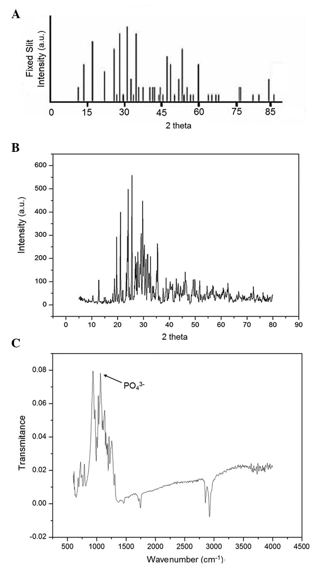
The XRD pattern and FTIR spectrum of the β-TCP
powder are presented in Fig. 1. The
powder exhibited sharp diffraction peaks and had a similar XRD
pattern to the standard pattern for β-TCP (43), indicative of a high crystallinity
(Fig. 1A and B). The mean
crystallite size of the particles was 31.5 nm. In the FTIR spectrum
of the powder, characteristic peaks of β-TCP were detected at 551,
609, 944 and 1,043 cm-1, and the characteristic absorption bands at
1,200 and 1,000 cm-1 were assigned to components of the PO4 group
(Fig. 1C).
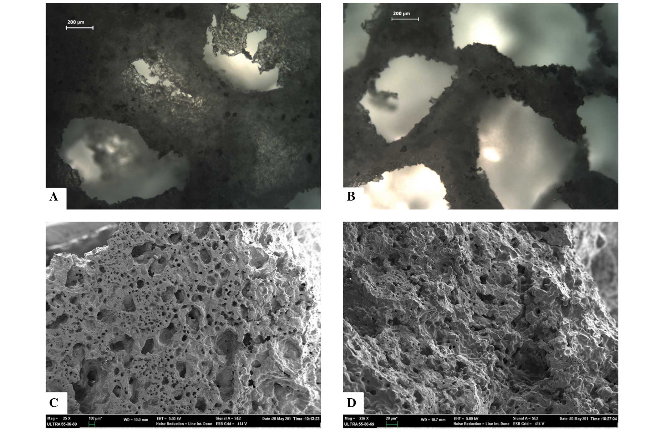
The β-TCP powder could be sintered together
effectively, and formed a porous structure containing
inter-connected pores, as observed under the CKX41 light microscope
(Fig. 2A and B). The SEM micrograph
suggested that the porous β-TCP scaffold had high pore
interconnectivity, abundant pore structures, including macropores
(diameter, 200–300 µm) and micropores (size, 60–100 µm), and a
large surface area (Fig. 2C and D).
In addition, the porosity and compressive strength of the scaffold
were 71.26±0.28% and 7.93±0.06 MPa, respectively.
Viability, attachment and morphology
of the rabbit ADSCs following incubation with the β-TCP
scaffold
Alterations to the viability of the ADSCs incubated
with various concentrations of the β-TCP extraction fluid were
assessed using the CCK-8 assay (Table
I). Following 72 h of incubation, 100% ADSCs had retained their
viability, suggesting that no significant loss in viability was
observed, as compared with the control. At the highest extract
concentration (100%, v/v) the cell viability was 107.48±10.32% of
the control. These results suggest that the β-TCP scaffold does not
exert toxic effects on ADSCs.
 | Table I.Viability of rabbit adipose-derived
stem cells treated with various concentrations of β-tricalcium
phosphate extraction fluid. |
Table I.
Viability of rabbit adipose-derived
stem cells treated with various concentrations of β-tricalcium
phosphate extraction fluid.
| Extract
concentration (v/v; %) | Cell viability (%
of control) |
|---|
| 100 | 107.48±10.32 |
| 50 | 105.63±9.85 |
| 10 | 108.51±12.06 |
|
1 | 109.04±8.17 |
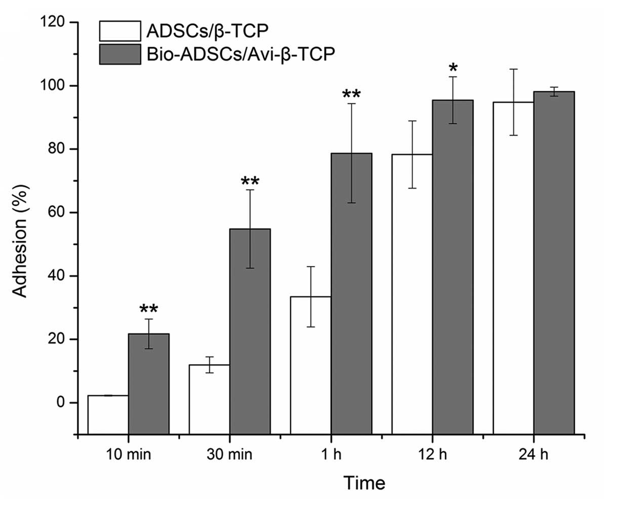
Bio-ADSCs attach to Avi-β-TCP
Following biotinylation, ADSCs were incubated with
avidin-FITC. All Bio-ADSCs were green in appearance under a
fluorescence microscope, whereas the untreated control cells did
not exhibit fluorescence (images not shown), thus suggesting that
the biotinylation procedure was successful. The degree of
biotinylation was calculated to be 95% of cells using fluorescence
correlation microscopy. Subsequently, the Bio-ADSCs and ADSCs were
incubated with the Avi-β-TCP and β-TCP scaffolds, respectively, in
order to evaluate cell adhesion (Fig.
3). In the first hour of incubation, the biotin-avidin
interaction significantly enhanced the rate of cell attachment, as
compared with the ADSCs/β-TCP mixture (P<0.01). After 12 h,
95.46±7.38% of Bio-ADSCs were attached to the Avi-β-TCP scaffold,
which was significantly increased, as compared with the ADSCs/β-TCP
group (78.29±10.63%; P<0.05). However, there was no significant
difference in adhesion between the two groups after 24 h of
incubation (P>0.05; Fig. 3).
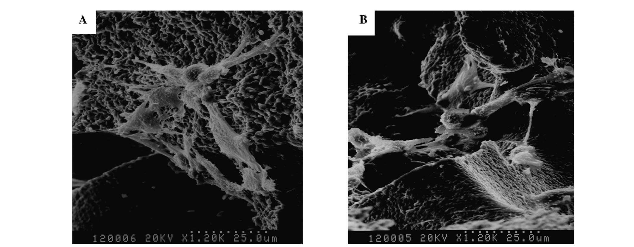
Morphology of ADSCs/β-TCP and
Bio-ADSCs/Avi-β-TCP constructs under SEM exhibits no visible
difference
All cells were attached and exhibited a
morphologically normal appearance on the surface of the scaffolds
following incubation for 1 day. A large number of ADSCs were firmly
attached to the surface and around the pores, and a few cells were
enclosed within the three-dimensional scaffolds following 4 days of
incubation. At 7 days, pseudopodia extending from the cells were
connected with each other and covered the pores. In addition, the
cells were embedded in the interspaces of the scaffolds and the
secretion of extracellular matrix (ECM) (Fig. 4A and B).
CT analysis
In order to analyze the three-dimensional structure
of the repaired mandibles, 3D-CT images were captured 4 weeks
post-operation. In group 1 rabbits, which received the
Bio-ADSCs/Avi-β-TCP construct, only a poor bony union was achieved
and the graft appeared to slide out of its groove (Fig. 5A). Conversely, in the group 2
rabbits, which received the Bio-ADSCs/Avi-β-TCP/PRP construct, a
good bony union was achieved and the mandible shape resembled that
of normal edentulous mandibles (Fig.
5B).
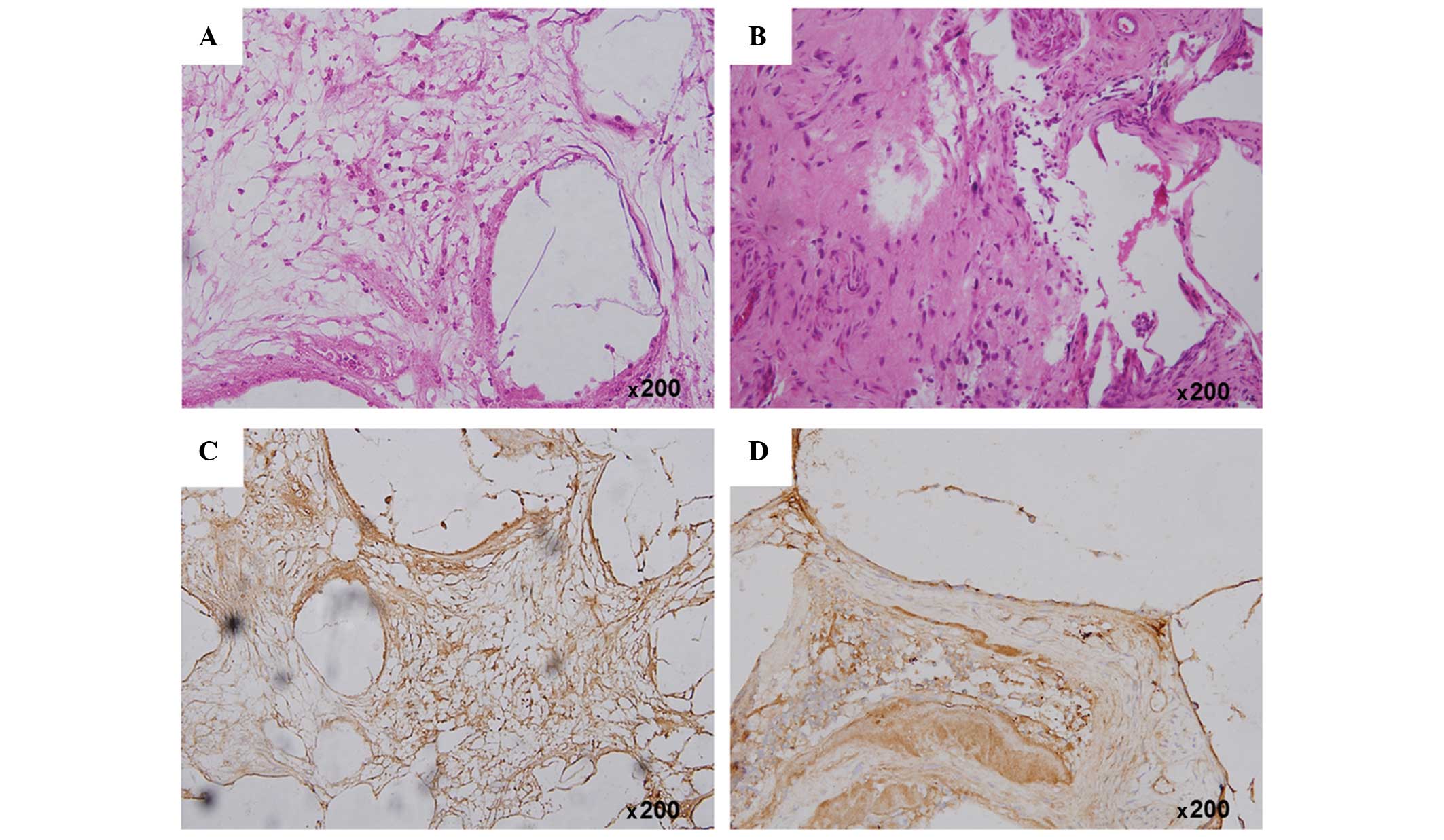
Histological analysis
Following implantation for 4 weeks, H&E staining
detected neogenetic bone-like structure within the area of the
Bio-ADSCs/Avi-β-TCP implant in group 1 rabbits (Fig. 6A). Conversely, in group 2 rabbits,
the amount of newly formed bone was markedly increased and was
shown to have integrated to form matured interwoven bone and
trabecular bone (Fig. 6B).
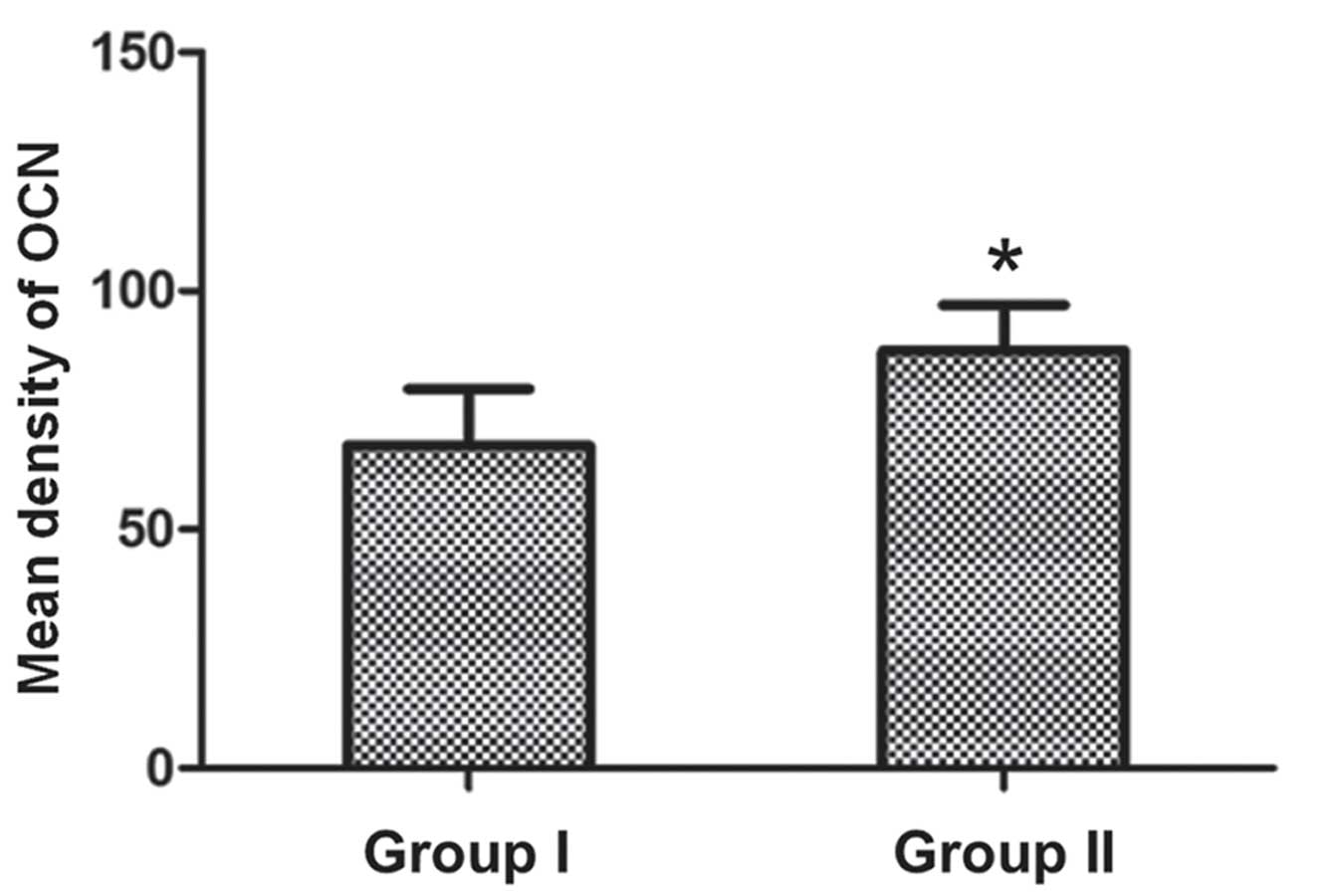
Immunohistochemical analysis of OCN protein
expression detected new bone formation within the
positively-stained area (brown color), and OCN protein expression
was markedly upregulated in the defect area at 4 weeks (Fig. 6C and D). In addition, the
quantitative analysis suggested that the OCN protein expression was
increased in the group 2 rabbits, as compared with the group 1
rabbits (87.41±9.65 vs. 67.53±11.87; P<0.05; Fig. 7).
Analysis of gene expression
The mRNA expression levels of the osteospecific
genes OCN and ALP in the tissues surrounding the two graft
constructs were analyzed at 4 weeks post-implantation. RT-qPCR
demonstrated that the relative expression levels of OCN and ALP
were significantly increased in group 2 rabbits, as compared with
group 1 rabbits (OCN: 26.38±7.17 vs. 17.49±3.52; ALP: 49.62±8.34
vs. 15.12±4.21; P<0.01; Fig. 8A and
B).
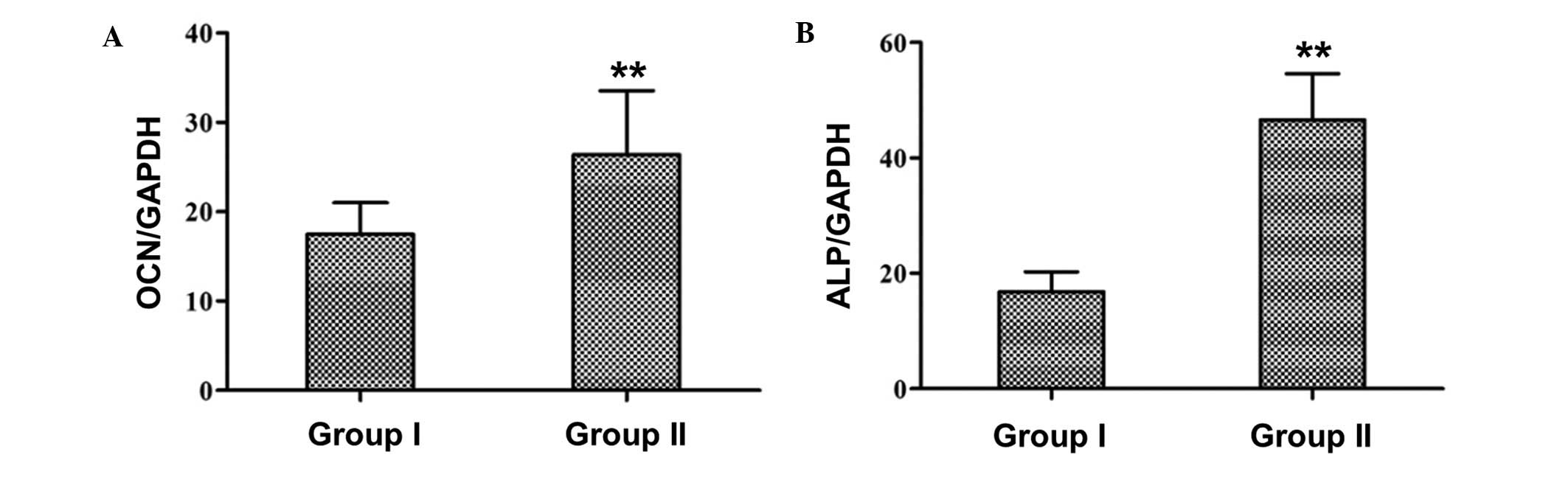 | Figure 8.Relative mRNA expression levels of
(A) OCN and (B) ALP in the group 1 and group 2 rabbits were
quantified using reverse transcription-quantitative polymerase
chain reaction. Expression levels were normalized to the
housekeeping gene GAPDH. Group 1 and group 2 rabbits received the
Bio-ADSCs/Avi-β-TCP and Bio-ADSCs/Avi-β-TCP/PRP constructs,
respectively. Data are presented as the mean ± standard deviation.
**P<0.01, vs. group 1 rabbits. OCN, osteocalcin; ALP, alkaline
phosphatase; ADSCs, adipose-derived stem cells; β-TCP, β-tricalcium
phosphate; Bio-ADSCs, biotinylated-ADSCs; Avi-β-TCP, avidin-coated
β-TCP; PRP, platelet-rich plasma; GAPDH, glyceraldehyde-3-phosphate
dehydrogenase. |
Discussion
Bone tissue engineering has been widely studied and
typically involves combining scaffolds with various growth factors
and cell sources (22). Searching
for an optimal scaffold for bone regeneration remains an unsolved
and interesting challenge for researchers. In the present study,
the porous β-TCP scaffold exhibited a high porosity, suitable pore
size and good mechanical strength. The high-affinity avidin-biotin
binding system increased the initial attachment of ADSCs to β-TCP.
In addition, the use of PRP significantly improved the quality of
bone healing in a rabbit mandibulofacial defect model. These
results suggested that the Bio-ADSCs/Avi-β-TCP/PRP composite may
have potential application in bone tissue engineering.
Various material properties, including composition,
geometry, porosity, size, and microstructure, are important
parameters to consider when evaluating appropriate bone formation
(44). Previous studies have
demonstrated that a high porosity is critical for bone
regeneration, as it allows sufficient void space for the ingrowth
of surrounding bone and vascularized tissue (27,45,46).
Furthermore, the osteogenic capability of composites has been shown
to increase with increasing scaffold porosity (45). In addition, small pores may induce
osteochondral formation prior to osteogenesis due to relative
hypoxic conditions, whereas large pores may lead to direct
osteogenesis by simultaneous vascularization due to a high oxygen
content (47). Therefore, the pore
size of the scaffold is an important parameter for bone tissue
engineering. A previous study reported that the minimal pore size
for bone regeneration was ~100 µm (48). In the present study, the β-TCP
scaffold had a high porosity (71.26%) and was able to provide a
suitable pore size, including 200–300 µm macropores and 60–100 µm
micropores, for bone regeneration, in accordance with the results
from previous studies (17,49).
The ability of seed cells to adhere to the
biomaterial is a prerequisite and crucial step in various tissue
engineering applications (50,51).
Numerous strategies have been applied in order to improve
affinities between cells and biomaterial surfaces, including ECM
adhesion proteins (52), conjugating
peptides containing cell-binding sequences (53), and antibodies targeting cell-membrane
antigens (54). In addition,
previous studies have suggested that the utilization of an
avidin-biotin binding system may enhance cell adhesion to
biomaterial surfaces (51,55). In the present study, biotinylated
ADSCs adhered to the avidin-coated β-TCP scaffold at a faster rate,
as compared with untreated cells. Furthermore, the binding system
markedly enhanced the initial cell adhesion in the first hour, as
previously reported (51,56). These results suggested that the
avidin-biotin system may be applied to bone tissue engineering.
Previous studies have demonstrated that PRP, which
is a platelet concentrate of autogenous blood, has an important
role in tissue repair mechanisms and may exert potential positive
effects in the course of bone formation (57–60). In
addition, PRP may improve osteochondral healing (61) and increase new bone formation
(57). Furthermore, PRP has
demonstrated success when applied to orthopedic and dental bone
reconstruction surgeries (62). The
present study demonstrated that the Bio-ADSCs/Avi-β-TCP graft
loaded with PRP exhibited enhanced bone regeneration and healing
properties, as compared with the scaffold untreated by PRP, based
on radiological, histological and gene expression analyses. These
results suggested that the addition of PRP may significantly
enhance new bone formation and improve the quality of bone
healing.
In conclusion, the fabricated porous β-TCP scaffold
had a high porosity and suitable pore size, and the utilization of
an avidin-biotin binding system increased the adherence of
Bio-ADSCs to the Avi-β-TCP scaffold. Furthermore, the addition of
PRP increased the rate of new bone formation and improved the
quality of bone healing. These results suggested that the novel
Bio-ADSCs/Avi-β-TCP/PRP composite has potential application in bone
repair and bone tissue engineering.
References
|
1
|
Damaraju S and Duncan NA: Stem cell-based
tissue engineering for bone repair. Tissue Engineering. Fernandes
PR and Bartolo PJ: 31:(Dordrecht, The Netherlands). Springer. 1–30.
2014. View Article : Google Scholar
|
|
2
|
Liu Y, Zhou Y, Feng H, Ma GE and Ni Y:
Injectable tissue-engineered bone composed of human adipose-derived
stromal cells and platelet-rich plasma. Biomaterials. 29:3338–3345.
2008. View Article : Google Scholar : PubMed/NCBI
|
|
3
|
Buser Z, Liu J, Thorne KJ, Coughlin D and
Lotz JC: Inflammatory response of intervertebral disc cells is
reduced by fibrin sealant scaffold in vitro. J Tissue Eng Regen
Med. 8:77–84. 2014. View Article : Google Scholar : PubMed/NCBI
|
|
4
|
Ravichandran R, Venugopal JR, Sundarrajan
S, Mukherjee S and Ramakrishna S: Precipitation of
nanohydroxyapatite on PLLA/PBLG/Collagen nanofibrous structures for
the differentiation of adipose derived stem cells to osteogenic
lineage. Biomaterials. 33:846–855. 2012. View Article : Google Scholar : PubMed/NCBI
|
|
5
|
Chang NJ, Lam CF, Lin CC, Chen WL, Li CF,
Lin YT and Yeh ML: Transplantation of autologous endothelial
progenitor cells in porous PLGA scaffolds create a microenvironment
for the regeneration of hyaline cartilage in rabbits.
Osteoarthritis Cartilage. 21:1613–1622. 2013. View Article : Google Scholar : PubMed/NCBI
|
|
6
|
Szpalski C, Barbaro M, Sagebin F and
Warren SM: Bone tissue engineering: Current strategies and
techniques - part. II Cell types. Tissue Eng Part B Rev.
18:258–269. 2012. View Article : Google Scholar : PubMed/NCBI
|
|
7
|
Khaled EG, Saleh M, Hindocha S, Griffin M
and Khan WS: Tissue engineering for bone production-stem cells,
gene therapy and scaffolds. Open Orthop J. 5(Suppl 2): S289–S295.
2011. View Article : Google Scholar
|
|
8
|
Cha C, Liechty WB, Khademhosseini A and
Peppas NA: Designing biomaterials to direct stem cell fate. ACS
Nano. 6:9353–9358. 2012. View Article : Google Scholar : PubMed/NCBI
|
|
9
|
Mosna F, Sensebé L and Krampera M: Human
bone marrow and adipose tissue mesenchymal stem cells: A user's
guide. Stem Cells Dev. 19:1449–1470. 2010. View Article : Google Scholar : PubMed/NCBI
|
|
10
|
Farzadi A, Solati-Hashjin M, Bakhshi F and
Aminian A: Synthesis and characterization of
hydroxyapatite/β-tricalcium phosphate nanocomposites using
microwave irradiation. Ceram Int. 37:65–71. 2011. View Article : Google Scholar
|
|
11
|
Kolk A, Handschel J, Drescher W, Rothamel
D, Kloss F, Blessmann M, Heiland M, Wolff KD and Smeets R: Current
trends and future perspectives of bone substitute materials - from
space holders to innovative biomaterials. J Craniomaxillofac Surg.
40:706–718. 2012. View Article : Google Scholar : PubMed/NCBI
|
|
12
|
Miyaji H, Yokoyama H, Kosen Y, Nishimura
H, Nakane K, Tanaka S, Otani K, Inoue K, Ibara A, Kanayama I, et
al: Bone augmentation in rat by highly porous β-TCP scaffolds with
different open-cell sizes in combination with fibroblast growth
factor-2. J Oral Tissue Engin. 10:172–181. 2013.
|
|
13
|
Zheng H, Bai Y, Shih MS, Hoffmann C,
Peters F, Waldner C and Hübner WD: Effect of a β-TCP collagen
composite bone substitute on healing of drilled bone voids in the
distal femoral condyle of rabbits. J Biomed Mater Res B Appl
Biomater. 102:376–383. 2014. View Article : Google Scholar : PubMed/NCBI
|
|
14
|
Rezwan K, Chen QZ, Blaker JJ and
Boccaccini AR: Biodegradable and bioactive porous polymer/inorganic
composite scaffolds for bone tissue engineering. Biomaterials.
27:3413–3431. 2006. View Article : Google Scholar : PubMed/NCBI
|
|
15
|
Choi D and Kumta PN: Mechano-chemical
synthesis and characterization of nanostructured β-TCP powder.
Materials Science and Engineering: C. 27:377–381. 2007. View Article : Google Scholar
|
|
16
|
Griffin M, Iqbal S and Bayat A: Exploring
the application of mesenchymal stem cells in bone repair and
regeneration. J Bone Joint Surg Br. 93:427–434. 2011. View Article : Google Scholar : PubMed/NCBI
|
|
17
|
Cao H and Kuboyama N: A biodegradable
porous composite scaffold of PGA/beta-TCP for bone tissue
engineering. Bone. 46:386–395. 2010. View Article : Google Scholar : PubMed/NCBI
|
|
18
|
Guasti L, Prasongchean W, Kleftouris G,
Mukherjee S, Thrasher AJ, Bulstrode NW and Ferretti P: High
plasticity of pediatric adipose tissue-derived stem cells: Too much
for selective skeletogenic differentiation? Stem Cells Transl Med.
1:384–395. 2012. View Article : Google Scholar : PubMed/NCBI
|
|
19
|
Levi B and Longaker MT: Concise review:
Adipose-derived stromal cells for skeletal regenerative medicine.
Stem Cells. 29:576–582. 2011. View
Article : Google Scholar : PubMed/NCBI
|
|
20
|
Zuk PA, Zhu M, Mizuno H, Huang J, Futrell
JW, Katz AJ, Benhaim P, Lorenz HP and Hedrick MH: Multilineage
cells from human adipose tissue: Implications for cell-based
therapies. Tissue Eng. 7:211–228. 2001. View Article : Google Scholar : PubMed/NCBI
|
|
21
|
Zuk PA, Zhu M, Ashjian P, De Ugarte DA,
Huang JI, Mizuno H, Alfonso ZC, Fraser JK, Benhaim P and Hedrick
MH: Human adipose tissue is a source of multipotent stem cells. Mol
Biol Cell. 13:4279–4295. 2002. View Article : Google Scholar : PubMed/NCBI
|
|
22
|
Liao HT, Chen JP and Lee MY: Bone tissue
engineering with adipose-derived stem cells in bioactive composites
of laser-sintered porous polycaprolactone scaffolds and
platelet-rich plasma. Materials. 6:4911–4929. 2013. View Article : Google Scholar
|
|
23
|
Fang X, Murakami H, Demura S, Hayashi K,
Matsubara H, Kato S, Yoshioka K, Inoue K, Ota T, Shinmura K and
Tsuchiya H: A novel method to apply osteogenic potential of adipose
derived stem cells in orthopaedic surgery. PloS One. 9:e888742014.
View Article : Google Scholar : PubMed/NCBI
|
|
24
|
Ahn HH, Kim KS, Lee JH, Lee JY, Kim BS,
Lee IW, Chun HJ, Kim JH, Lee HB and Kim MS: In vivo osteogenic
differentiation of human adipose-derived stem cells in an
injectable in situ-forming gel scaffold. Tissue Eng Part A.
15:1821–1832. 2009. View Article : Google Scholar : PubMed/NCBI
|
|
25
|
Grottkau BE and Lin Y: Osteogenesis of
adipose-derived stem cells. Bone Res. 1:133–145. 2013. View Article : Google Scholar : PubMed/NCBI
|
|
26
|
Rehman J, Traktuev D, Li J, Merfeld-Clauss
S, Temm-Grove CJ, Bovenkerk JE, Pell CL, Johnstone BH, Considine RV
and March KL: Secretion of angiogenic and antiapoptotic factors by
human adipose stromal cells. Circulation. 109:1292–1298. 2004.
View Article : Google Scholar : PubMed/NCBI
|
|
27
|
Liao HT and Chen CT: Osteogenic potential:
Comparison between bone marrow and adipose-derived mesenchymal stem
cells. World J Stem Cells. 6:288–295. 2014. View Article : Google Scholar : PubMed/NCBI
|
|
28
|
Nurden AT: Platelets, inflammation and
tissue regeneration. Thromb Haemost. 105(Suppl 1): S13–S33. 2011.
View Article : Google Scholar : PubMed/NCBI
|
|
29
|
Nurden AT, Nurden P, Sanchez M, Andia I
and Anitua E: Platelets and wound healing. Front Biosci.
13:3532–3548. 2008.
|
|
30
|
Marx RE: Platelet-rich plasma: Evidence to
support its use. J Oral Maxillofac Surg. 62:489–496. 2004.
View Article : Google Scholar : PubMed/NCBI
|
|
31
|
Marx RE, Carlson ER, Eichstaedt RM,
Schimmele SR, Strauss JE and Georgeff KR: Platelet-rich plasma:
Growth factor enhancement for bone grafts. Oral Surg Oral Med Oral
Pathol Oral Radiol Endod. 85:638–646. 1998. View Article : Google Scholar : PubMed/NCBI
|
|
32
|
Niemeyer P, Fechner K, Milz S, Richter W,
Suedkamp NP, Mehlhorn AT, Pearce S and Kasten P: Comparison of
mesenchymal stem cells from bone marrow and adipose tissue for bone
regeneration in a critical size defect of the sheep tibia and the
influence of platelet-rich plasma. Biomaterials. 31:3572–3579.
2010. View Article : Google Scholar : PubMed/NCBI
|
|
33
|
Mazzocca AD, McCarthy MB, Chowaniec DM,
Dugdale EM, Hansen D, Cote MP, Bradley JP, Romeo AA, Arciero RA and
Beitzel K: The positive effects of different platelet-rich plasma
methods on human muscle, bone and, tendon cells. Am J Sports Med.
40:1742–1749. 2012. View Article : Google Scholar : PubMed/NCBI
|
|
34
|
El Backly RM, Zaky SH, Muraglia A,
Tonachini L, Brun F, Canciani B, Chiapale D, Santolini F, Cancedda
R and Mastrogiacomo M: A platelet-rich plasma-based membrane as a
periosteal substitute with enhanced osteogenic and angiogenic
properties: properties = A platelet-rich plasma-based membrane as a
periosteal substitute with enhanced osteogenic and angiogenic
properties: A new concept for bone repair. Tissue Eng Part A.
19:152–165. 2013. View Article : Google Scholar : PubMed/NCBI
|
|
35
|
Wang C, Zhong D, Zhou X, Yin K, Liao Q,
Kong L and Liu A: Preparation of a new composite combining
strengthened β-tricalcium phosphate with platelet-rich plasma as a
potential scaffold for the repair of bone defects. Exp Ther Med.
8:1081–1086. 2014.PubMed/NCBI
|
|
36
|
Mirhadi B, Mehdikhani B and Askari N:
Synthesis of nano-sized β-tricalcium phosphate via wet
precipitation. Processing and Application of Ceramics. 5:193–198.
2011. View Article : Google Scholar
|
|
37
|
Monshi A, Foroughi MR and Monshi MR:
Modified Scherrer equation to estimate more accurately
nano-crystallite size using XRD. World J Nano Sci Eng. 2:1542012.
View Article : Google Scholar
|
|
38
|
Tran RT, Thevenot P, Zhang Y, Gyawali D,
Tang L and Yang J: Scaffold sheet design strategy for soft tissue
engineering. Nat Mater. 3:1375–1389. 2010. View Article : Google Scholar : PubMed/NCBI
|
|
39
|
American Society of the International
Association for Testing and Materials (ASTM) International: ASTM
Standard D695-91: Standard test method for compressive properties
of rigid plastics. Annual Book of ASTM Standards (8th). (West
Conshohocken, PA). ASTM International. 204–210. 1993.
|
|
40
|
Jeong SM, Lee CU, Son JS, Oh JH, Fang Y
and Choi BH: Simultaneous sinus lift and implantation using
platelet-rich fibrin as sole grafting material. J Craniomaxillofac
Surg. 42:990–994. 2014. View Article : Google Scholar : PubMed/NCBI
|
|
41
|
Schneider CA, Rasband WS and Eliceiri KW:
NIH Image to ImageJ: 25 years of image analysis. Nat Methods.
9:671–675. 2012. View Article : Google Scholar : PubMed/NCBI
|
|
42
|
Schmittgen TD and Livak KJ: Analyzing
real-time PCR data by the comparative C(T) method. Nat Protoc.
3:1101–1108. 2008. View Article : Google Scholar : PubMed/NCBI
|
|
43
|
Frondel C: Mineralogy of the calcium
phosphates in insular phosphate rock. Am Mineral. 28:215–232.
1943.
|
|
44
|
Yuan H, Yang Z, De Bruij JD, De Groot K
and Zhang X: Material-dependent bone induction by calcium phosphate
ceramics: A 2.5-year study in dog. Biomaterials. 22:2617–2623.
2001. View Article : Google Scholar : PubMed/NCBI
|
|
45
|
Okamoto M, Dohi Y, Ohgushi H, Shimaoka H,
Ikeuchi M, Matsushima A, Yonemasu K and Hosoi H: Influence of the
porosity of hydroxyapatite ceramics on in vitro and in vivo bone
formation by cultured rat bone marrow stromal cells. J Mater Sci
Mater Med. 17:327–336. 2006. View Article : Google Scholar : PubMed/NCBI
|
|
46
|
Annaz B, Hing K, Kayser M, Buckland T and
Di Silvio L: Porosity variation in hydroxyapatite and osteoblast
morphology: A scanning electron microscopy study. J Microsc.
215:100–110. 2004. View Article : Google Scholar : PubMed/NCBI
|
|
47
|
Karageorgiou V and Kaplan D: Porosity of
3D biomaterial scaffolds and osteogenesis. Biomaterials.
26:5474–5491. 2005. View Article : Google Scholar : PubMed/NCBI
|
|
48
|
Hulbert SF, Young FA, Mathews RS,
Klawitter JJ, Talbert CD and Stelling FH: Potential of ceramic
materials as permanently implantable skeletal prostheses. J Biomed
Mater Res. 4:433–456. 1970. View Article : Google Scholar : PubMed/NCBI
|
|
49
|
Yuan J, Cui L, Zhang WJ, Liu W and Cao YL:
Repair of canine mandibular bone defects with bone marrow stromal
cells and porous β-tricalcium phosphate. Biomaterials. 6:1005–1013.
2007. View Article : Google Scholar
|
|
50
|
Kasemo B: Biological surface science. Surf
Sci. 500:656–677. 2002. View Article : Google Scholar
|
|
51
|
Tsai WB and Wang MC: Effects of an
avidin-biotin binding system on chondrocyte adhesion and growth on
biodegradable polymers. Macromol Biosci. 5:214–221. 2005.
View Article : Google Scholar : PubMed/NCBI
|
|
52
|
Balcells M and Edelman ER: Effect of
pre-adsorbed proteins on attachment, proliferation, and function of
endothelial cells. J Cell Physiol. 191:155–161. 2002. View Article : Google Scholar : PubMed/NCBI
|
|
53
|
Massia SP and Hubbell JA: Human
endothelial cell interactions with surface-coupled adhesion
peptides on a nonadhesive glass substrate and two polymeric
biomaterials. J Biomed Mater Res. 25:223–242. 1991. View Article : Google Scholar : PubMed/NCBI
|
|
54
|
Dekker A, Poot AA, van Mourik JA, Workel
MP, Beugeling T, Bantjes A, Feijen J and Van Aken WG: Improved
adhesion and proliferation of human endothelial cells on
polyethylene precoated with monoclonal antibodies directed against
cell membrane antigens and extracellular matrix proteins. Thromb
Haemost. 66:715–724. 1991.PubMed/NCBI
|
|
55
|
Sinclair J and Salem AK: Rapid localized
cell trapping on biodegradable polymers using cell surface
derivatization and microfluidic networking. Biomaterials.
27:2090–2094. 2006. View Article : Google Scholar : PubMed/NCBI
|
|
56
|
Bhat V, Truskey G and Reichert WM: Using
avidin-mediated binding to enhance initial endothelial cell
attachment and spreading. J Biomed Mater Res. 40:57–65. 1998.
View Article : Google Scholar : PubMed/NCBI
|
|
57
|
Poeschl PW, Ziya-Ghazvini F, Schicho K,
Buchta C, Moser D, Seemann R, Ewers R and Schopper C: Application
of platelet-rich plasma for enhanced bone regeneration in grafted
sinus. J Oral Maxillofac Surg. 70:657–664. 2012. View Article : Google Scholar : PubMed/NCBI
|
|
58
|
Albanese A, Licata ME, Polizzi B and
Campisi G: Platelet-rich plasma (PRP) in dental and oral surgery:
From the wound healing to bone regeneration. Immun Ageing.
10:232013. View Article : Google Scholar : PubMed/NCBI
|
|
59
|
Khairy N, Shendy E, Askar N and El-Rouby
DH: Effect of platelet rich plasma on bone regeneration in
maxillary sinus augmentation (randomized clinical trial). Int J
Oral Maxillofac Surg. 42:249–255. 2013. View Article : Google Scholar : PubMed/NCBI
|
|
60
|
Mifune Y, Matsumoto T, Takayama K, Ota S,
Li H, Meszaros LB, Usas A, Nagamune K, Gharaibeh B, Fu FH and Huard
J: The effect of platelet-rich plasma on the regenerative therapy
of muscle derived stem cells for articular cartilage repair.
Osteoarthritis Cartilage. 21:175–185. 2013. View Article : Google Scholar : PubMed/NCBI
|
|
61
|
Sun Y, Feng Y, Zhang CQ, Chen SB and Cheng
XG: The regenerative effect of platelet-rich plasma on healing in
large osteochondral defects. Int Orthop. 34:589–597. 2010.
View Article : Google Scholar : PubMed/NCBI
|
|
62
|
Intini G: The use of platelet-rich plasma
in bone reconstruction therapy. Biomaterials. 30:4956–4966. 2009.
View Article : Google Scholar : PubMed/NCBI
|