Spandidos Publications Logo
  • About
    • About Spandidos
    • Aims and Scopes
    • Abstracting and Indexing
    • Editorial Policies
    • Reprints and Permissions
    • Job Opportunities
    • Terms and Conditions
    • Contact
  • Journals
    • All Journals
    • Oncology Letters
      • Oncology Letters
      • Information for Authors
      • Editorial Policies
      • Editorial Board
      • Aims and Scope
      • Abstracting and Indexing
      • Bibliographic Information
      • Archive
    • International Journal of Oncology
      • International Journal of Oncology
      • Information for Authors
      • Editorial Policies
      • Editorial Board
      • Aims and Scope
      • Abstracting and Indexing
      • Bibliographic Information
      • Archive
    • Molecular and Clinical Oncology
      • Molecular and Clinical Oncology
      • Information for Authors
      • Editorial Policies
      • Editorial Board
      • Aims and Scope
      • Abstracting and Indexing
      • Bibliographic Information
      • Archive
    • Experimental and Therapeutic Medicine
      • Experimental and Therapeutic Medicine
      • Information for Authors
      • Editorial Policies
      • Editorial Board
      • Aims and Scope
      • Abstracting and Indexing
      • Bibliographic Information
      • Archive
    • International Journal of Molecular Medicine
      • International Journal of Molecular Medicine
      • Information for Authors
      • Editorial Policies
      • Editorial Board
      • Aims and Scope
      • Abstracting and Indexing
      • Bibliographic Information
      • Archive
    • Biomedical Reports
      • Biomedical Reports
      • Information for Authors
      • Editorial Policies
      • Editorial Board
      • Aims and Scope
      • Abstracting and Indexing
      • Bibliographic Information
      • Archive
    • Oncology Reports
      • Oncology Reports
      • Information for Authors
      • Editorial Policies
      • Editorial Board
      • Aims and Scope
      • Abstracting and Indexing
      • Bibliographic Information
      • Archive
    • Molecular Medicine Reports
      • Molecular Medicine Reports
      • Information for Authors
      • Editorial Policies
      • Editorial Board
      • Aims and Scope
      • Abstracting and Indexing
      • Bibliographic Information
      • Archive
    • World Academy of Sciences Journal
      • World Academy of Sciences Journal
      • Information for Authors
      • Editorial Policies
      • Editorial Board
      • Aims and Scope
      • Abstracting and Indexing
      • Bibliographic Information
      • Archive
    • International Journal of Functional Nutrition
      • International Journal of Functional Nutrition
      • Information for Authors
      • Editorial Policies
      • Editorial Board
      • Aims and Scope
      • Abstracting and Indexing
      • Bibliographic Information
      • Archive
    • International Journal of Epigenetics
      • International Journal of Epigenetics
      • Information for Authors
      • Editorial Policies
      • Editorial Board
      • Aims and Scope
      • Abstracting and Indexing
      • Bibliographic Information
      • Archive
    • Medicine International
      • Medicine International
      • Information for Authors
      • Editorial Policies
      • Editorial Board
      • Aims and Scope
      • Abstracting and Indexing
      • Bibliographic Information
      • Archive
  • Articles
  • Information
    • Information for Authors
    • Information for Reviewers
    • Information for Librarians
    • Information for Advertisers
    • Conferences
  • Language Editing
Spandidos Publications Logo
  • About
    • About Spandidos
    • Aims and Scopes
    • Abstracting and Indexing
    • Editorial Policies
    • Reprints and Permissions
    • Job Opportunities
    • Terms and Conditions
    • Contact
  • Journals
    • All Journals
    • Biomedical Reports
      • Information for Authors
      • Editorial Policies
      • Editorial Board
      • Aims and Scope
      • Abstracting and Indexing
      • Bibliographic Information
      • Archive
    • Experimental and Therapeutic Medicine
      • Information for Authors
      • Editorial Policies
      • Editorial Board
      • Aims and Scope
      • Abstracting and Indexing
      • Bibliographic Information
      • Archive
    • International Journal of Epigenetics
      • Information for Authors
      • Editorial Policies
      • Editorial Board
      • Aims and Scope
      • Abstracting and Indexing
      • Bibliographic Information
      • Archive
    • International Journal of Functional Nutrition
      • Information for Authors
      • Editorial Policies
      • Editorial Board
      • Aims and Scope
      • Abstracting and Indexing
      • Bibliographic Information
      • Archive
    • International Journal of Molecular Medicine
      • Information for Authors
      • Editorial Policies
      • Editorial Board
      • Aims and Scope
      • Abstracting and Indexing
      • Bibliographic Information
      • Archive
    • International Journal of Oncology
      • Information for Authors
      • Editorial Policies
      • Editorial Board
      • Aims and Scope
      • Abstracting and Indexing
      • Bibliographic Information
      • Archive
    • Medicine International
      • Information for Authors
      • Editorial Policies
      • Editorial Board
      • Aims and Scope
      • Abstracting and Indexing
      • Bibliographic Information
      • Archive
    • Molecular and Clinical Oncology
      • Information for Authors
      • Editorial Policies
      • Editorial Board
      • Aims and Scope
      • Abstracting and Indexing
      • Bibliographic Information
      • Archive
    • Molecular Medicine Reports
      • Information for Authors
      • Editorial Policies
      • Editorial Board
      • Aims and Scope
      • Abstracting and Indexing
      • Bibliographic Information
      • Archive
    • Oncology Letters
      • Information for Authors
      • Editorial Policies
      • Editorial Board
      • Aims and Scope
      • Abstracting and Indexing
      • Bibliographic Information
      • Archive
    • Oncology Reports
      • Information for Authors
      • Editorial Policies
      • Editorial Board
      • Aims and Scope
      • Abstracting and Indexing
      • Bibliographic Information
      • Archive
    • World Academy of Sciences Journal
      • Information for Authors
      • Editorial Policies
      • Editorial Board
      • Aims and Scope
      • Abstracting and Indexing
      • Bibliographic Information
      • Archive
  • Articles
  • Information
    • For Authors
    • For Reviewers
    • For Librarians
    • For Advertisers
    • Conferences
  • Language Editing
Login Register Submit
  • This site uses cookies
  • You can change your cookie settings at any time by following the instructions in our Cookie Policy. To find out more, you may read our Privacy Policy.

    I agree
Search articles by DOI, keyword, author or affiliation
Search
Advanced Search
presentation
Experimental and Therapeutic Medicine
Join Editorial Board Propose a Special Issue
Print ISSN: 1792-0981 Online ISSN: 1792-1015
Journal Cover
March-2016 Volume 11 Issue 3

Full Size Image

Sign up for eToc alerts
Recommend to Library

Journals

International Journal of Molecular Medicine

International Journal of Molecular Medicine

International Journal of Molecular Medicine is an international journal devoted to molecular mechanisms of human disease.

International Journal of Oncology

International Journal of Oncology

International Journal of Oncology is an international journal devoted to oncology research and cancer treatment.

Molecular Medicine Reports

Molecular Medicine Reports

Covers molecular medicine topics such as pharmacology, pathology, genetics, neuroscience, infectious diseases, molecular cardiology, and molecular surgery.

Oncology Reports

Oncology Reports

Oncology Reports is an international journal devoted to fundamental and applied research in Oncology.

Experimental and Therapeutic Medicine

Experimental and Therapeutic Medicine

Experimental and Therapeutic Medicine is an international journal devoted to laboratory and clinical medicine.

Oncology Letters

Oncology Letters

Oncology Letters is an international journal devoted to Experimental and Clinical Oncology.

Biomedical Reports

Biomedical Reports

Explores a wide range of biological and medical fields, including pharmacology, genetics, microbiology, neuroscience, and molecular cardiology.

Molecular and Clinical Oncology

Molecular and Clinical Oncology

International journal addressing all aspects of oncology research, from tumorigenesis and oncogenes to chemotherapy and metastasis.

World Academy of Sciences Journal

World Academy of Sciences Journal

Multidisciplinary open-access journal spanning biochemistry, genetics, neuroscience, environmental health, and synthetic biology.

International Journal of Functional Nutrition

International Journal of Functional Nutrition

Open-access journal combining biochemistry, pharmacology, immunology, and genetics to advance health through functional nutrition.

International Journal of Epigenetics

International Journal of Epigenetics

Publishes open-access research on using epigenetics to advance understanding and treatment of human disease.

Medicine International

Medicine International

An International Open Access Journal Devoted to General Medicine.

Journal Cover
March-2016 Volume 11 Issue 3

Full Size Image

Sign up for eToc alerts
Recommend to Library

  • Article
  • Citations
    • Cite This Article
    • Download Citation
    • Create Citation Alert
    • Remove Citation Alert
    • Cited By
  • Similar Articles
    • Related Articles (in Spandidos Publications)
    • Similar Articles (Google Scholar)
    • Similar Articles (PubMed)
  • Download PDF
  • Download XML
  • View XML
Case Report

Transient severe left ventricular dysfunction following percutaneous patent ductus arteriosus closure in an adult with bicuspid aortic valve: A case report

  • Authors:
    • Hui‑Jeong Hwang
    • Kyung Lim Yoon
    • Il Suk Sohn
  • View Affiliations / Copyright

    Affiliations: Department of Cardiology, Kyung Hee University School of Medicine, Kyung Hee University Hospital at Gangdong, Seoul 05278, Republic of Korea, Department of Pediatrics, Kyung Hee University School of Medicine, Kyung Hee University Hospital at Gangdong, Seoul 05278, Republic of Korea
  • Pages: 969-972
    |
    Published online on: January 25, 2016
       https://doi.org/10.3892/etm.2016.3024
  • Expand metrics +
Metrics: Total Views: 0 (Spandidos Publications: | PMC Statistics: )
Metrics: Total PDF Downloads: 0 (Spandidos Publications: | PMC Statistics: )
Cited By (CrossRef): 0 citations Loading Articles...

This article is mentioned in:


Abstract

The present study reported the case of a 60-year-old female with patent ductus arteriosus (PDA) and a bicuspid aortic valve, who presented with transient severe left ventricular (LV) dysfunction following percutaneous closure of PDA, as identified by speckle tracking analysis. Transient LV dysfunction following PDA closure has previously been reported; however, severe LV dysfunction is rare. In the present case, the combination of a large PDA size, large amount of shunting, LV remodeling and bicuspid aortic valve may have induced serious deterioration of LV function following PDA closure. Furthermore, speckle‑tracking echocardiography may be useful in the estimation of functional alterations in the myocardium of the LV following PDA closure. The observations detailed in the present study may improve the understanding of the pathophysiology and myocardial patterns of transient left ventricular dysfunction following PDA closure in adult humans.

Introduction

Patent ductus arteriosus (PDA) is a congenital heart condition characterized by persistent patency between the proximal left pulmonary artery and the descending aorta, which leads to left-to-right shunting from the aorta to the pulmonary artery, and a subsequent increase of left ventricular (LV) preload (1,2). Prior to the commercialization of echocardiography, the incidence of PDA was demonstrated to be approximately 1 in 2,000 births, with the exception of complex defects with associated PDA (3,4). Recently however, the incidence of PDA may be as high as 1 in 500 births as asymptomatic PDA is frequently identified by echocardiography (2,3). PDA presents with a broad spectrum of clinical manifestations ranging from asymptomatic cardiac murmur to severe complications, including congestive heart failure or Eisenmenger syndrome, which are predominantly associated with the amount of left-to-right shunting and the size of PDA (3,5). Treatment of the majority of PDAs is accomplished by surgical or catheter closure with a complete closure rate of 90–95% (3,6–9). PDA closure is required in patients with signs of LV volume overload, pulmonary arterial hypertension or symptoms of heart failure (3). Transient LV dysfunction following PDA closure has previously been reported, although severe complications are rare (7–10). The present study reported the case of an adult patient with PDA and bicuspid aortic valve (AV) who experienced transient severe LV dysfunction following percutaneous closure of PDA.

Case report

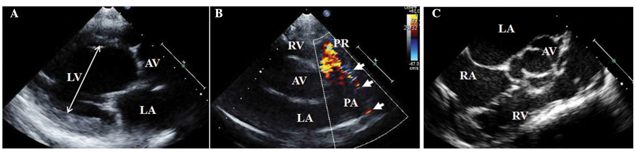
A 60-year-old woman underwent transthoracic echocardiography at the Kyung Hee University Hospital at Gangdong (Seoul, Korea) in October 2010 due to incidental systolic cardiac murmur. The patient was diagnosed with a PDA with LV dilatation (Fig. 1A and B) and mild pulmonary hypertension based on the following observations: (LV end-diastolic dimension/body surface area) : (LV end-systolic dimension/body surface area), 5.0:3.3 cm/m2 (reference value, ≤3.1:2.1 cm/m2); ejection fraction (EF), 60% (reference value, ≥55%); estimated systolic pulmonary arterial pressure (sPAP), 55 mmHg (reference value, <35 mmHg). Echocardiography also detected bicuspid AV (Fig. 1C) without significant flow obstruction [maximal velocity on AV, 2.2 m/s (reference value <2 m/s); mean pressure gradient on AV, 10 mmHg (reference value <20 mmHg)]. A normal sinus rhythm was demonstrated via an electrocardiogram (ECG). The patient refused to undergo PDA closure and was followed-up at an outpatient clinic.

Figure 1.

Echocardiography performed at the time of diagnosis. (A) Parasternal long axis view demonstrating a dilated left ventricle (double-headed arrow); (B) parasternal right ventricular outflow view demonstrating left to right flow (arrows) from the descending aorta to the pulmonary artery through the PDA; and (C) transesophageal echocardiography demonstrating the bicuspid aortic valve. PDA, patent ductus arteriosus; LV, left ventricle; LA, left atrium; RA, right atrium; AV, (bicuspid) aortic valve; RV, right ventricle; PA, pulmonary artery; PR, pulmonary regurgitation.

The patient was hospitalized two years later on October 2012 with diarrhea and dyspnea, and atrial fibrillation was detected following an ECG. Echocardiography demonstrated moderate pulmonary hypertension (sPAP, 77 mmHg) without LV dysfunction (EF, 62%). Following electric cardioversion, the ECG was normalized to a sinus rhythm without T wave inversion. The patient completely recovered following fluid therapy for acute gastritis. The patient agreed to a transcatheter procedure for PDA closure prior to 
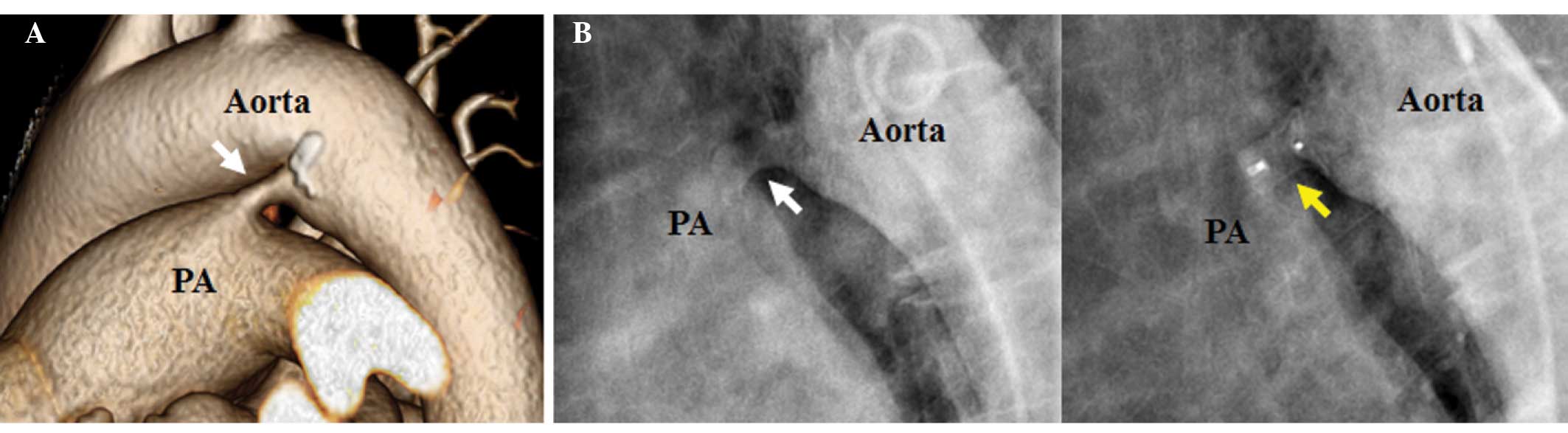
discharge. Thoracic aortic computed tomography revealed that the PDA width was 10×6 mm and length was 7 mm (Fig. 2A). Coronary arteries were normal, as detected using angiography. Subsequent cardiac catheterization demonstrated systolic, diastolic and mean PAP of 52, 29 and 40 mmHg, respectively (reference value, ≤30, 12, and 19 mmHg, respectively), and a Qp/Qs ratio of 3.9 (reference value, 1:1). Following aortography, an Amplatzer duct occluder (AGA Medical Corp., Golden Valley, MN, USA) was successfully implanted (Fig. 2B). The following day, the patient 
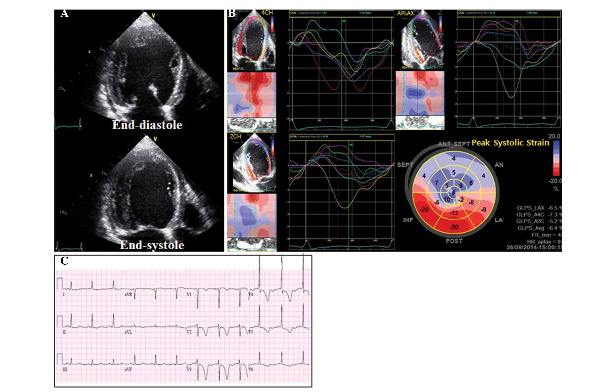
complained of mild dyspnea. Conventional echocardiography demonstrated global LV hypokinesia with decreased LV systolic function (EF, 32%; Fig. 3A). Abnormal shunt flow was not detected and the occluder was well-positioned, as detected by echocardiography. Speckle-tracking echocardiography demonstrated dyskinetic movement in the septal and anterior segments with low global longitudinal peak systolic strain (GLS) at −6.4% (Fig. 3B). The brain natriuretic peptide measured using a Triage BNP assay (Alere, Inc., San Diego, CA, USA) and a Beckman Coulter DXI 800 analyzer (Beckman Coulter, Inc., Brea, CA, USA) was found to be 821 pg/ml (reference value, <100 pg/ml) and creatine-kinase MB and cardiac troponin I measured using a Beckman Coulter DC-800 analyzer (Beckman Coulter, Inc.) were within the reference range. An ECG revealed new deep T-wave inversions in the precordial leads V2-V6 (Fig. 3C). Subsequently, the patient received medical treatment, including angiotensin-converting enzyme inhibitor perindopril (4mg acertil®; Servier Inc., Neuilly-sur-Seine, France) and was then discharged one day later.

Figure 2.

Images demonstrating PDA (white arrow) and PDA closure (yellow arrow) in the present patient. (A) Computed tomography three-dimensional reconstruction of a PDA. (B) Aortography of PDA (left) and percutaneous deployment of Amplatzer ductal occluder on PDA (right) PDA, patent ductus arteriosus; PA, pulmonary artery.

Figure 3.

Representative echocardiography and electrocardiogram (ECG) 1 day after PDA closure. (A) Conventional echocardiography demonstrated severe left ventricular (LV) systolic dysfunction. (B) Segmental longitudinal strain curves and bull's-eye plot demonstrated dyskinetic movement and post-systolic shortening, which was dominant in the septal and anterior segments (presented in blue). (C) ECG demonstrating T-wave inversions of LV strain pattern.

Echocardiography performed 3 weeks after PDA closure demonstrated improved LV function (EF, 60%; GLS, −15%; Fig. 4A and B). ST-segments and T-wave abnormalities appeared to be completely resolved after 9 months, as detected by an ECG (Fig. 4C).

Figure 4.

Follow-up echocardiography 3 weeks after PDA closure and electrocardiogram 9 months later. (A) Echocardiography demonstrating normal left ventricular (LV) systolic function. (B) Bull's-eye plot demonstrating high and homogeneous longitudinal peak systolic strain for the entire LV. (C) Electrocardiogram demonstrating the resolution of T-wave inversions in the present patient.

The patient interrupted perindopril medication one year following the procedure as her LV function was preserved and her blood pressure was low (systolic and diastolic blood pressure, 95 and 55 mmHg). At the three-year follow up following the procedure (October 2015), echocardiography revealed that the patient's LV systolic function (EF, 66%) was normal, and LV chamber size (LV end-systolic dimension/body surface area, 4.1: 2.7 cm/m2) and pulmonary hypertension (sPAP, 38 mmHg) were decreased. At a follow up on December 2015 the patient showed normal sinus rhythm following an ECG.

Discussion

Abrupt interruption of pulmonary recirculation following PDA closure induces a decrease in LV end-diastolic volume. If decreased LV end-diastolic volume does not accompany a decrease in LV end-systolic volume, the EF will decrease via several possible mechanisms including decreased myocardial contractility or increased afterload (3,10). Previous studies have demonstrated that an overall transient decrease of LV function following PDA closure occurs in 50–70% of patients, whereas significant transient decrease of LV ejection fraction of <55% or fractional shortening of <29% was observed in 8–43% of patients (5–7). In addition, transient LV dysfunction following PDA closure was found to be associated with large PDAs (7–9), large amount of shunting (9), presence of high pulmonary hypertension (9) or low EF (8–10) prior to PDA closure, and an increased age of patients (8). Furthermore, the presence of anomalies associated with PDA, such as mitral regurgitation or aortic coarctation, has been reported to be a risk factor that can provoke or deteriorate LV dysfunction following PDA closure (8).

In the present case, the patient presented a large PDA, a high Qp/Qs ratio and increased pulmonary hypertension. The patient's prolonged LV volume overload is suggested to have caused the LV remodeling into a spherical shape. Furthermore, combined bicuspid AV anomaly may have contributed to the deterioration of LV function, as it is a potential risk factor for increased afterload.

Speckle-tracking echocardiography data gathered from the present patient following PDA closure was consistent with a previous study in infants, which observed a global reduction of peak systolic strain following PDA closure, thus mimicking systolic heart failure (11). However, the dyskinetic movement in the septal and anterior segments accompanied by abnormal LV geometry and severe LV systolic dysfunction in the present case was not concordant with the results from a previous study conducted on infants (11).

In conclusion, the combination of large PDA size, large amount of shunting, LV remodeling and bicuspid aortic valve may have induced severe deterioration of LV function following PDA closure in the present case. Speckle-tracking echocardiography may also be a useful tool in the detection of decreased LV myocardial function and altered LV strain pattern following PDA closure.

References

1 

Daniel B: Congenital heart disease: Patent ductus arteriosus. Nelson Textbook of Pediatrics. Kliegman RM, Stanton BF, St. Geme JW, Schor NF and Behrman RE: (19th). (Philadelphia, PA). Elsevier Saunders. 1559–1560. 2011.

2 

Lloyd TR and Beekman RH III: Clinically silent patent ductus arteriosus. Am Heart J. 127:1664–1665. 1994. View Article : Google Scholar : PubMed/NCBI

3 

Schneider DJ and Moore JW: Patent ductus arteriosus. Circulation. 114:1873–1882. 2006. View Article : Google Scholar : PubMed/NCBI

4 

Mitchell SC, Korones SB and Berendes HW: Congenital heart disease in 56,109 births: Incidence and natural history. Circulation. 43:323–332. 1971. View Article : Google Scholar : PubMed/NCBI

5 

Schneider DJ: The patent ductus arteriosus in term infants, children and adults. Semin Perinatol. 36:146–153. 2012. View Article : Google Scholar : PubMed/NCBI

6 

Rao PS and Kern MJ: Summary and comparison of patent ductus arteriosus closure methods. Catheter Based devices for the Treatment of Non-Coronary Cardiovascular Disease in Adults and Children (Philadelphia, PA). Lippincott, Williams, and Wilkins. 219–228. 2003.

7 

Galal MO, Amin M, Hussein A, Kouatli A, Al-Ata J and Jamjoom A: Left ventricular dysfunction after closure of large patent ductus arteriosus. Asian Cardiovasc Thorac Ann. 13:24–29. 2005. View Article : Google Scholar : PubMed/NCBI

8 

Tilahun B and Tefera E: Transient left ventricular systolic dysfunction following surgical closure of large patent ductus arteriosus among children and adolescents operated at the cardiac centre, Ethiopia. J Cardiothorac Surg. 8:1392013. View Article : Google Scholar : PubMed/NCBI

9 

Kim YH, Choi HJ, Cho Y, Lee SB and Hyun MC: Transient left ventricular dysfunction after percutaneous patent ductus arteriosus closure in children. Korean Circ J. 38:596–600. 2008. View Article : Google Scholar

10 

Jeong YH, Yun TJ, Song JM, Park JJ, Seo DM, Koh JK, Lee SW, Kim MJ, Kang DH and Song JK: Left ventricular remodeling and change of systolic function after closure of patent ductus arteriosus in adults: Device and surgical closure. Am Heart J. 154:436–440. 2007. View Article : Google Scholar : PubMed/NCBI

11 

El-Khuffash AF, Jain A, Dragulescu A, McNamara PJ and Mertens L: Acute changes in myocardial systolic function in preterm infants undergoing patent ductus arteriosus ligation: A tissue Doppler and myocardial deformation study. J Am Soc Echocardiogr. 25:1058–1067. 2012. View Article : Google Scholar : PubMed/NCBI

Related Articles

  • Abstract
  • View
  • Download
  • Twitter
Copy and paste a formatted citation
Spandidos Publications style
Hwang HJ, Yoon KL and Sohn IS: Transient severe left ventricular dysfunction following percutaneous patent ductus arteriosus closure in an adult with bicuspid aortic valve: A case report. Exp Ther Med 11: 969-972, 2016.
APA
Hwang, H., Yoon, K.L., & Sohn, I.S. (2016). Transient severe left ventricular dysfunction following percutaneous patent ductus arteriosus closure in an adult with bicuspid aortic valve: A case report. Experimental and Therapeutic Medicine, 11, 969-972. https://doi.org/10.3892/etm.2016.3024
MLA
Hwang, H., Yoon, K. L., Sohn, I. S."Transient severe left ventricular dysfunction following percutaneous patent ductus arteriosus closure in an adult with bicuspid aortic valve: A case report". Experimental and Therapeutic Medicine 11.3 (2016): 969-972.
Chicago
Hwang, H., Yoon, K. L., Sohn, I. S."Transient severe left ventricular dysfunction following percutaneous patent ductus arteriosus closure in an adult with bicuspid aortic valve: A case report". Experimental and Therapeutic Medicine 11, no. 3 (2016): 969-972. https://doi.org/10.3892/etm.2016.3024
Copy and paste a formatted citation
x
Spandidos Publications style
Hwang HJ, Yoon KL and Sohn IS: Transient severe left ventricular dysfunction following percutaneous patent ductus arteriosus closure in an adult with bicuspid aortic valve: A case report. Exp Ther Med 11: 969-972, 2016.
APA
Hwang, H., Yoon, K.L., & Sohn, I.S. (2016). Transient severe left ventricular dysfunction following percutaneous patent ductus arteriosus closure in an adult with bicuspid aortic valve: A case report. Experimental and Therapeutic Medicine, 11, 969-972. https://doi.org/10.3892/etm.2016.3024
MLA
Hwang, H., Yoon, K. L., Sohn, I. S."Transient severe left ventricular dysfunction following percutaneous patent ductus arteriosus closure in an adult with bicuspid aortic valve: A case report". Experimental and Therapeutic Medicine 11.3 (2016): 969-972.
Chicago
Hwang, H., Yoon, K. L., Sohn, I. S."Transient severe left ventricular dysfunction following percutaneous patent ductus arteriosus closure in an adult with bicuspid aortic valve: A case report". Experimental and Therapeutic Medicine 11, no. 3 (2016): 969-972. https://doi.org/10.3892/etm.2016.3024
Follow us
  • Twitter
  • LinkedIn
  • Facebook
About
  • Spandidos Publications
  • Careers
  • Cookie Policy
  • Privacy Policy
How can we help?
  • Help
  • Live Chat
  • Contact
  • Email to our Support Team