Introduction
Ischemia/reperfusion (I/R) can occur following renal
surgery or transplantation, often resulting in acute kidney injury,
chronic renal failure and kidney transplantation failure (1,2). It has
been demonstrated that inflammation and apoptosis serve crucial
functions in I/R-induced renal injury (3), therefore the development of effective
drugs to prevent inflammation and apoptosis in I/R-induced renal
injury is required.
Gynostemma pentaphyllum is a traditional
Chinese medicine that has previous been used for the treatment of
renal diseases (4,5). The major component of G.
pentaphyllum is gypenoside (GP), which exhibits
anti-inflammatory, antitumor and anti-oxidative properties
(6–8). In particular, the protective effect of
GP against I/R-induced injury has been reported. For example, Qi
et al (9) reported that GP
can protect neuronal DNA against the damage resulting from
I/R-induced cerebral injury. Furthermore, it has been demonstrated
in mice that GP exerts a therapeutic effect on chronic renal
injury, fibrosis and fatty renal disease induced by alcohol
consumption and a diet high in fat and cholesterol (10,11). In
addition, the authors of the present study previously observed that
GP attenuated hepatic I/R injury in mice via anti-oxidative and
anti-apoptotic bioactive characteristics (12). However, it remains unclear whether GP
is able to exert a protective effect against I/R-induced renal
injury, and the molecular mechanisms potentially underlying this
process are as yet unknown.
Previous studies demonstrated that the inflammatory
response was significantly upregulated following I/R (13). In addition, the inflammatory response
to hypoxia has been shown to contribute to the resulting renal
tissue injury (13).
Inflammation-associated factors include complement proteins and
chemokines, including tumor necrosis factor-α (TNF-α), interferon
(IFN)-γ, interleukin (IL)-1β and IL-6 (14). Inhibition of their production has
been shown to attenuate I/R-induced tissue injury (15); in a previous study, catalpol
protected mice against renal I/R injury by suppressing the
phosphoinositide-3-kinase/Akt-endothelial nitric oxide synthase
signaling pathway, as well as the expression of TNF-α, IL-1β, IL-6
and IL-10 (16). Furthermore,
gypenoside was shown to exert suppressive effects on the expression
of inflammatory molecules (8), and
thus, may have an inhibitory role in I/R-induced renal injury.
In the present study, the protective effect of GP
against I/R-induced renal injury was evaluated using a renal I/R
injury model in C57BL/6 mice. In addition, the underlying molecular
mechanisms involving inflammation and apoptosis in renal I/R injury
were investigated.
Materials and methods
Renal I/R injury model
A total of 30 male C57BL/6 mice (age, 12 weeks;
weight, 20–25 g) were obtained from the Animal Center of Central
South University (Changsha, China). The mice were housed at the
Xiangya Medical Experimental Animal Center of the Central South
University in a laminar flow, temperature-controlled, pathogen-free
environment with a 12 h light/dark cycle and with ad libitum
access to food and water. Mice were fasted for 24 h prior to the
renal I/R procedure, then an intraperitoneal injection of
pentobarbital (50 mg/kg) was administered to anesthetize the
animals. The animal experiment was approved by the Ethics Committee
of The Third Xiangya Hospital of Central South University.
Administration of GP
GP was purchased from the China National
Pharmaceutical Group Corporation (Beijing, China) and dissolved in
saline in accordance with the manufacturer's instructions. Mice
were divided into three groups, with each group containing 5 mice.
In the sham (control) group, the mice underwent anesthesia with 4%
chloral hydrate (8 µl/g; Dalian Meilun Biology Technology Co.,
Ltd., Dalian, China) and the renal I/R procedure, which included
bilateral flank incisions and a right nephrectomy. In the
GP-treated I/R (I/R+GP) group, mice were intravenously injected
with 50 mg/kg GP using an infusion pump 1 h prior to the renal I/R
procedure. In the saline-treated I/R group (I/R), mice were
administered 50 mg/kg saline using an infusion pump 1 h prior to
the renal I/R procedure.
Assessment of kidney function
Renal function was assessed by measuring serum
creatinine (Cr) and blood urea nitrogen (BUN) levels. These tests
were performed by technicians at the Clinical Laboratory at The
Third Xiangya Hospital, Central South University.
Assessment of renal malondialdehyde
(MDA) expression levels
Homogenization buffer (Cayman Chemical Company, Ann
Arbor, MI, USA), which contained 0.32 mmol/l sucrose, 20 mmol/l
N-(2-hydroxyethyl)piperazine-N-(2-ethanesulfonic acid), 0.5 mmol/l
ethylenediaminetetraacetic acid, 1 mmol/l 1,4-dithio-DL-threitol
and 1 mmol/l phenylmethanesulfonyl fluoride, was added to renal
tissue (1 ml/0.1 g). Following homogenization, renal tissue was
centrifuged at 1,500 × g for 10 min. According to Esterbauer and
Cheeseman (17), MDA in tissue
reacts with thiobarbituric acid in the sample, and thus, the
changes in fluorescence as a result of this were detected at a
wavelength of 535 nm in the present study. Recorded concentrations
of MDA were divided by 1,000, and the results were expressed in
µmol/g wet tissue.
Assessment of renal tissue superoxide
dismutase (SOD) activity
SOD activity was measured using a Superoxide
Dismutase Activity Assay kit (BioVision, Inc., Milpitas, CA, USA),
based on the inhibition of adenochrome production by SOD by
adenochrome production during epinephrine auto-oxidation. Changes
in fluorescence were detected at a wavelength of 480 nm using the
721S Spectrophotometer (Shanghai Lengguang Industrial Co., Ltd.,
Shanghai, China).
Reverse transcription-quantitative
polymerase chain reaction (RT-qPCR) assay
Total RNA was extracted using TRIzol® reagent
(Invitrogen; Thermo Fisher Scientific, Inc., Carlsbad, CA, USA).
The RevertAid Reverse Transcription kit (Thermo Fisher Scientific,
Inc.,) was used to convert RNA (1 µg) into cDNA, according to the
manufacturer's instructions. qPCR was then conducted using an
Applied Biosystems 7500 thermocycler (Thermo Fisher Scientific,
Inc.) and 0.33 µl cDNA solution, 10 µl SYBR Green PCR Master Mix
(Thermo Fisher Scientific, Inc.), 2 µl primers (Sangon Biotech Co.,
Ltd., Shanghai, China) and 7.67 µl H2O, to obtain a
final reaction volume of 20 µl. The primers used for the qPCR were
as follows: TNF-α forward, 5′-CAGGCGGTGCCTATGTCTC-3′ and reverse,
5′-CGATCACCCCGAAGTTCAGTAG-3′; IFN-γ forward,
5′-GCCACGGCACAGTCATTGA-3′ and reverse, 5′-TGCTGATGGCCTGATTGTCTT-3′;
IL-1β forward, 5′-GAAATGCCACCTTTTGACAGTG-3′ and reverse,
5′-TGGATGCTCTCATCAGGACAG-3′; IL-6 forward,
5′-CTGCAAGAGACTTCCATCCAG-3′ and reverse,
5′-AGTGGTATAGACAGGTCTGTTGG-3′; and GAPDH forward,
5′-TGACCTCAACTACATGGTCTACA-3′ and reverse,
5′-CTTCCCATTCTCGGCCTTG-3′. The cycling conditions were as follows:
95°C for 10 min, followed by 40 cycles of denaturation at 95°C for
15 sec and annealing/elongation at 60°C for 60 sec. GAPDH was used
as an internal reference. The relative expression levels were
analyzed by the Applied Biosystems 7500 Fast Real-Time PCR System
(Thermo Fisher Scientific, Inc.), using the 2−ΔΔCq
method (18).
Measurement of serum proinflammatory
cytokine levels
Enzyme-linked immunosorbent assay (ELISA) kits
(Sigma-Aldrich, St. Louis, MO, USA) were used to determine the
serum concentration levels of various key proinflammatory
cytokines, including TNF-α (RAB0477), IFN-γ (RAB0224), IL-1β
(RAB0274) and IL-6 (RAB0308), in accordance with the manufacturer's
instructions.
Western blot analysis
The NE-PER® Nuclear and Cytoplasmic Extraction
reagents (Pierce Biotechnology, Inc., Rockford, IL, USA) were used
to extract cytosolic proteins from mice renal tissue, in accordance
with the manufacturer's protocol. Protein assay reagents (Beyotime
Institute of Biotechnology, Haimen, China) were used to determine
protein concentration, after which 20 µg protein was separated by
10% SDS-PAGE (initially 80 V and then 150 V; Beyotime Institute of
Biotechnology), transferred to nitrocellulose membranes (Thermo
Fisher Scientific, Inc.) and maintained at room temperature for 1 h
in a buffer solution containing 5% dried skim milk. The membrane
was then incubated at room temperature for 3 h with the following
antibodies from Abcam (Cambridge, MA, USA): Polyclonal rabbit
anti-human p-ERK (1:100; ab65142), ERK (1:200; ab32537), heme
oxygenase-1 (HO-1; 1:200; ab13248), Bcl-2 (1:100; ab117115), Bax
(1:50; ab79459) and GAPDH (1:50; ab181602). Subsequently, the
samples were incubated for 1 h with horseradish
peroxidase-conjugated goat anti-rabbit IgG (1:20,000; ab6721). The
signals on the membranes were detected using enhanced
chemiluminescence reagent (Pierce Biotechnology, Inc.) and
densitometry was conducted using Image-Pro Plus software, version
6.0 (Media Cybernetics, Inc., Rockville, MD, USA).
Apoptosis analysis
Cell apoptosis was analyzed using the Annexin V-FITC
Apoptosis Detection kit (BD Biosciences, Franklin Lakes, NJ, USA),
according to the manufacturer's protocol. Briefly, at 24 h
post-transfection, the cells were harvested and washed twice with
cold PBS. Subsequently, 106 cells were resuspended in
200 µl binding buffer added to 10 µl Annexin-V-FITC and 5 µl PI-PE,
followed by incubation in the dark for 30 min. Finally, 300 µl
binding buffer was added, followed by the flow cytometry assay.
Statistical analysis
All data are presented as the mean ± standard error,
and analyzed using one-way analysis of variance. Statistical
analyses were conducted using SPSS software, version 16.0 (SPSS,
Inc., Chicago, IL, USA). P<0.05 was considered to indicate a
statistically significant difference.
Results
GP exerted a protective effect on
I/R-induced renal injury in mice
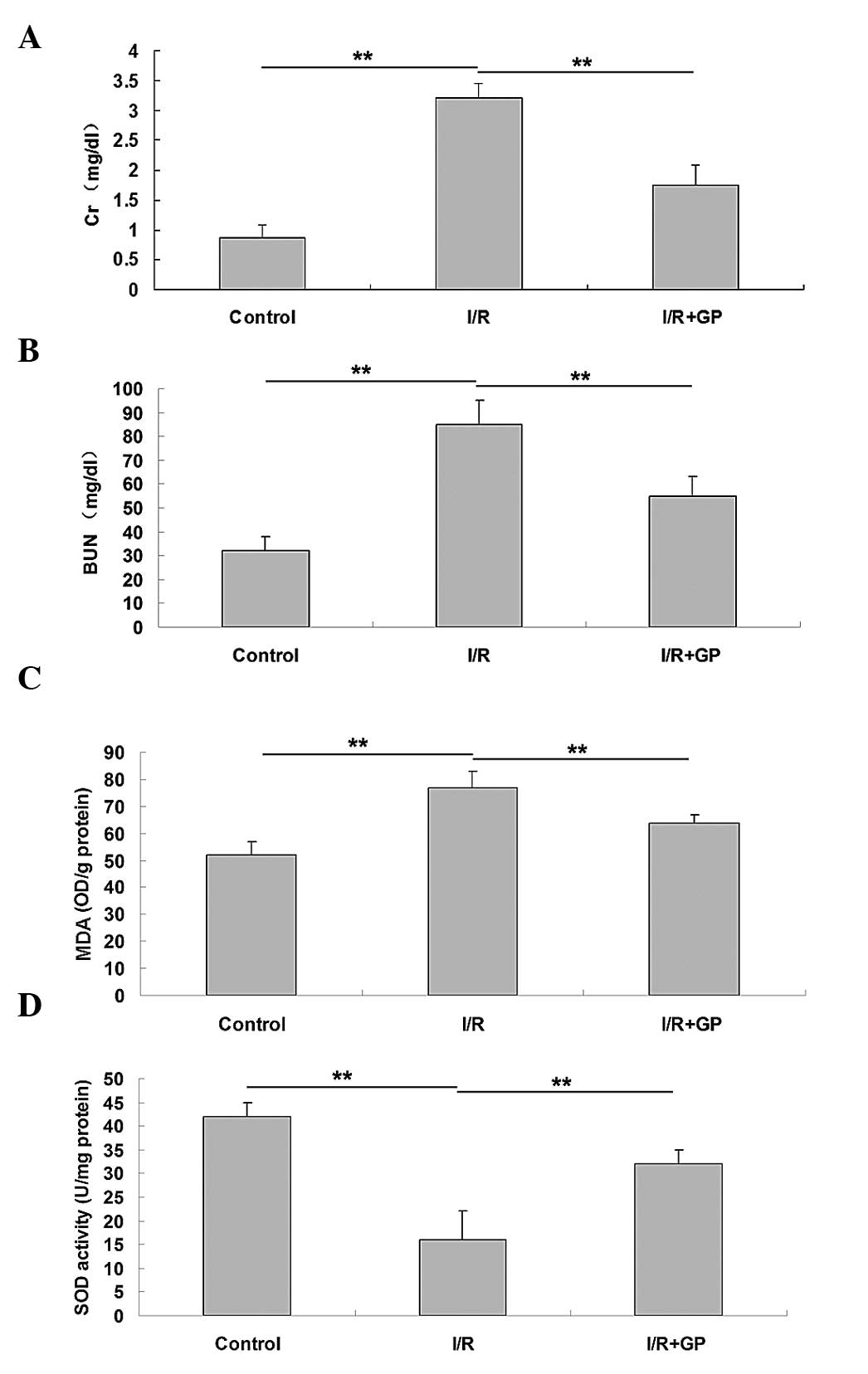
Serum concentrations of Cr and BUN were measured
following I/R-induced renal injury, and it was observed that mice
in the I/R group presented significantly higher serum levels of Cr
and BUN compared with the control group (P<0.01; Fig. 1A and B), indicating the occurrence of
I/R-induced renal injury. In the I/R+GP group, serum concentration
levels of Cr and BUN were significantly reduced compared with the
I/R group (P<0.01; Fig. 1A and
B), suggesting that pretreatment with GP has a protective
effect against I/R-induced renal injury. However, the MDA
concentration in the renal tissue at 6 h after reperfusion was
significantly increased in the I/R group compared with the control
and I/R+GP groups (Fig. 1C). In
addition, SOD activity was higher in the I/R+GP group compared with
the I/R group (Fig. 1D).
Pretreatment with GP attenuated
I/R-induced inflammatory responses in the kidney
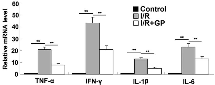
The inflammatory responses resulting from I/R in
kidney tissue were examined in mice. It was observed that the mRNA
expression levels of the inflammatory cytokines TNF-α, IFN-γ, IL-1β
and IL-6 were higher in renal tissue following I/R compared with
the control and I/R+GP groups (Fig.
2). To further confirm these results, an ELISA was performed,
which demonstrated that the secretion levels of TNF-α, IFN-γ, IL-1β
and IL-6 were significantly increased in renal tissue following
I/R, but reduced in the GP treatment group (P<0.01; Fig. 3).
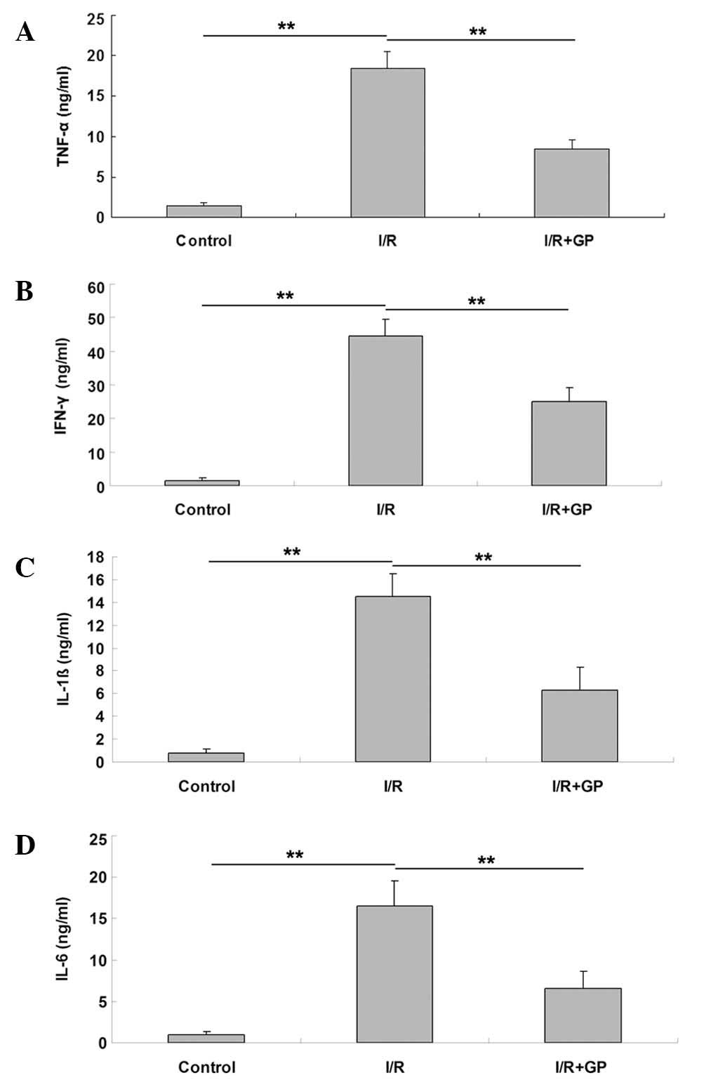 | Figure 3.Renal tissue expression levels of
proinflammatory cytokines, including (A) TNF-α, (B) IFN-γ, (C)
IL-1β and (D) IL-6. Data are presented as the mean ± standard
error. **P<0.01, comparisons as shown by brackets. TNF-α, tumor
necrosis factor-α; IFN-γ, interferon-γ; IL-1β, interleukin-1β;
IL-6, interleukin-6; I/R, saline-treated ischemia/reperfusion
group; I/R+GP, geniposide-treated ischemia/reperfusion group. |
Pretreatment with GP attenuated
I/R-induced oxidative damage in the kidney
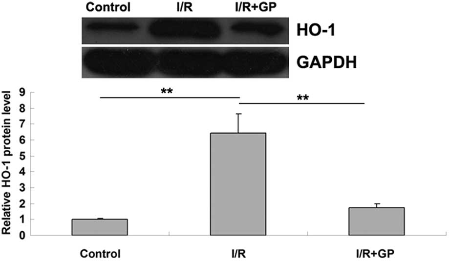
Heme oxygenase-1 (HO-1) is crucial in the defense
against oxidative damage, and the expression of HO-1 can be induced
by I/R (19). In the present study,
HO-1 protein expression was detected using western blot analysis.
The results indicated that HO-1 expression was significantly
increased in the I/R group compared with the control group
(P<0.01; Fig. 4), suggesting that
oxidative damage had occurred. However, this I/R-induced
upregulation of HO-1 was attenuated by pretreatment with GP; the
IR+GP group exhibited significantly reduced levels of HO-1 protein,
compared with the IR group (P<0.01; Fig. 4). It may therefore be suggested that
pretreatment with GP attenuates I/R-induced oxidative damage in
mice kidney tissue.
Pretreatment with GP attenuated
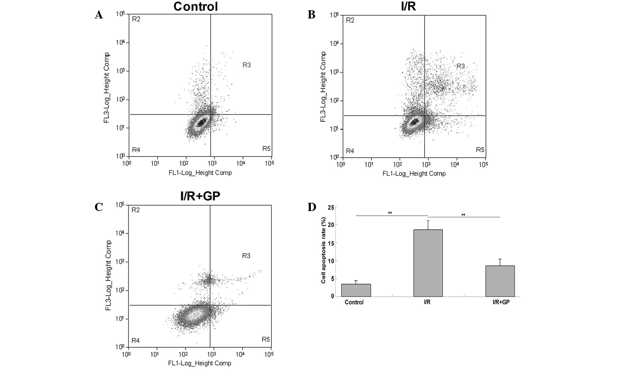
I/R-induced apoptosis of renal cells
The effect of GP on I/R-induced renal cell injury
was investigated using an apoptosis assay. As presented in Fig. 5, the level of apoptosis in kidney
tissue following I/R was significantly increased compared with the
control group (P<0.01); however, pretreatment with GP
significantly attenuated I/R-induced renal cell apoptosis
(P<0.01 vs. I/R group).
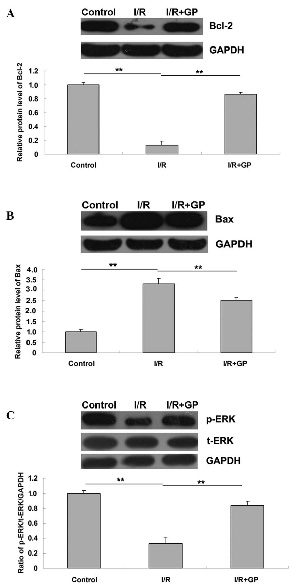
Molecular mechanisms of GP
The expression levels of the apoptosis-associated
Bcl-2 and Bax proteins were examined. As demonstrated in Fig. 6A, the protein expression levels of
anti-apoptotic Bcl-2 were significantly reduced in the I/R group
compared with the control group (P<0.01). However, the
expression level was higher in the I/R+GP group compared with the
I/R group (P<0.01). In addition, it was observed that the
protein level of pro-apoptotic Bax was increased in the I/R group
compared with the control group, and that Bax protein expression
was attenuated by pretreatment with GP (P<0.01; Fig. 6B). This suggests that the protective
effect of GP against I/R-induced renal injury may be attributed to
the inhibition of cell apoptosis in the kidney.
GP suppresses I/R-induced
downregulation of ERK signaling in renal tissues
As presented in Fig.
6C, the expression level of phosphorylated ERK (p-ERK) in the
I/R group was significantly reduced compared with the control
group, and p-ERK was significantly attenuated following
pretreatment with GP in the I/R+GP group (P<0.01). This suggests
that the administration of GP suppressed I/R-induced the
downregulation of ERK signaling in renal tissue, which may be
associated with the protective effect of GP against I/R-induced
renal cell apoptosis.
Discussion
It has previously been demonstrated that GP
possesses various bioactive characteristics including
anti-oxidative, anti-inflammatory, anti-apoptotic and antitumor
properties (9,11,20). In
the present study, kidney tissue that had been pretreated with GP
exhibited significantly inhibited I/R-induced upregulation of serum
Cr and BUN protein expression. In addition, the I/R+GP group
demonstrated significantly lower expression levels of MDA and
higher SOD activity compared with the I/R group. Furthermore,
pretreatment with GP inhibited the I/R-induced production of
pro-inflammatory cytokines, oxidative damage and apoptosis.
Investigation of the molecular mechanisms involved suggested that
pretreatment with GP inhibits the I/R-induced activation of ERK
signaling.
Inflammation has been recognized as a key mechanism
underlying the pathogenesis of renal I/R injury (21). In response to I/R, neutrophils,
lymphocytes and macrophages infiltrate the damaged tissue, which
can lead to microcirculatory failure as a result of the reduction
in renal blood flow (22,23). Other proinflammatory factors,
including TNF-α, IFN-γ, IL-1β and IL-6 further promote the
inflammatory response in renal tissue (24).
GP has been demonstrated to produce a suppressive
effect on the I/R-induced inflammatory response. Quan et al
(8) observed that treatment with GP
inhibited the expression of inflammatory molecules, including
intercellular adhesion molecule 1, monocyte chemotactic protein-1
and nuclear factor-κB, in atherosclerotic rats that were induced by
a high-fat diet (8). Another
previous study demonstrated that treatment with GP protects against
I/R-induced hepatic injury by inhibiting neutrophil infiltration
(12). In the present study, it was
observed that the secretion of TNF-α, IFN-γ, IL-1β and IL-6 was
significantly reduced in the I/R+GP group compared with the I/R
group. Based on current knowledge, it may be hypothesized that the
protective effect of GP against I/R-induced renal injury is partly
caused by the suppression of inflammatory responses.
Cell death is frequently controlled by the Bcl-2
protein family, which contains pro-apoptotic and anti-apoptotic
proteins (25). The Bcl-2 family
serves a number of crucial functions in the control of cell
survival during developmental and pathophysiological processes,
including ischemic injury, neuronal Ca2+ homeostasis,
trophic factor withdrawal, oxidative stress, excitotoxicity and
energy stress (26). As an
anti-apoptotic protein, Bcl-2 is able to inhibit lipid peroxide
formation, Ca2+ release from endoplasmic reticulum and
free radical production (27). Bax,
another member of the Bcl-2 family, is an endogenous antagonist of
Bcl-2 that inhibits the protein by directly binding via an
associated protein homologue, thereby promoting cell apoptosis
(28). However, Bcl-2 can reversely
suppress the pro-apoptotic effect of Bax by binding to Bcl-xL and
forming a heterodimer (29). The
expression of Bcl-2 and Bax are carefully maintained and balanced
in a healthy physiological state. However, the current study
observed that I/R activates renal cell apoptosis by increasing the
protein expression levels of Bax and reducing the protein levels of
Bcl-2. In addition, the present results demonstrated that Bcl-2 can
be attenuated by pretreating renal tissue with GP, suggesting that
the protective effect of GP on I/R-induced renal cell apoptosis
occurs via a Bcl-2-dependent mechanism.
Mitogen-activated protein kinases (MAPKs) have been
demonstrated to be key factors in I/R-induced tissue damage. MAPK
compounds, including ERK, stress-activated protein kinases/c-Jun
NH2-terminal kinases and p38 MAPKs have been implicated
in inflammatory signaling mechanisms in I/R injury (30,31).
Additionally, ERK signaling serves a crucial function in
I/R-induced cell apoptosis (32,33). Ban
et al (34) reported that the
inhibition of ERK signaling worsened intestinal I/R injury. In
addition, Yang et al (35)
observed that Apelin-13 protects the brain against I/R injury by
activating ERK signaling pathways. Furthermore, GP has been
demonstrated to upregulate ERK signaling and inhibit JNK signaling
in local cerebral I/R injury rats (36). In the present study, it was
demonstrated that GP significantly suppresses the downregulation of
p-ERK protein expression levels induced by I/R, indicating that GP
activates ERK signaling. It may therefore be suggested that, in
mice, the protective effect of GP on renal cell apoptosis occurs as
a result of the upregulation of ERK signaling in I/R-induced kidney
injury.
In conclusion, the current study demonstrates that
the administration of GP exerts protective effects against
I/R-induced renal injury in mice by attenuating inflammatory
responses and suppressing apoptosis. As a result, it can be
suggested that GP may be used in the treatment of renal I/R
injury.
Acknowledgements
The current study was supported by the Scientific
and Technological Brainstorm Project of Wuhan City (grant no.
201161038344-01), the Natural Science Fund of Hubei Province (grant
no. 2012FFA044) and the Public Service Platform Construction
Projects of Wuhan Technology Bureau (grant no.
2013060705010326).
References
|
1
|
Zhou M, Tang W, Fu Y, Xu X, Wang Z, Lu Y,
Liu F, Yang X, Wei X, Zhang, Y, et al: Progranulin protects against
renal ischemia/reperfusion injury in mice. Kidney Int. 87:918–929.
2015. View Article : Google Scholar : PubMed/NCBI
|
|
2
|
Menke J, Sollinger D, Schamberger B,
Heemann U and Lutz J: The effect of ischemia/reperfusion on the
kidney graft. Curr Opin Organ Transplant. 19:395–400. 2014.
View Article : Google Scholar : PubMed/NCBI
|
|
3
|
Kaczorowski DJ, Tsung A and Billiar TR:
Innate immune mechanisms in ischemia/reperfusion. Front Biosci
(Elite Ed). 1:91–98. 2009.PubMed/NCBI
|
|
4
|
Quan Y, Yang Y, Wang H, Shu B, Gong QH and
Qian M: Gypenosides attenuate cholesterol-induced DNA damage by
inhibiting the production of reactive oxygen species in human
umbilical vein endothelial cells. Mol Med Rep. 11:2845–2851.
2015.PubMed/NCBI
|
|
5
|
Deng Q and Yang X: Protective effects of
Gynostemma pentaphyllum polysaccharides on PC12 cells
impaired by MPP(+). Int J Biol Macromol. 69:171–175. 2014.
View Article : Google Scholar : PubMed/NCBI
|
|
6
|
Li K, Du Y, Fan Q, Tang CY and He JF:
Gypenosides might have neuroprotective and immunomodulatory effects
on optic neuritis. Med Hypotheses. 82:636–638. 2014. View Article : Google Scholar : PubMed/NCBI
|
|
7
|
Yan H, Wang X, Niu J, Wang Y, Wang P and
Liu Q: Anti-cancer effect and the underlying mechanisms of
gypenosides on human colorectal cancer SW-480 cells. PLoS One.
9:e956092014. View Article : Google Scholar : PubMed/NCBI
|
|
8
|
Quan Y and Qian MZ: Effect and mechanism
of gypenoside on the inflammatory molecular expression in high-fat
induced atherosclerosis rats. Zhong Guo Zhong Xi Yi Jie He Za Zhi.
30:403–406. 2010.(In Chinese).
|
|
9
|
Qi G, Zhang L, Xie WL, Chen XY and Li JS:
Protective effect of gypenosides on DNA and RNA of rat neurons in
cerebral ischemia-reperfusion injury. Acta Pharmacol Sin.
21:1193–1196. 2000.PubMed/NCBI
|
|
10
|
Zhang Y, Zhang JE, Xiao HQ, Wu PY and Bai
SJ: Gypenosides inhibit renal fibrosis by regulating expression of
related genes in rats with unilateral ureteral obstruction. J
Nephrol. 24:112–118. 2011. View Article : Google Scholar : PubMed/NCBI
|
|
11
|
Qin R, Zhang J, Li C, Zhang X, Xiong A,
Huang F, Yin Z, Li K, Qin W, Chen M, et al: Protective effects of
gypenosides against fatty liver disease induced by high fat and
cholesterol diet and alcohol in rats. Arch Pharm Res. 35:1241–1250.
2012. View Article : Google Scholar : PubMed/NCBI
|
|
12
|
Zhao J, Ming Y, Wan Q, Ye S, Xie S, Zhu Y,
Wang Y, Zhong Z, Li L and Ye Q: Gypenoside attenuates hepatic
ischemia/reperfusion injury in mice via anti-oxidative and
anti-apoptotic bioactivities. Exp Ther Med. 7:1388–1392.
2014.PubMed/NCBI
|
|
13
|
Daemen MA, de Vries B and Buurman WA:
Apoptosis and inflammation in renal reperfusion injury.
Transplantation. 73:1693–1700. 2002. View Article : Google Scholar : PubMed/NCBI
|
|
14
|
Cao XZ, Ma H, Wang JK, Liu F, Wu BY, Tian
AY, Wang LL and Tan WF: Postoperative cognitive deficits and
neuroinflammation in the hippocampus triggered by surgical trauma
are exacerbated in aged rats. Prog Neuropsychopharmacol Biol
Psychiatry. 34:1426–1432. 2010. View Article : Google Scholar : PubMed/NCBI
|
|
15
|
Facio FN Jr, Sena AA, Araújo LP, Mendes
GE, Castro I, Luz MA, Yu L, Oliani SM and Burdmann EA: Annexin 1
mimetic peptide protects against renal ischemia/reperfusion injury
in rats. J Mol Med (Berl). 89:51–63. 2011. View Article : Google Scholar : PubMed/NCBI
|
|
16
|
Zhu J, Chen X, Wang H and Yan Q: Catalpol
protects mice against renal ischemia/reperfusion injury via
suppressing PI3K/Akt-eNOS signaling and inflammation. Int J Clin
Exp Med. 8:2038–2044. 2015.PubMed/NCBI
|
|
17
|
Esterbauer H and Cheeseman KH:
Determination of aldehydic lipid peroxidation products:
Malonaldehyde and 4-hydroxynonenal. Methods Enzymol. 186:407–421.
1990. View Article : Google Scholar : PubMed/NCBI
|
|
18
|
Livak KJ and Schmittgen TD: Analysis of
relative gene expression data using real-time quantitative PCR and
the 2(−Delta Delta C(T)) Method. Methods. 25:402–408. 2001.
View Article : Google Scholar : PubMed/NCBI
|
|
19
|
Sass G, Barikbin R and Tiegs G: The
multiple functions of heme oxygenase-1 in the liver. Z
Gastroenterol. 50:34–40. 2012. View Article : Google Scholar : PubMed/NCBI
|
|
20
|
Chen JC, Tsai CC, Chen LD, Chen HH and
Wang WC: Therapeutic effect of gypenoside on chronic liver injury
and fibrosis induced by CCl4 in rats. Am J Chin Med. 28:175–185.
2000. View Article : Google Scholar : PubMed/NCBI
|
|
21
|
El Morsy EM, Ahmed MA and Ahmed AA:
Attenuation of renal ischemia/reperfusion injury by açaí extract
preconditioning in a rat model. Life Sci. 123:35–42. 2014.
View Article : Google Scholar : PubMed/NCBI
|
|
22
|
Ysebaert DK, De Greef KE, Vercauteren SR,
Ghielli M, Verpooten GA, Eyskens EJ and De Broe ME: Identification
and kinetics of leukocytes after severe ischaemia/reperfusion renal
injury. Nephrol Dial Transplant. 15:1562–1574. 2000. View Article : Google Scholar : PubMed/NCBI
|
|
23
|
Contaldo C, Meier C, Elsherbiny A, Harder
Y, Trentz O, Menger MD and Wanner GA: Human recombinant
erythropoietin protects the striated muscle microcirculation of the
dorsal skinfold from postischemic injury in mice. Am J Physiol
Heart Circ Physiol. 293:H274–H283. 2007. View Article : Google Scholar : PubMed/NCBI
|
|
24
|
Domański L, Pawlik A, Safranow K, Rozański
J, Myślak M, Sulikowski T, Romanowski M, Ostrowski M, Wiśniewska M,
Domański M, et al: Changes in cytokine concentrations in graft
renal vein during reperfusion in patients with and without delayed
graft function. Ann Acad Med Stetin. 54:49–52. 2008.PubMed/NCBI
|
|
25
|
Akl H, Vervloessem T, Kiviluoto S,
Bittremieux M, Parys JB, De Smedt H and Bultynck G: A dual role for
the anti-apoptotic Bcl-2 protein in cancer: Mitochondria versus
endoplasmic reticulum. Biochim Biophys Acta. 1843:2240–2252. 2014.
View Article : Google Scholar : PubMed/NCBI
|
|
26
|
Anilkumar U and Prehn JH: Anti-apoptotic
BCL-2 family proteins in acute neural injury. Front Cell Neurosci.
8:2812014. View Article : Google Scholar : PubMed/NCBI
|
|
27
|
Saitoh Y, Ouchida R, Kayasuga A and Miwa
N: Anti-apoptotic defense of bcl-2 gene against
hydroperoxide-induced cytotoxicity together with suppressed lipid
peroxidation, enhanced ascorbate uptake, and upregulated Bcl-2
protein. J Cell Biochem. 89:321–334. 2003. View Article : Google Scholar : PubMed/NCBI
|
|
28
|
Renault TT and Chipuk JE: Death upon a
kiss: Mitochondrial outer membrane composition and organelle
communication govern sensitivity to BAK/BAX-dependent apoptosis.
Chem Biol. 21:114–123. 2014. View Article : Google Scholar : PubMed/NCBI
|
|
29
|
Renault TT, Teijido O, Antonsson B, Dejean
LM and Manon S: Regulation of Bax mitochondrial localization by
Bcl-2 and Bcl-x(L): Keep your friends close but your enemies
closer. Int J Biochem Cell Biol. 45:64–67. 2013. View Article : Google Scholar : PubMed/NCBI
|
|
30
|
Jiang M, Li J, Peng Q, Liu Y, Liu W, Luo
C, Peng J, Li J, Yung KK and Mo Z: Neuroprotective effects of
bilobalide on cerebral ischemia and reperfusion injury are
associated with inhibition of pro-inflammatory mediator production
and down-regulation of JNK1/2 and p38 MAPK activation. J
Neuroinflammation. 11:1672014. View Article : Google Scholar : PubMed/NCBI
|
|
31
|
Jun JH, Jun NH, Shim JK, Shin EJ and Kwak
YL: Erythropoietin protects myocardium against ischemia-reperfusion
injury under moderate hyperglycemia. Eur J Pharmacol. 745:1–9.
2014. View Article : Google Scholar : PubMed/NCBI
|
|
32
|
Ottani A, Galantucci M, Ardimento E, Neri
L, Canalini F, Calevro A, Zaffe D, Novellino E, Grieco P, Giuliani
D and Guarini S: Modulation of the JAK/ERK/STAT signaling in
melanocortin-induced inhibition of local and systemic responses to
myocardial ischemia/reperfusion. Pharmacol Res. 72:1–8. 2013.
View Article : Google Scholar : PubMed/NCBI
|
|
33
|
Wu X, Xu T, Li D, Zhu S, Chen Q, Hu W, Pan
D, Zhu H and Sun H: ERK/PP1a/PLB/SERCA2a and JNK pathways are
involved in luteolin-mediated protection of rat hearts and
cardiomyocytes following ischemia/reperfusion. PLoS One.
8:e829572013. View Article : Google Scholar : PubMed/NCBI
|
|
34
|
Ban K, Peng Z and Kozar RA: Inhibition of
ERK1/2 worsens intestinal ischemia/reperfusion injury. PLoS One.
8:e767902013. View Article : Google Scholar : PubMed/NCBI
|
|
35
|
Yang Y, Zhang X, Cui H, Zhang C, Zhu C and
Li L: Apelin-13 protects the brain against ischemia/reperfusion
injury through activating PI3K/Akt and ERK1/2 signaling pathways.
Neurosci Lett. 568:44–49. 2014. View Article : Google Scholar : PubMed/NCBI
|
|
36
|
Wang QY, Liu F, Wu FJ and Li JL: Effects
of ginsenoside Rg1 on the expressions of p-eRK1/2 and p-JNK in
local cerebral ischemia/reperfusion injury rats. Zhong Guo Zhong Xi
Yi Jie He Za Zhi. 33:229–234. 2013.(In Chinese).
|