Open Access

MRI measurement on intercondylar notch after anterior cruciate ligament rupture and its correlation

  • Authors:
    • Xiao Ouyang
    • Yu Hao Wang
    • Jian Wang
    • Shi Dong Hong
    • Feng Xin
    • Lin Wang
    • Xiao Wei Yang
    • Jing Rong Wang
    • Li Ming Wang
    • Bo Wei
    • Qing Wang
    • Wei Ding Cui
    • Xing Li Fu
  • View Affiliations

  • Published online on: February 16, 2016     https://doi.org/10.3892/etm.2016.3078
  • Pages: 1275-1278
  • Copyright: © Ouyang et al. This is an open access article distributed under the terms of Creative Commons Attribution License.

Metrics: Total Views: 0 (Spandidos Publications: | PMC Statistics: )
Total PDF Downloads: 0 (Spandidos Publications: | PMC Statistics: )


Abstract

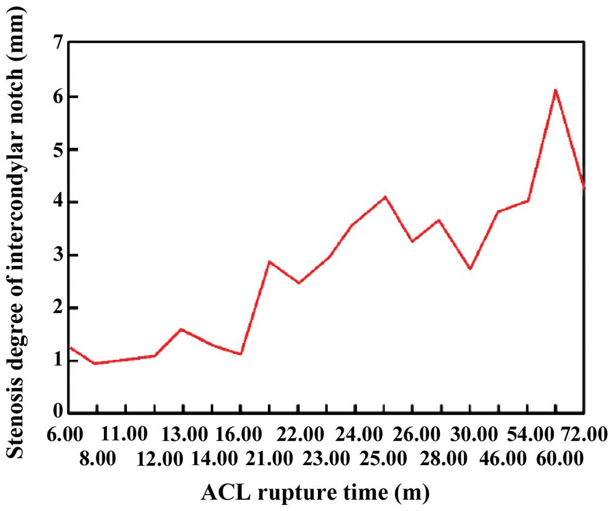
The knee joint is extremely susceptible to injury, which is usually identified by magnetic resonance imaging (MRI). In the present study, MRI was applied to quantitatively detect the association between anterior cruciate ligament (ACL) rupture and anatomic morphologic changes of the intercondylar notch. Forty patients with unilateral ACL rupture who were treated between July, 2013 and October, 2014 were enrolled in the present study. The patients were divided into the observation (affected side) and control (healthy side) groups. MRI measurements were undertaken based on parameters associated with intercondylar notch of double knee joints. The results showed that intercondylar notch width (ICW) in the observation group was significantly smaller than that in the control group, and differences were statistically significant (P<0.05). Differences on the intercondylar notch height and femoral condyle width [epicondylar width (EW)] between the two groups were not statistically significant (P>0.05). Notch width index (NWI) and notch shape index (NSI) in the observation group were significantly less than those in the control group and differences were statistically significant (P<0.05). Differences of Lysholm and Tegner scoring between the two groups were not statistically significant (P>0.05). The differential value of ICW in the observation group was 2.6±1.3 mm and the ACL rupture time of the affected knee was 20.4±1.3 months on average. The correlation was statistically significant (P<0.05). The correlation of Lysholm scoring, Tegner scoring and intercondylar notch stenosis degree on the affected knee was not statistically significant (P>0.05). In conclusion, after ACL rupture, ICW on the affected knee had significant stenosis, NSI and NWI were significantly reduced and the stenosis degree was aggravated with the prolongation of course. By contrast, Lysholm and Tegner scoring of patients with different degrees of stenosis had no correlation.
View Figures
View References

Related Articles

Journal Cover

April-2016
Volume 11 Issue 4

Print ISSN: 1792-0981
Online ISSN:1792-1015

Sign up for eToc alerts

Recommend to Library

Copy and paste a formatted citation
x
Spandidos Publications style
Ouyang X, Wang YH, Wang J, Hong SD, Xin F, Wang L, Yang XW, Wang JR, Wang LM, Wei B, Wei B, et al: MRI measurement on intercondylar notch after anterior cruciate ligament rupture and its correlation. Exp Ther Med 11: 1275-1278, 2016
APA
Ouyang, X., Wang, Y.H., Wang, J., Hong, S.D., Xin, F., Wang, L. ... Fu, X.L. (2016). MRI measurement on intercondylar notch after anterior cruciate ligament rupture and its correlation. Experimental and Therapeutic Medicine, 11, 1275-1278. https://doi.org/10.3892/etm.2016.3078
MLA
Ouyang, X., Wang, Y. H., Wang, J., Hong, S. D., Xin, F., Wang, L., Yang, X. W., Wang, J. R., Wang, L. M., Wei, B., Wang, Q., Cui, W. D., Fu, X. L."MRI measurement on intercondylar notch after anterior cruciate ligament rupture and its correlation". Experimental and Therapeutic Medicine 11.4 (2016): 1275-1278.
Chicago
Ouyang, X., Wang, Y. H., Wang, J., Hong, S. D., Xin, F., Wang, L., Yang, X. W., Wang, J. R., Wang, L. M., Wei, B., Wang, Q., Cui, W. D., Fu, X. L."MRI measurement on intercondylar notch after anterior cruciate ligament rupture and its correlation". Experimental and Therapeutic Medicine 11, no. 4 (2016): 1275-1278. https://doi.org/10.3892/etm.2016.3078