Introduction
Aggressive angiomyxoma (AAM) and
angiomyofibroblastoma (AMFB) are rare types of mesenchymal
neoplasms, with a marked predilection for the female genital tract,
in particular, the vulva and vagina. There has been a limited
number of reports concerning AAM or AMFB individually since AAM was
initially described by Steeper and Rosai in 1983 (1–5). The
prevalence rate of AAM and AMFB is unclear due to the rarity of the
condition. AMFB may be treated by surgical removal of the tumor
without reported recurrence (6,7). While
AAM is a slow-growing, but locally invasive, uncapsulated neoplasm,
relapse may still occur following wide excision of the tumor
(4,8). AMFB may be treated by surgical removal
of the tumor, while relapse may occur in patients with AAM even
after a wider excision of the tumor (4). Hence, differential diagnosis between
the two entities is crucial for surgical planning. In certain rare
cases, this differential diagnosis is difficult even for
experienced pathologists due to the overlapping morphological
features. The present study reported a case of well-circumscribed
AAM misdiagnosed as cellular AFMB, which was treated with a simple
resection and the patient presented a local relapse after 2
years.
Case report
A 25-year-old woman was initially admitted to the
Department of Gynecology, Women's Hospital, School of Medicine
Zhejiang University (Hangzhou, China) in April 2010, and again in
March 2012. The patient presented with a painless swelling in the
anterior wall of the vagina, incidentally detected during a routine
gynecological examination. A palpable, non-tender, solid mass was
identified on the anterior wall of the vagina upon the patient's
initial admission. The mass was freely movable in relation to the
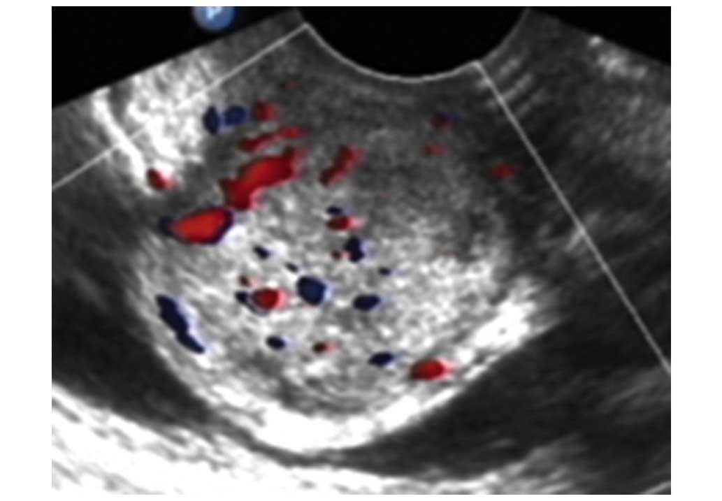
adjacent tissues. Ultrasound examination (Voluson 730 Ultrasound
System; GE Healthcare Life Sciences, Chalfont, UK) revealed that
the mass was a well-circumscribed soft tissue mass with
heterogeneous internal echoes. The mass was measured to be
~3.3×3.7×3.4 cm in size and protruded toward the bladder, with rich
intralesional vascularization observed by color Doppler (Fig. 1). The tumor was removed in full
without damage to the adjacent structures; the tumor was diagnosed
as leiomyoma. Upon sectioning, the tumor was well-demarcated with a
gelatinous appearance, with no foci of hemorrhage or necrosis. The
patient received no further treatment as the pathological diagnosis
at the time was AMFB.
After 2 years, the patient was admitted to the
Women's Hospital, School of Medicine Zhejiang University (Hangzhou,
China), with a chief complaint of frequent micturition. A
gynecological examination revealed a diffused mass in the anterior
wall of the vagina. The mass was rubbery to firm in consistency and
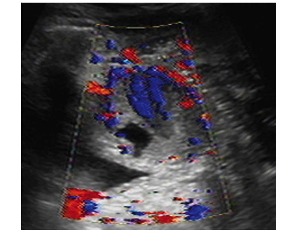
mildly tender on palpation. Ultrasonography indicated that the
hypoechoic mass, 4.9×3.6×3.0 cm in size, was circumscribed but not
well-demarcated between the paries anterior vagina and posterior
bladder wall. Color Doppler flow imaging revealed that the arterial
blood flow was of predominantly low resistance (Fig. 2). The patient underwent a wide tumor
excision and repair of the vesicovaginal fistula, as part of the
adhesive posterior bladder wall was incised during the surgery.
Upon gross examination, the excised specimen was irregular and
partially capsulated. The tumor appeared to focally extend into the
surrounding normal tissues and displayed a rubbery, glistening
grey-white surface. The final pathological diagnosis was AAM. The
patient remained free from tumor throughout the 3-year follow-up
period; the final follow-up was in May 2015.
The specimens from both surgeries were spliced
grossly into samples 2×2×0.2 cm, cut (4 µm) using a rotary
microtome (Micron HMB40E; Thermo Fisher Scientific, Inc., Waltham,
MA, USA), and stained with hematoxylin and eosin (Shenzhen Boao
Biotechnology Co., Ltd., Shenzen, China). The slides were observed
under a microscope (DMR 2000; Leica Microsystems, Wetzlar,
Germany). The typical histopathological changes in the initial and
second surgeries are displayed in Fig.
3, respectively. Common morphological features including
cellular spindle cells and abundant small blood vessels in the
slightly myxoid milieu were evident. The well-demarcated mass,
which did not show evidence of involving the surrounding normal
tissue and inconspicuous myxoid stroma, prompted the initial
diagnosis of AMFB, although the classical concentrated pattern
around the vessel was not prominent. The recurrence of the tumor,
invasion into the surrounding tissue and the evident myxoid
components in the second surgery provided substantial evidence for
the diagnosis of AAM by the pathologists.
Discussion
AMFB is a relatively rare soft tissue tumor
characterized by benign biological behavior. Previously published
studies concerning patients with AMFB have presented benign tumors
without local recurrence or metastatic potential, even after a
prolonged follow-up period of 5 years (6–8). In
contrast to AMFB, AAM is a more infiltrative neoplasm and has a
high propensity for local recurrence. The local recurrence of AAM
varies in the range of 36–72% according to the extent of resection
during the initial surgery or the location of the tumor (9,10).
Differential diagnosis for the two lesions is
important for the subsequent treatment, as the prognoses are
different. AAM and AMFB are associated neoplasms in a spectrum of
tumors displaying a myofibroblastic origin (11). Furthermore, immunohistochemical
examination is of limited value in discriminating between the two
tumors, while histomorphological analysis is the principal method
of distinction (1,12,13). The
histopathological appearance of AMFB includes a prominent
thin-walled vessel and round-to-spindle cells in a background of
abundant loose edematous stroma. The tumor cells are typically
concentrated to the areas surrounding the vessels. By contrast, AAM
is composed of stellate, spindle cells in a loosely collagenous
myxoedematous matrix with scattered vessels. However, the two
lesions also display overlapping histopathological features when
the vessels are less prominent in AMFB. Under such circumstances,
gross examination is the key to differential diagnosis. AMFB is
well-circumscribed, while AAM frequently adheres to fatty, muscle
and regional structures.
In the present case, the pathological morphology was
not typical of AAM in the initial surgical specimen.
Histopathological features including cellular spindle cells and
small blood vessels in the somewhat myxoid matrix did not provide
sufficient evidence to identify AAM or AMFB. The boarders in the
resected tumor was unable to adequately indicate the invasion
characteristics under the microscope due to the absence of
surrounding tissue. AMFB is much more cellular, and has a less
conspicuous myxoid stroma, than typical AAM. The aforementioned
peculiarities observed in the present case were similar to those
observed in AMFB, which lead to the misdiagnosis. However, the
biological behavior of local recurrence and histopathological
features of relatively larger, thin-wall blood vessels resulted in
the final diagnosis of AAM.
In conclusion, AAM and AMFB are rare mesenchymal
neoplasms with overlapping features. Histological delimitation is
the predominant marker for differential diagnosis, which is
difficult when the vessels are less prominent in AMFB. In the
present study, gross examination with local invasion served a
significant role in the diagnosis of AAM. Therefore, the present
study suggests that wide excision is necessary for decreasing local
recurrence and providing the pathologist with sufficient
information to achieve a correct diagnosis of AAM or AMFB.
References
|
1
|
Steeper TA and Rosai J: Aggressive
angiomyxoma of the female pelvis and perineum. Report of nine cases
of a distinctive type of gynecologic soft-tissue neoplasm. Am J
Surg Pathol. 7:463–475. 1983. View Article : Google Scholar : PubMed/NCBI
|
|
2
|
Bakhtiar UJ and Awan AS: Aggressive
angiomyxoma of vulva. J Coll Physicians Surg Pak. 23:507–508.
2013.PubMed/NCBI
|
|
3
|
Ki EY, Park JS, Lee A and Hur SY:
Aggressive angiomyxoma of the female genital tract: Report of two
cases. Eur J Gynaecol Oncol. 35:465–468. 2014.PubMed/NCBI
|
|
4
|
Sengupta SK, Bhattacharyya SK, Saha SP,
Roy H and Sarkar AN: Recurrent aggressive angiomyxoma of the
vulva-a rare presentation. J Clin Diagn Res. 8:OD01–OD02.
2014.PubMed/NCBI
|
|
5
|
Ducarme G, Valentin M, Davitian C,
Felce-Dachez M and Luton D: Angiomyofibroblastoma: A rare vulvar
tumor. Arch Gynecol Obstet. 281:161–162. 2010. View Article : Google Scholar : PubMed/NCBI
|
|
6
|
Tzanakis NE, Giannopoulos GA, Efstathiou
SP, Rallis GE and Nikiteas NI: Angiomyofibroblastoma of the
spermatic cord: A case report. J Med Case Rep. 4:792010. View Article : Google Scholar : PubMed/NCBI
|
|
7
|
Kairi-Vassilatou E, Dastamani C, Vouza E,
Mavrigiannaki P, Hasiakos D and Kondi-Pafiti A:
Angiomyofibroblastoma of the vulva: A clinicopathological and
immunohistochemical analysis of a rare benign mesenchymal tumor.
Eur J Gynaecol Oncol. 32:353–355. 2011.PubMed/NCBI
|
|
8
|
Kanda M, Sonoyama A, Hirano H, Kizaki T
and Ohara N: Angiomyofibroblastoma of the vulva. Eur J Gynaecol
Oncol. 35:77–80. 2014.PubMed/NCBI
|
|
9
|
Fetsch JF, Laskin WB, Lefkowitz M,
Kindblom LG and Meis-Kindblom JM: Aggressive angiomyxoma: A
clinicopathologic study of 29 female patients. Cancer. 78:79–90.
1996. View Article : Google Scholar : PubMed/NCBI
|
|
10
|
Begin LR, Clement PB, Kirk ME, Jothy S,
McCaughey WT and Ferenczy A: Aggressive angiomyxoma of pelvic soft
parts: A clinicopathologic study of nine cases. Hum Pathol.
16:621–628. 1985. View Article : Google Scholar : PubMed/NCBI
|
|
11
|
Granter SR, Nucci MR and Fletcher CD:
Aggressive angiomyxoma: Reappraisal of its relationship to
angiomyofibroblastoma in a series of 16 cases. Histopathology.
30:3–10. 1997. View Article : Google Scholar : PubMed/NCBI
|
|
12
|
Fletcher CD, Tsang WY, Fisher C, Lee KC
and Chan JK: Angiomyofibroblastoma of the vulva. A benign neoplasm
distinct from aggressive angiomyxoma. The Am J Surg Pathol.
16:373–382. 1992. View Article : Google Scholar : PubMed/NCBI
|
|
13
|
Mathlouthi N, Slimani O, Soumaya R, Ben
Jilani Sarra B, Ben Temime R, Makhlouf T, Abdelhamid K, Attia L and
Chachia A: Aggressive angiomyxoma of the female pelvic and
perineum. Tunis Med. 91:76–77. 2013.(In French). PubMed/NCBI
|