Introduction
Vascular endothelial growth factor (VEGF) is a
signal protein produced by endothelial cells, smooth muscle cells
and macrophages, which is able to increase vascular permeability,
stimulate the expression of vascular endothelial cells and induce
angiogenesis (1). Independently of
the physiological or pathological state, VEGF is crucially involved
in vascular endothelial function repairing and vascular
regeneration (2,3). Previous studies have demonstrated that
plasma VEGF levels are significantly elevated in patients with
acute myocardial infarction (AMI) compared with those in healthy
individuals (4–7). Furthermore, studies involving AMI in
animal models (8,9) have shown that VEGF promotes
angiogenesis in infarct regions and reduces AMI area. Yin et
al (10) and Hojo et al
(11) observed that hypoxia and
ischemia stimulate VEGF secretion in patients with AMI, and
suggested that the marked increase in VEGF indicates a protective
effect on patients as a result of angiogenesis and endothelial cell
proliferation. According to its biological effects, VEGF may be
able to improve the long-term prognosis of patients with AMI
(11). However, the correlation
between plasma VEGF levels and long-term prognosis in patients with
AMI remains controversial. Matsudaira et al (12) observed that low plasma VEGF levels
following the onset of AMI are associated with a significantly
increased risk for MACE during 6 months of follow-up. In addition,
Heeschen et al (13)
indicated that high VEGF levels following the onset of acute
coronary syndrome (ACS) are associated with a poor outcome.
Therefore, in the present study, the correlation between plasma
VEGF levels in the same infarct zone at 7 days after the onset of
AMI following successful revascularization by percutaneous coronary
intervention (PCI) and the long-term prognosis were evaluated in
patients with AMI.
Materials and methods
Subjects
A total of 124 patients (76 females and 48 males;
mean age, 59.1 years) with AMI were recruited from Laiwu People's
Hospital (Laiwu, China) between June 2010 and February 2014. The
present study was conducted in accordance with the declaration of
Helsinki and was approved by the Ethics Committee of the Laiwu
People's Hospital. Written informed consent was obtained from all
participants.
Inclusion criteria
According to the standard diagnosis for AMI
(14,15), patients were required to meet minimum
two out of the three of the following criteria to be included in
the study: i) Clinical history of ischemic chest pain; ii) dynamic
electrocardiogram (ECG) changes; iii) dynamic changes in plasma
levels of markers of myocardial necrosis. Furthermore, according to
coronary artery segmentation (16),
lesions involve segment 6 of left anterior descending (LAD) artery
but not in left circumflex and right coronary arteries. In
addition, PCI and stent implantation performed within 12 h from
symptom onset to achieve a thrombolysis in MI (TIMI) flow grade
3.
Exclusion criteria
Patients were excluded if they had a history of any
of the following: i) Angina following PCI; ii) previous MI
indicated by past medical history, ECG or Color Doppler
echocardiography; iii) prior PCI or coronary artery bypass
grafting; iv) history of heart failure; v) left ventricular
hypertrophy; vi) atrial fibrillation; vii) pacemaker implantation;
viii) renal insufficiency; ix) digitalis administration; x)
valvular heart disease; xi) severe lung disease; xii) acute or
chronic infection; xiii) anemia; xiv) acute or chronic liver
diseases, such as chronic hepatitis and liver cirrhosis; xv)
cancer; xvi) organ or bone marrow transplantation.
Control subjects
A total of 30 subjects were recruited into the
control group, over the same time period from the same hospital.
Subjects in the control group were selected according to the
following criteria: i) No hypertension or diabetes or other organic
diseases; ii) normal blood routine, liver and kidney function test
results, and normal abdomen and cardiac ultrasound, ECG and chest
radiograph; and iii) no cancer or pregnancy.
Treatment
According to the guidelines for the management of
patients with ST-elevation MI (14,15), all
the patients received conventional drug therapy, and underwent PCI
and stent implantation (EXCEL drug-eluting stent; JW Medical
Systems Co., Weihai, China) within 12 h following symptom onset, to
achieve thrombolysis in TIMI flow grade 3. The conventional drug
therapy involved administration of aspirin enteric-coated tablets
(100 mg orally, once a day; Bayer Health Care AG, Beijing, China),
clopidogrel hydrogen sulphate tablets (75 mg orally, once a day;
Sanofi Pharmaceuticals Co., Ltd., Hangzhou, China), rosuvastatin
calcium tablets (10 mg orally, once a day; AstraZeneca
Pharmaceutical Co., Ltd., Wuxi, China), metoprolol succinate
sustained-release tablets (11.875 mg orally, once a day;
AstraZeneca Pharmaceutical Co., Ltd.).
VEGF measurement
Fasting venous blood samples (2 ml) were obtained
from patients 7 days after the onset of AMI and centrifuged at
3,000 rpm for 10 min. Plasma was isolated and stored at −20°C for
further analysis. VEGF levels were determined using VEGF-specific
enzyme-linked immunosorbent assay (R&D Systems, Minneapolis,
MN, USA), and measurements were performed according to the
manufacturer's instructions. The antiserum containing the mouse
anti-human monoclonal VEGF antibody (cat. no. MAB286; dilution,
1:100; R&D Systems) was adsorbed into the wells of a microtiter
plate (BrandTech Scientific, Inc., Essex, CT, USA) and then washed
three times with phosphate-buffered saline. Next, the blood sample
was added, and binding occurred if the antiserum antibody matched
the blood sample antigen. Subsequently, the excessive antibody was
washed away, and the rabbit anti-mouse IgG secondary antibody (cat.
no. G-202-C; dilution 1:400; R&D Systems) was added, which
would specifically react with the test antigen to form the
‘sandwich’. Tetramethyl benzidine (Sigma-Aldrich, St. Louis, MO,
USA) was then added and the production of colored enzymatic
products indicated that the corresponding antigen was present on
the well walls. The optical density of the end-product was measured
using an F-7000 fluorescence spectrophotometer (Hitachi
High-Technologies Corp., Tokyo, Japan).
Echocardiography
Echocardiography was performed to determine left
ventricular ejection fraction (LVEF) and evaluate the cardiac
systolic function in patients with AMI during hospital stay and
follow-ups, as well as 1, 6 and 12 months after PCI. A Vivid E9
Doppler ultrasonic scanner (GE Healthcare Life Sciences, Shanghai,
China) was used at a scanning speed of 50 mm/sec and frequency of
2.0–2.5 MHz. The left ventricular end systolic and diastolic
volumes (LVESV and LVEDV, respectively) were measured according to
the modified Simpson's rule using the apical two-chamber and
four-chamber views recommended by the American Society of
Echocardiography (17). LVEF =
(LVEDV-LVESV)/LVEDV. An LVEF of <40% was diagnosed as heart
failure.
Follow-up
All patients with AMI were followed up every 2
months for an average of 12 months. The endpoint of the study was
the incidence of major adverse cardiovascular events (MACE) during
hospitalization period or follow-up, including cardiac mortality,
heart failure, severe arrhythmia, cardiac shock and post-infarction
angina.
Statistical analysis
Statistical analysis was performed using SPSS
software, version 19.0 (IBM SPSS, Armonk, NY, USA). Data were
presented as mean ± standard deviation and were analyzed using the
t-test and χ2 test. VEGF was numerical variable
following skewed distribution and presented as a median. The
Wilcoxon rank-sum test was employed for the comparison of two
independent samples. The comparison among multiple samples was
performed using the χ2 test. P<0.05 was considered to
indicate a statistically significant difference.
Results
General patient data and VEGF
levels
According to their plasma VEGF levels, the patients
were divided into the L (≤190 pg/ml) and H (>190 pg/ml) groups.
General data and plasma VEGF levels of all groups are presented in
Table I. No significant differences
were identified in the general data of patients, including gender,
age, systolic blood pressure (SBP), body mass index (BMI), presence
of diabetes, smoking habits, and angiotensin-converting enzyme
inhibitors (ACEI) and β-blocker administration among the control, H
and L groups (P>0.05). By contrast, the concentrations of serum
VEGF in the H and L groups was significantly different compared
with control group (P<0.05).
 | Table I.General data and plasma VEGF levels
among the three groups. |
Table I.
General data and plasma VEGF levels
among the three groups.
| Characteristic | Control group
(n=30) | H group (n=62) | L group (n=62) | P-value |
|---|
| Males, n | 18 | 25 | 23 | 0.100 |
| Age, years | 57.3±8.7 | 59.3±9.8 | 58.9±8.8 | 0.683 |
| SBP, mmHg | 145±21 | 156±18 | 150±20 | 0.248 |
| Diabetes, n | 4 | 13 | 19 | 0.156 |
| Smoking, n | 12 | 20 | 18 | 0.574 |
| BMI | 25.5±2.1 | 27.8±3.0 | 26.1±2.3 | 0.053 |
| ACEI administration,
n | 10 | 18 | 16 | 0.751 |
| β-blocker
administration, n | 5 | 10 | 8 | 0.843 |
| Median VEGF,
pg/ml | 82.15 | 265.42 | 121.60 | 0.033 |
Follow-up
At the 12-month follow-up, 18/124 patients with MI
presented with MACE, including 2 cases of mortality, 7 cases of
heart failure and 9 cases of recurrent angina or MI.
Correlation between VEGF levels and
clinical outcomes
Based on the incidence of MACE, the 124 patients
were divided into the MACE (n=18) and non-MACE (N-MACE; n=106)
groups. The median plasma VEGF levels in the MACE group were found
to be significantly reduced (153.23 pg/ml) compared with those in
the N-MACE group (208.05 pg/ml; P<0.001).
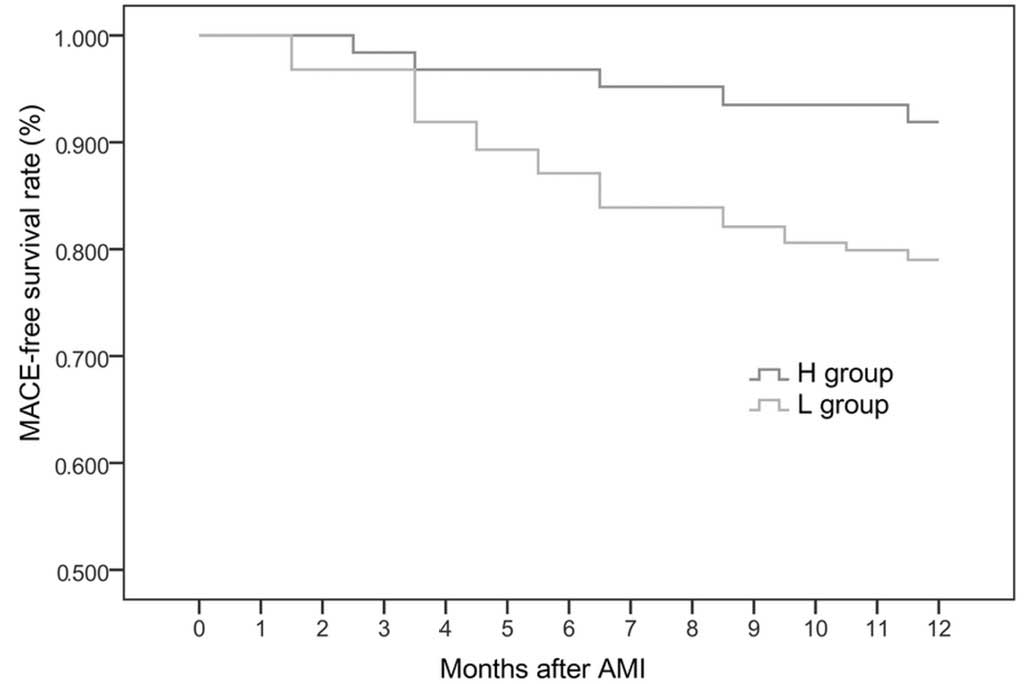
Incidence of MACE
At 6 months after the onset of AMI, the incidence of
MACE was markedly higher in the L group compared with the H group;
MACE-free survival rate was markedly higher in the H group compared
with the L group (Table II and
Fig. 1).
| Table II.Incidence of major adverse
cardiovascular events in the H and L groups (n=62 per group). |
Table II.
Incidence of major adverse
cardiovascular events in the H and L groups (n=62 per group).
| Time after AMI
(months) | H group, % (n) | L group, % (n) | P-value |
|---|
| 0 | 0.0 (0) | 0.0
(0) | – |
| 2 | 0.0 (0) | 3.2
(2) | 0.154 |
| 4 | 3.2 (2) | 8.1
(5) | 0.243 |
| 6 | 3.2 (2) | 12.9 (8) | 0.048 |
| 8 | 4.8 (3) | 16.1
(10) | 0.040 |
| 10 | 6.5 (4) | 19.4
(12) | 0.032 |
| 12 | 8.1 (5) | 21.0
(13) | 0.041 |
Logistic regression analysis
Logistic regression analysis was performed with MACE
as the dependent variable (yes=1, no=0) and gender, age, SBP,
diabetes, smoking, BMI, ACEI and β-blocker administration and VEGF
levels as the independent variables. The results suggested that a
low VEGF level (≤190 pg/ml) was an independent risk factor for MACE
(β=1.243; 95% CI, 1.018–1.326; P=0.026).
Discussion
The present study involved 124 patients with AMI
with single-vessel, proximal LAD disease, in which
revascularization was performed within 12 h. Plasma VEGF levels
were evaluated at 7 days after the onset of AMI. At the 12-month
follow-up, the incidence of MACE in the patients, including cardiac
mortality, heart failure, severe arrhythmia, cardiac shock and
post-infarction angina was recorded. The results showed that the
incidence of MACE in the H group was significantly higher compared
with the L group at the 6-12-month follow-ups, and that the plasma
VEGF levels were markedly elevated in the N-MACE group, as compared
with those in the MACE group. The correlation between plasma VEGF
levels and long-term prognosis in patients with AMI was
investigated using logistic regression analysis, which revealed
that a low VEGF level (β, 1.243; 95% CI, 1.018–1.326; P=0.026) was
an independent risk factor for MACE, while high VEGF levels
facilitated long-term prognosis in patients with AMI.
VEGF is a dimeric glycoprotein composed of two
identical polypeptide chains, which is produced by endothelial
cells, smooth muscle cells and macrophages. VEGF is able to promote
vascular endothelial cell mitosis, proliferation and migration,
increase vascular permeability, stimulate the expression of
vascular endothelial cells and induce angiogenesis (1). VEGF expression has been shown to be
promoted by several factors, including hypoxia, angiotensin,
advanced glycation end products, endotoxin and high glucose
(18,19). VEGF is known to be crucially involved
in the induction of vasculogenesis and angiogenesis, exerting a
marked effect on angiogenesis by binding to kinase insert
domain-containing receptor (20).
Furthermore, VEGF may exert a positive effect in the
revascularization of ischemic tissues, specifically through the
promotion of vascular endothelial cell proliferation and
neovascularization, increase of vascular permeability, and
improvement of collateral circulation (20). Sluimer et al (21) performed carotid endarterectomy in
patients with carotid atherosclerosis and observed an increase in
the mRNA and protein expression of hypoxia-inducible factor 1
(HIF-1α), VEGF expression and microvessel density in the early
stages of atherosclerosis. In addition, hypoxia correlated with
thrombosis, angiogenesis, HIF-1α and VEGF expression; therefore,
VEGF and HIF-1α were associated with atherosclerotic plaque
instability and were considered to be an index indicating plaque
instability in patients with ACS. Mao et al (22) used a rat model of myocardial ischemia
and ligated the left descending coronary artery. After 3 h the VEGF
expression in the ischemic myocardium had peaked, subsequently
decreasing 6 h later. Thus, the VEGF expression level was able to
assist in determining the degree of severity of ischemic injury.
Wang et al (23) showed that
short-term intermittent hypoxia has a cardioprotective effect,
which may result from the upregulation of VEGF and HIF-1α
expression in a rat model of ischemia/reperfusion injury. In 2003,
Heeschen et al (13) measured
the plasma VEGF levels of 1,090 patients with ACS at 8.7 h after
the onset of MI, and observed that the incidence of MACE was
increased in patients that exhibited significantly elevated plasma
VEGF levels at the 6-month follow-up. In 2012, Matsudaira et
al (12) detected the plasma
VEGF levels in 873 patients with AMI 7 days after the onset of MI
and found that the incidence of MACE was higher in patients with
low plasma VEGF levels (≤190 pg/ml) at the 6-month follow-up. The
present study included patients with AMI with single-vessel,
proximal LAD disease, with a similar myocardial ischemia and injury
area, in order to more accurately indicate the association between
VEGF and MACE. In 2012, Devaux et al (24) recruited 290 patients with AMI and
revealed that the risk of left ventricular remodeling was higher in
patients with lower VEGF levels. Zentilin et al (25) reported that the long-term expression
of VEGF-A165 and VEGF-B167 in the myocardium may reduce myocardial
fibrosis and enhance myocardial contractility to protect the viable
myocardium and improve ventricular remodeling. Furthermore,
previous studies have indicated that the plasma VEGF levels in
patients with AMI peaked on days 7–14 after the onset of AMI, and
in patients that underwent successful percutaneous
revascularization on day 7 (26–28).
Collectively, the aforementioned and present results indicate that
the increase in VEGF levels serves a crucial function in
cardiovascular repair.
In addition, the results of the aforementioned
studies suggest that due to the ischemia and hypoxia stimulation,
patients with AMI may be expected to secrete increased quantities
of VEGF to promote the angiogenesis and endothelial cell
proliferation, therefore promoting self-repair and cardiac
remodeling, which indicates the cardioprotective effect of VEGF
expression. In the present study, increased VEGF levels were shown
to facilitate long-term prognosis in patients with AMI, while low
VEGF levels were found to be independent risk factors for MACE,
which is consistent with the results from the study by Matsudaira
et al (12). By contrast,
Shimokawahara et al (29)
observed that in the acute period, the left ventricle volume index
of the high-serum-VEGF-concentration group of patients with MI was
significantly elevated compared with the
low-serum-VEGF-concentration group, while the intergroup
differences observed in the chronic period were not statistically
significant. In conclusion, high plasma VEGF levels at 7 days after
AMI onset facilitate the long-term prognosis in the same infarct
zone in patients with AMI, while low plasma VEGF levels are
independent risk factors for MACE. However, the correlation between
plasma VEGF levels at different stages, genetic factors and
prognosis in patients with AMI requires further investigation.
Acknowledgements
This study was supported by a grant from the
Shandong Science and Technology Development in Health and Medical
Care (grant no. 2013WS0072).
References
|
1
|
Giatromanolaki A, Sivridis E, Athanassou
N, Zois E, Thorpe PE, Brekken RA, Gatter KC, Harris AL, Koukourakis
IM and Koukourakis MI: The angiogenic pathway ‘vascular endothelial
growth factor/flk-1(KDR)-receptor’ in rheumatoid arthritis and
osteoarthritis. J Pathol. 194:101–108. 2001. View Article : Google Scholar : PubMed/NCBI
|
|
2
|
Gerber HP, McMurtrey A, Kowalski J, Yan M,
Keyt BA, Dixit V and Ferrara N: Vascular endothelial growth factor
regulates endothelial cell survival through the
phosphatidylinositol 3′-kinase/Akt signal transduction pathway:
Requirement for Flk-1/KDR activation. J Biol Chem. 273:30336–30343.
1998. View Article : Google Scholar : PubMed/NCBI
|
|
3
|
Deuse T, Peter C, Fedak PW, Doyle T,
Reichenspurner H, Zimmermann WH, Eschenhagen T, Stein W, Wu JC,
Robbins RC and Schrepfer S: Hepatocyte growth factor or vascular
endothelial growth factor gene transfer maximizes mesenchymal stem
cell-based myocardial salvage after acute myocardial infarction.
Circulation. 120(Suppl 11): S247–S254. 2009. View Article : Google Scholar : PubMed/NCBI
|
|
4
|
Soeki T, Tamura Y, Shinohara H, Tanaka H,
Bando K and Fukuda N: Serial changes in serum VEGF and HGF in
patients with acute myocardial infarction. Cardiology. 93:168–174.
2000. View Article : Google Scholar : PubMed/NCBI
|
|
5
|
Ogawa H, Suefuji H, Soejima H, Nishiyama
K, Misumi K, Takazoe K, Miyamoto S, Kajiwara I, Sumida H, Sakamoto
T, et al: Increased blood vascular endothelial growth factor levels
in patients with acute myocardial infarction. Cardiology. 93:93–99.
2000. View Article : Google Scholar : PubMed/NCBI
|
|
6
|
Lee KW, Lip GY and Blann AD: Plasma
angiopoietin-1, angiopoietin-2, angiopoietin receptor tie-2 and
vascular endothelial growth factor levels in acute coronary
syndromes. Circulation. 110:2355–2360. 2004. View Article : Google Scholar : PubMed/NCBI
|
|
7
|
Soeki T, Tamura Y, Shinohara H, Sakabe K,
Onose Y and Fukuda N: Serum hepatocyte growth factor predicts
ventricular remodeling following myocardial infarction. Circ J.
66:1003–1007. 2002. View Article : Google Scholar : PubMed/NCBI
|
|
8
|
Hagikura K, Fukuda N, Yokoyama S, Yuxin L,
Kusumi Y, Matsumoto T, Ikeda Y, Kunimoto S, Takayama T, Jumabay M,
et al: Low invasive angiogenic therapy for myocardial infarction by
retrograde transplantation of mononuclear cells expressing the VEGF
gene. Int J Cardiol. 142:56–64. 2010. View Article : Google Scholar : PubMed/NCBI
|
|
9
|
Ye L, Zhang W, Su LP, Haider HK, Poh KK,
Galupo MJ, Songco G, Ge RW, Tan HC and Sim EK: Nanoparticle based
delivery of hypoxia-regulated VEGF transgene system combined with
myoblast engraftment for myocardial repair. Biomaterials.
32:2424–2431. 2011. View Article : Google Scholar : PubMed/NCBI
|
|
10
|
Yin R, Feng J, Chen D and Wu H: Serum
levels of vascular endothelial growth factor in patients with
anginapectoris and acute myocardial infarct. Chin Med Sci J.
15:205–209. 2000.PubMed/NCBI
|
|
11
|
Hojo Y, Ikeda U, Zhu Y, Okada M, Ueno S,
Arakawa H, Fujikawa H, Katsuki T and Shimada K: Expression of
vascular endothelial growth factor in patients with acute
myocardial infarct. J Am Coll Cardiol. 35:968–973. 2000. View Article : Google Scholar : PubMed/NCBI
|
|
12
|
Matsudaira K, Maeda K, Okumura N,
Yoshikawa D, Morita Y, Mitsuhashi H, Ishii H, Kondo T and Murohara
T: Nagoya Acute Myocardial Infarction Study (NAMIS) Group: Impact
of low levels of vascular endothelial growth factor after
myocardial infarction on 6-month clinical outcome: Results from the
Nagoya Acute Myocardial Infarction Study. Circ J. 76:1509–1516.
2012. View Article : Google Scholar : PubMed/NCBI
|
|
13
|
Heeschen C, Dimmeler S, Hamm CW, Boersma
E, Zeiher AM and Simoons ML: CAPTURE (c7E3 Anti-Platelet Therapy in
Unstable REfractory angina) Investigators: Prognostic significance
of angiogenic growth factor serum levels in patients with acute
coronary syndromes. Circulation. 107:524–530. 2003. View Article : Google Scholar : PubMed/NCBI
|
|
14
|
Canadian Cardiovascular Society; American
Academy of Family Physicians; American College of Cardiology;
American Heart Association. Antman EM, Hand M, Armstrong PW, Bates
ER, Green LA, Halasyamani LK, Hochman JS, Krumholz HM, Lamas GA,
Mullany CJ, et al: 2007 focused update of the ACC/AHA 2004
guidelines for the management of patients with ST-elevation
myocardial infarction: A report of the American College of
Cardiology/American Heart Association Task Force on Practice
Guidelines. J Am Coll Cardiol. 51:210–247. 2008. View Article : Google Scholar : PubMed/NCBI
|
|
15
|
Braunwald E, Antman EM, Beasley JW, Califf
RM, Cheitlin MD, Hochman JS, Jones RH, Kereiakes D, Kupersmith J,
Levin TN, et al: American College of Cardiology; American Heart
Association. Committee on the Management of Patients With Unstable
Angina: ACC/AHA 2002 guideline update for the management of
patients with unstable angina and non-ST-segment elevation
myocardial infarction - summary article: A report of the American
College of Cardiology/American Heart Association task force on
practice guidelines (Committee on the Management of Patients with
Unstable Angina). J Am Coll Cardiol. 40:1366–1374. 2002. View Article : Google Scholar : PubMed/NCBI
|
|
16
|
Austen WG, Edwards JE, Frye RL, Gensini
GG, Gott VL, Griffith LS, McGoon DC, Murphy ML and Roe BB: A
reporting system on patients evaluated for coronary artery disease.
Report of the Ad Hoc Committee for Grading of Coronary Artery
Disease, Council on Cardiovascular Surgery, American Heart
Association. Circulation. 51(Suppl 4): 5–40. 1975. View Article : Google Scholar : PubMed/NCBI
|
|
17
|
Poulsen SH: Clinical aspects of left
ventricular diastolic function assessed by Doppler echocardiography
following acute myocardial infarction. Dan Med Bull. 48:199–210.
2001.PubMed/NCBI
|
|
18
|
Bates DO, Hillman NJ, Williams B, Neal CR
and Pocock TM: Regulation of microvascular permeability by vascular
endothelial growth factors. J Anat. 200:581–597. 2002. View Article : Google Scholar : PubMed/NCBI
|
|
19
|
Maeda K, Chung YS, Ogawa Y, Takatsuka S,
Kang SM, Ogawa M, Sawada T and Sowa M: Prognostic value of vascular
endothelial growth factor expression in gastric carcinoma. Cancer.
77:858–863. 1996. View Article : Google Scholar : PubMed/NCBI
|
|
20
|
Hoberg M, Schmidt EL, Tuerk M, Stark V,
Aicher WK and Rudert M: Induction of endostatin expression in
meniscal fibrochondrocytes by co-culture with endothelial cells.
Arch Orthop Trauma Surg. 129:1137–1143. 2009. View Article : Google Scholar : PubMed/NCBI
|
|
21
|
Sluimer JC, Gasc JS, van Wanroij JL,
Kisters N, Groeneweg M, Sollewijn Gelpke MD, Cleutjens JP, van den
Akker LH, Corvol P, Wouters BG, et al: Hypexia, hypoxia-inducible
transcription factor, and macrophages in human atherosclerotic
plaques are correlated with intraplaque angiogenesis. J Am Coll
Cardial. 51:1258–1265. 2008. View Article : Google Scholar
|
|
22
|
Mao RM, Du ZB, Gao WM, Mi L and Zhu BL:
Time-dependent expression of vascular endothelial growth factor
after acute myocardial ischemia in rats. Fa Yi Xue Za Zhi.
28:179–184. 2012.(In Chinese). PubMed/NCBI
|
|
23
|
Wang Z and Si LY: Hypoxia-inducible
factor-1α and vascular endothelial growth factor in the
cardioprotective effects of intermittent hypoxia in rats. Ups J Med
Sci. 118:65–74. 2013. View Article : Google Scholar : PubMed/NCBI
|
|
24
|
Devaux Y, Vausort M, Azuaje F, Vaillant M,
Lair ML, Gayat E, Lassus J, Ng LL, Kelly D, Wagner DR and Squire
IB: Low levels of vascular endothelial growth factor B predict left
ventricular remodeling after acute myocardial infarction. J Card
Fail. 18:330–337. 2012. View Article : Google Scholar : PubMed/NCBI
|
|
25
|
Zentilin L, Puligadda U, Lionetti V,
Zacchigna S, Collesi C, Pattarini L, Ruozi G, Camporesi S, Sinagra
G, Pepe M, et al: Cardiomyocyte VEGFR-1 activation by VEGF-B
induces compensatory hypertrophy and preserves cardiac function
after myocardial infarction. FASEB J. 24:1467–1478. 2010.
View Article : Google Scholar : PubMed/NCBI
|
|
26
|
Ferrara N, Gerber HP and LeCouter J: The
biology of VEGF and its receptors. Nat Med. 9:669–676. 2003.
View Article : Google Scholar : PubMed/NCBI
|
|
27
|
Gruchala M, Roy H, Bhardwaj S and
Ylä-Herttuala S: Gene therapy for cardiovascular diseases. Curr
Pharm Des. 10:407–423. 2004. View Article : Google Scholar : PubMed/NCBI
|
|
28
|
Ratner M: Genentech discloses safety
concerns over Avastin. Nat Biotechnol. 22:11982004. View Article : Google Scholar : PubMed/NCBI
|
|
29
|
Shimokawahara H, Jougasaki M, Setoguchi M,
Ichiki T, Sonoda M, Nuruki N, Nakashima H, Murohara T and Tsubouchi
H: Relationship between vascular endothelial growth factor and left
ventricular dimension in patients with acute myocardial infarction.
J Cardiol. 64:360–365. 2014. View Article : Google Scholar : PubMed/NCBI
|