Introduction
Sepsis, one of the main issues encountered in the
majority of health care centers, is a life-threatening disease that
causes widespread mortality worldwide (1). The mortality rate of uncomplicated
sepsis is ~25%, increasing to 80% in patients that proceed to
develop multiple organ failure (2).
Sepsis is associated with the presence of pathogenic microorganisms
or their toxins in the bloodstream. Oxidative stress has been
consistently reported in patients with sepsis, and thus
antioxidants may be used as a potential therapy (3). However, the effect of antioxidants
administrated in septic shock is limited (4); therefore, the expression levels of
certain antioxidants (such as the glutathione S-transferases gene
family), oxidative stress responsive genes (including oxidases,
peroxidases, catalase and superoxide dismutase) or
anti-inflammatory genes (such as interleukins) can be detected in
the tissues in response to sepsis or any curative treatment
(5,6). Oxidative stress is indicated by
increased levels of lipid peroxides, direct detection of
circulating radicals and decreased antioxidant concentrations
(7). Mitochondrial dysfunction
resulting from oxidative stress has been suggested to serve a role
in the development of multiorgan failure in sepsis, including liver
and lung failure (3,8). Approximately 50% of patients with
severe sepsis also develop acute lung injury (9,10).
Pericytes in lung tissue have been shown to produce an increase in
pro-inflammatory cytokines in response to bacterial
lipopolysaccharide (LPS) (11).
Tumour necrosis factor (TNF) and interferon-γ
(IFN-γ) are of particular importance in the development of septic
shock (12–15). Therefore, the development of
individual sepsis treatments has focused on the regulation of TNF
expression (16,17). However, previous clinical trials
investigating specific anti-TNF sepsis treatments have demonstrated
the complexity of this disease and the involvement of various
cytokines with overlapping functions (17). IFN-γ is also a crucial regulator of
LPS-induced pathology (18,19). Treatment with IFN-γ or a neutralizing
antibody against IFN-γ has been found to alter the lethal outcomes
of several types of Gram-negative bacterial infections and
endotoxic shock (19). IFN-γ
receptor-deficient mice are relatively resistant to LPS-induced
septic shock (20,21).
INF-γ serves an important role in the regulation of
innate and acquired antimicrobial immunity. The expression of INF-γ
is regulated by a set of complex interactions between accessory
cells, such as macrophages, dendritic cells, T lymphocytes and
natural killer cells (22,23). INF-γ amplifies antimicrobial immune
responses by stimulating macrophage functions such as phagocytosis,
respiratory burst activity, antigen presentation and cytokine
secretion (18,21,24).
Previous studies have suggested that IFN-γ may increase the
responsiveness to LPS by altering the signal transduction pathway,
in particular through the upregulation of the Toll-like receptor 4
gene expression (25), or promotion
of IL-1 receptor-associated kinase expression and its association
with myeloid differentiation primary response gene 88 (26).
Despite recent advances in antibiotic therapy, no
significant improvements have been accomplished in the treatment of
sepsis. Thus, there is an urgent demand for the development of a
novel therapy for sepsis management. The increase of
antibiotic-resistant pathogenic bacteria has stimulated the search
for antimicrobial agents from alternative sources (27,28).
Crude extract from the sea cucumber, also known as Holothuria
atra (H. atra), contains cytostatic, antifungal,
hemolytic, anticancer and antioxidant phenolic compounds, while it
also has immunomodulatory effects. This extract has been found to
have a potential hepatoprotective activity against
thioacetamide-induced liver injury in a rat model (29). In addition, the curative effects of
the sea cucumber extract was reported against DMBA-induced
hepatorenal diseases in rats (30,31).
Apoptosis, another prominent feature of sepsis,
involves a mechanism of closely-regulated disassembly of cells
resulting from the activation of caspases, which are specialized
proteases. In septic animal models, increased apoptotic cell death
has also been reported in parenchymal cells, including intestinal
and lung epithelial cells (32,33).
Furthermore, in situ localization of cleaved caspase-3 may
have an application in the histological labeling of cells in
apoptosis (34). Lysophospholipids
from H. atra were also shown to inhibit
H2O2-induced apoptosis in macrophages
(35).
Cecum ligation and puncture (CLP) is currently the
most widely used animal model of sepsis (36,37). In
the CLP rat model, autophagy was also induced in multiple organs,
including the lung and liver (38,39). In
the present study, the potent in vivo efficacy of sea
cucumber body wall extract against induced sepsis in a CLP rat
model was investigated at molecular and histopathological
levels.
Materials and methods
Sample collection and preparation of
H. atra extract (HaE)
Sea cucumbers (H. atra; n=50) were collected
from the Thuwal area on Saudi Arabia's Red Sea coast. The taxonomic
identity of the samples was confirmed based on the studies of
Purcell et al (40). The
animals were transported to the Medical Laboratory of Applied
Medical Sciences, Taif University, (Turabah, Saudi Arabia), in an
ice box. They were rinsed thoroughly, removing any internal organs
and body fluids, and then the animals' body wall was soaked in
appropriate amounts of methanol-water mixture (50:50) and stirred
using a magnetic stirrer for 16 h. The mixture was filtered twice.
Finally, the two extracts were pooled together and concentrated in
a rotary evaporator during which the extract was evaporated at low
pressure in a double boiler at 30°C using a LABROTA 4001 efficient
(Heidolph Instruments GmbH & Co., Schwabach, Germany) to avoid
degradation of compounds, for 2 h. The powdered extract was
obtained by freeze drying and was stored at −20°C until further use
(41).
Animals
All animal procedures were approved by the Ethical
Committee Office of the Scientific Dean of Taif University (Taif,
Saudi Arabia).
In total, 30 adult male albino rats (Rattus
norvegicus; age, 6–8 weeks) weighing 150–170 g were obtained
from the King Fahd Research Unit at King Abdulaziz University
(Jeddah, Saudi Arabia). The rats were housed in polypropylene cages
in an air conditioned room at a temperature of 25±2°C and under
natural light cycle. They were fed standard chow pellets and had
access to water ad libitum. The rats were kept for 1 week
for acclimatization and then divided into three groups (10 animals
in each) as follows: Sham (Sh) group, which was used as a negative
control (received distilled water and underwent surgery along with
cecal manipulations, but without ligation and puncture); sepsis
(Se) group, which was surgically subjected to CLP [sepsis was
achieved in rats by cecal ligation at a point ~1 cm from the cecal
tip and punctured with a 20-gauge needle (36)], and was used as positive control; and
the Ho group, in which animals were subjected to CLP and orally
administered 200 mg/kg body weight HaE, once daily for 7 days. All
groups were handled under sterile and antiseptic conditions. All
animals were sacrificed by inhalation of diethyl ether, and
dissected after 7 days. The animals were observed daily subsequent
to the surgery. The mortality rate was calculated in all groups
after the incidence of the initial case of mortality. The death
rate was scored each day for 7 days until the end of the
experiment. The mortality rate was presented as a percentage.
The rat organs (liver and lung) were collected and
applied for RNA extraction and histopathological examinations.
Mortality rate study
Following CLP surgery, the mortality rate and
symptoms of sepsis were calculated over the subsequent 7 days for
the 3 experimental groups. Rat mortality was recorded every 24 h
until the 7th day and is expressed as a percentage.
RNA extraction and quantification
Total RNA was extracted from the liver and lungs of
all rat groups according to the method described by Attia et
al (42). RNA samples were
diluted in diethylpyrocarbonate water to 40 µg/ml according to
spectroscopy quantification using a Bio-Rad SmartSpec Plus
UV/Visible Spectrophotometer (Bio-Rad Laboratories, Inc., Hercules,
CA, USA).
Primers
All primers were designed (based on the gene
sequences published in the GenBank database) using the Primer3Plus
online software (http://primer3plus.com/). The primers were
manufactured by Bioron GmbH (Ludwigshafen, Germany). Primers
sequences and polymerase chain reaction (PCR) product sizes are
presented in Table I. The
investigated genes included Caspase-3, glutathione
S-transferase α2 (Gsta2), catalase (Cat), superoxide
dismutase 1 (Sod1), TNF, IFN-γ, interleukin 1a
(Il1a) and interleukin 1b (Il1b).
 | Table I.Primers sequence used to amplify a
partial sequence of the target gene. |
Table I.
Primers sequence used to amplify a
partial sequence of the target gene.
| Gene | Accession no. | Primer
sequence | Primer
position | Band size (bp) |
|---|
|
Caspase-3 | NM_012922 | F:
5′-TTGGCTTGTTGAAGGCTACC-3′ | 1,540 | 400 |
|
|
| R:
5′-GCAGGAGCTTCTGATCTGGT-3′ | 1,939 |
|
| Gsta2 | NM_017013 | F:
5′-GGCAAAAGACAGGACCAAAA-3′ |
432 | 231 |
|
|
| R:
5′-GGCTGCAGGAACTTCTTCAC-3′ |
662 |
|
| Cat | NM_012520 | F:
5′-GACACATCCGGGCTCACTAT-3′ | 1,130 | 238 |
|
|
| R:
5′-GAGCCTAAGCCTGAATGCAC-3′ | 1,367 |
|
| Sod1 | NM_017050 | F:
5′-CCACTGCAGGACCTCATTTT-3′ |
269 | 216 |
|
|
| R:
5′-CACCTTTGCCCAAGTCATCT-3′ |
484 |
|
| TNF | NM_012675 | F:
5′-ATGGGCTCCCTCTCATCAGT-3′ |
341 | 547 |
|
|
| R:
5′-GGCTGGGTAGAGAACGGATG-3′ |
887 |
|
| IFN-γ | AH002184 | F:
5′-TCCCTCCCCACTCCATTAGG-3′ | 1,130 | 526 |
|
|
| R:
5′-ATTCCTCTGGTCAGCAGCAC-3′ | 1,655 |
|
| Il1a | NM_017019 | F:
5′-CATGCAGCTCATCATGCTTT-3′ | 1,726 | 172 |
|
|
| R:
5′-CTTGGGCTCAAAAATGTGGT-3′ | 1,897 |
|
| Il1b | NM_031512 | F:
5′-AGGCTTCCTTGTGCAAGTGT-3′ |
26 | 230 |
|
|
| R:
5′-TGAGTGACACTGCCTTCCTG-3′ |
255 |
|
| Gapdh | NM_017008 | F:
5′-AGACAGCCGCATCTTCTTGT-3′ |
28 | 323 |
|
|
| R:
5′-TACTCAGCACCAGCATCACC-3′ |
350 |
|
Semi-quantitative reverse
transcription-PCR (RT-PCR) analysis
Total RNA (2 µg) was reverse transcribed into cDNA
using RevertAid First Strand cDNA Synthesis kit (Fermentas; Thermo
Fisher Scientific Inc., Waltham, MA, USA). The reaction was
incubated for 60 min at 42°C and terminated by heating at 70°C for
5 min. Next, 1 ml cDNA was used for PCR analysis, which was
performed using a Perkin Elmer GeneAmp 9600 system (PerkinElmer,
Inc., Waltham, MA, USA). The PCR cycling conditions were as
follows: Initial cycle of 10 min at 95°C, 45 sec at 54°C and 1 min
at 72°C; followed by 30 cycles of denaturation at 95°C for 1 min,
annealing at 54°C for 45 sec and extension at 72°C for 1 min; and
then a final extension step at 72°C for 7 min. The total volume of
reactions mixture was 25 µl and contained 1 unit of AmpliTaq Gold
(Applied Biosystems; Thermo Fisher Scientific Inc.), 1X AmpliTaq
buffer, 1.5 mM MgCl2, 2.5 mM dNTPs and 10 pmol of
forward and reverse primers. The expression of Gapdh was
detected as a reference value using specific primers (Table I). A negative control containing RNA
was used to rule out genomic DNA contamination. The PCR products
were confirmed by 2% agarose gel electrophoresis, and the band
density was measured using ImageJ version 1.48 software (http://imagej.nih.gov/ij/).
Histopathological examination
Tissue samples collected from the liver and lungs of
rats were fixed in 10% neutral buffer formalin solution, washed in
tap water, dehydrated through an upgraded series of ethanol (50,
70, 80, 90 and 95%, followed by absolute ethanol)., cleared by
xylene and then embedded in paraffin. The paraffin-embedded samples
were cut into 5 µm sections, which were then routinely stained with
hematoxylin and eosin (Sigma-Aldrich, St. Louis, MO, USA) as
previously described (43).
Immunohistochemical analysis
The paraffin-embedded samples were cut into 3-µm
sections and mounted on positively charged slides for caspase-3
immunohistochemical examination. Sections were dewaxed, rehydrated
and autoclaved at 95°C for 20 min in antigen retrieval buffer (10
mM citrate buffer, pH 6). After washing with phosphate-buffered
saline (PBS), endogenous peroxidase was blocked using 3%
H2O2 in methanol for 15 min. A primary
rat-specific antibody for caspase-3 (cat.no. RB-1197-B0,-B1; Thermo
Fisher Scientific Inc.) was added following dilution in PBS
(1:100), and incubated for 30 min. The slides were then washed
three times for 3 min each with PBS. Subsequently, a horseradish
peroxidase-conjugated goat anti-mouse IgG secondary antibody (cat.
no. 32230; Thermo Fisher Scientific Inc.) was applied to the tissue
sections and co-incubated for 30 min. The slides were washed three
times for 3 min each with PBS, and then visualized by adding metal
enhanced DAB substrate working solution (Thermo Fisher Scientific
Inc.) to the tissues and incubating for 10 min. Next, the slides
were washed two times with PBS (3 min each time) and then
counterstained by adding an adequate amount of hematoxylin to the
slide to cover the entire tissue surface (44). The immune reactivity score used to
evaluate the intensity of immunohistochemical staining and the
proportion of the stained cells was classified as i) absent (0);
ii) mild, 25–50%; and iii) strong, >50%.
Statistical analysis
The results are expressed as the mean ± standard
error of 10 different rats per group. Statistical analysis was
performed with analysis of variance and Fisher's post-hoc test,
with P<0.05 considered to indicate statistically significant
differences.
Results
Mortality rate following CLP
surgery
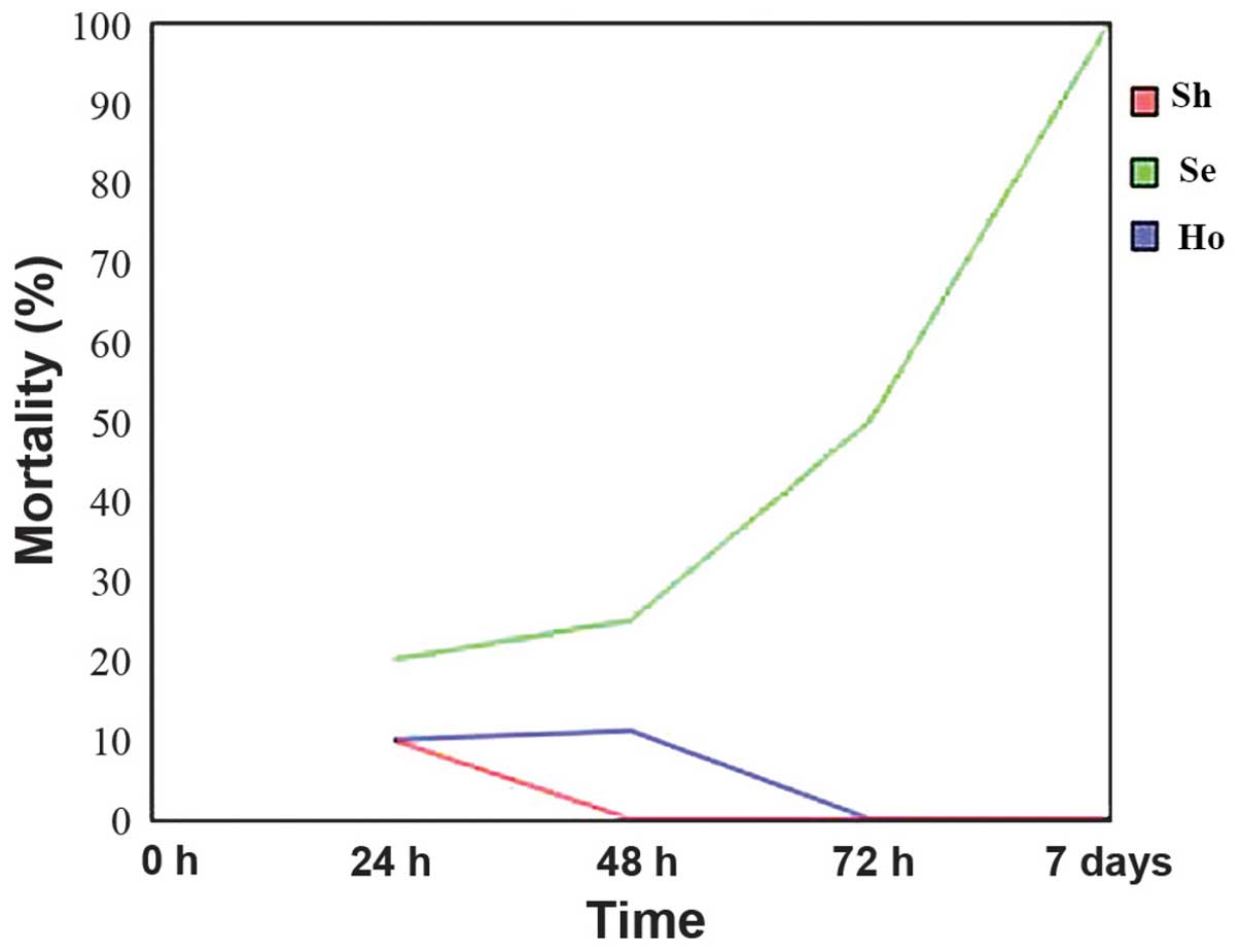
At 24 h after CLP surgery, the rats clearly
displayed the sepsis symptoms, such as decreased motor activities,
ocular exudates and ruffled fur. In the Sh group, the mortality
rate was 10% at 24 h decreasing to 0% after 48 h. In the Se group,
the mortality rate was 20% after 24 h from CLP surgery, increasing
to 100% after 7 days in comparison with the negative group (Sh).
Although the mortality rate was 10% at 48 h after the CLP surgery
in the Ho-treated group, it decreased to 0% after 72 h and remained
at this rate until the end of the experiment (Fig. 1).
Molecular detection of gene
expression
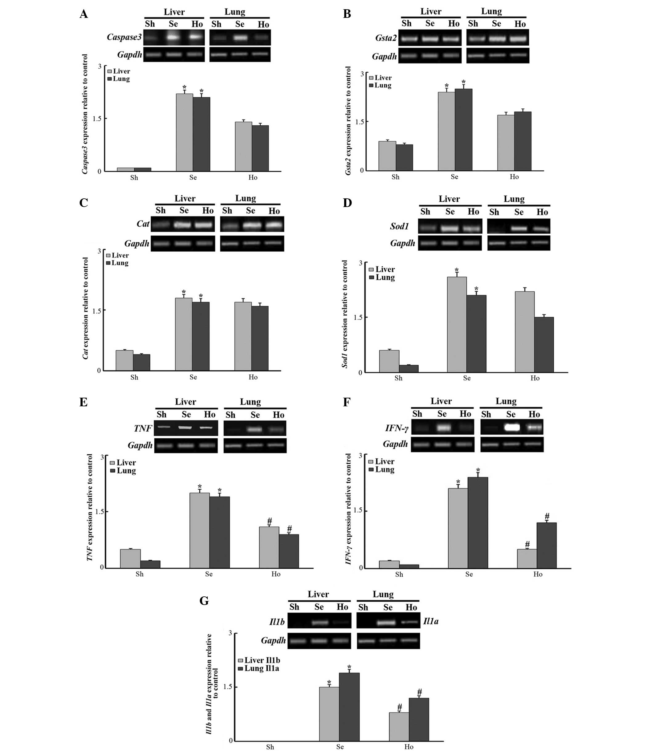
Eight genes, including Caspase-3,
Gsta2, Cat, Sod1, TNF, IFN-γ,
Il1b and Il1a, were tested for their expression in
the liver and lungs of the adult male albino rats in the three
groups at 7 days after CLP (Fig. 2),
with the expression of Gapdh used as an internal control. As shown
in Fig. 2A, the expression of
Caspase-3 was found to be significantly increased (P<0.05) in
the liver and lung tissues of the Se group in response to sepsis
when compared with the negative control (Sh) tissues; however, the
expression was reduced in the Ho group compared with the Se group.
In addition, the expression of the antioxidant Gsta2 gene was
significantly increased (P<0.05) in the organs of the Se
compared with the Sh group, whilst the Ho group did not display a
significant difference compared with the Se group (Fig. 2B), but it was lower in the Ho group.
Expression levels of the oxidative stress responsive genes, Cat and
Sod1, were increased in the two organs in response to sepsis stress
in the Se and Ho groups (Fig. 2C and
D). Expression was significantly increased in Se group compared
with the Sh group (P<0.05), whilst the Ho group did not display
any significance compared with Se group. Furthermore, the
expression levels of anti-inflammatory genes, TNF and IFN-γ, as
well as of the liver tissue-specific Il1b and the lung
tissue-specific Il1a, were significantly increased in the Se group
in response to sepsis when compared with the Sh group, whereas the
expression of the aforementioned genes was significantly reduced
(P<0.05) in the Ho group, when compared with Se group (Fig. 2E–G). The results indicated that HaE
increased the expression of oxidative stress as well as antioxidant
genes, while decreased the expression of apoptotic (Caspase-3),
anti-inflammatory (TNF and IFN-γ), interleukins (1a and 1b) genes
in septic rats. This suggests that the extract of sea cucumber
H. atra may posses antiseptic and anti-inflammatory
properties.
 | Figure 2.Expression of the responsive genes
(A) Caspase-3, (B) Gsta2, (C) Cat, (D)
Sod1, (E) TNF and (F) IFN-γ in the liver and
lung tissues, as well as of (G) Il1b in the liver and
Il1a in the lung tissues, in Sh, Se and Ho rat groups. Data
represent the mean ± standard error. *P<0.05 vs. Sh group;
#P<0.05 vs. Se group. Sh, sham (negative control)
group; Se, CLP-induced sepsis group; Ho, sepsis and Holothuria
atra extract-treated group; CLP, cecal ligation and puncture;
Gsta2, glutathione S-transferase α2; Cat, catalase;
Sod1, superoxide dismutase 1; TNF, tumor necrosis
factor; IFN, interferon; Il, interleukin. |
Histopathological examination
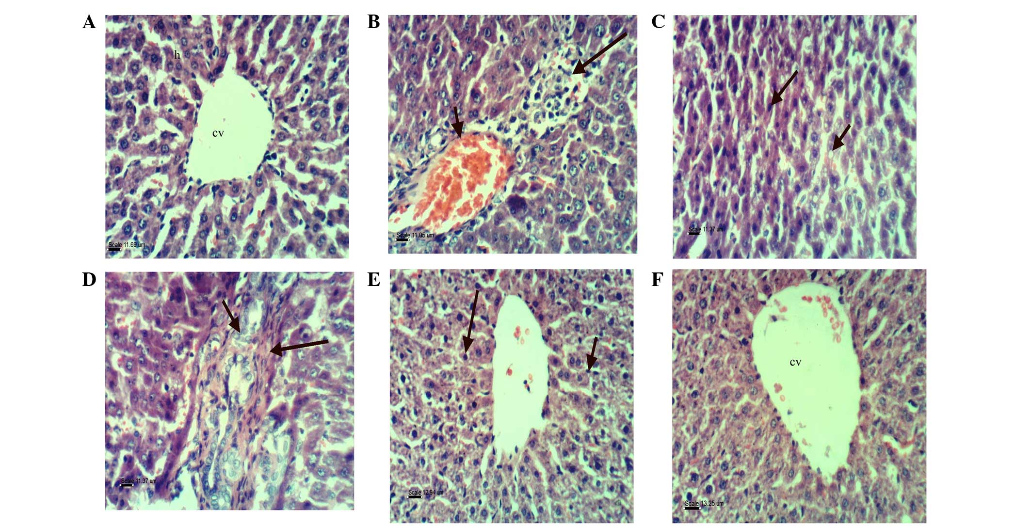
The negative control (Sh) group showed normal
architecture of the liver, with hepatic lobules around the central
vein and each lobule consisting of hepatic cords. The hepatocytes
represented the hepatic cords and consisted of polygonal cells with
centrally basophilic nuclei and clear acidophilic cytoplasm
(Fig. 3A). However, the liver of the
Se group presented congestion of the hepatoportal blood vessel,
focal hepatic necrosis associated with inflammatory cell
infiltration (Fig. 3B), fatty change
of hepatocytes, sporadic necrosis of hepatocytes (Fig. 3C), hyperplasia of the epithelial
lining of bile duct and fibroplasia in the portal tract (Fig. 3D). In addition, certain examined
sections from the Se group showed Küpffer cell activation and
necrosis of sporadic hepatocytes (Fig.
3E). By contrast, sections from the Ho group revealed apparent
normal hepatic parenchyma (Fig.
3F).
 | Figure 3.Photomicrograph of rat livers in the
various groups: (A) Sham group, showing the normal histological
structure of hepatic lobule (bar, 11.69 µm); (B) Se group, showing
the congestion of hepatoportal blood vessel, focal hepatic necrosis
(short arrow) associated with inflammatory cells infiltration (long
arrow; H&E; bar, 11.05 µm); (C) Se group, showing fatty change
of hepatocytes (short arrow) and sporadic necrosis of hepatocytes
(long arrow; bar, 11.37 µm); (D) Se group, showing hyperplasia of
epithelial lining of the bile duct (short arrow) and fibroplasia in
portal tract (long arrow; bar, 11.37 µm); (E) Se group, showing
Küpffer cell activation (short arrow) and necrosis of sporadic
hepatocytes (long arrow; bar, 12.94 µm); and (F) Sepsis and
Holothuria atra extract treatment group, showing apparent
normal hepatic parenchyma (bar, 13.25 µm). All stained with
hematoxylin-eosin. Se, sepsis group; cv, central vein; h,
hepatocyte. |
The lung histological examination revealed no
evidence of sepsis in the Sh group, which appeared to have a normal
structure of alveoli and alveolar sacs with thin alveolar septum
(Fig. 4A). By contrast, the lung
tissues of the Se group showed marked edema, hemorrhage, leukocyte
infiltration and alveolar septal thickening (Fig. 4B), whereas tissue sections from the
Ho group presented slight interstitial pneumonia (Fig. 4C).
Immunohistochemistry
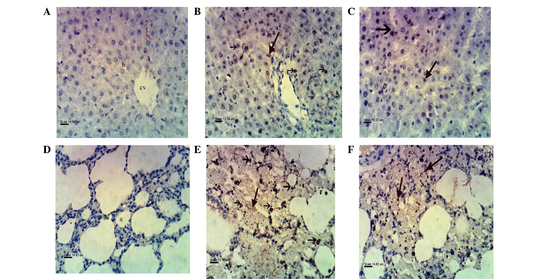
Immunohistochemical staining of Caspase-3 was
localized in the nuclei of hepatocytes. The liver tissues of the Sh
group showed no expression of Caspase-3 (Fig. 5A), whereas strong expression was
observed in the Se group (Fig. 5B)
and moderate expression in the Ho group (Fig. 5C). Upon investigation of lung
tissues, the expression of Caspase-3 was found to be localized in
the nuclei of pulmonary cells. The lung tissues of the Sh group
showed no expression of Caspase-3 (Fig.
5D), strong expression in the Se group (Fig. 5E) and moderate expression in the Ho
group (Fig. 5F).
Discussion
H. atra is one of the most important species
in the sea cucumber family, and it is found worldwide, including in
the Red Sea region (40,45,46). The
extract of H. atra has been evaluated for the presence of
bioactive compounds and its various biological activities (47). In the present study, the
administration of HaE was found to reduce the mortality rate in the
CLP rats (Ho group), which was evidently elevated in the CLP rats
that did not receive HaE (Se group). The decrease in sepsis-induced
mortality upon HaE treatment can be explained according to the
findings of Dhinakaran and Lipton (47), who stated that the extract of sea
cucumber has antimicrobial activity. The Se group exhibited
molecular and histopathological changes in the liver and lung
tissues, which were characterized by increased expression of
oxidative stress, antioxidant and anti-inflammatory genes, as well
as a congestion of the hepatoportal blood vessel and focal hepatic
necrosis associated with inflammatory cell infiltration. The Se rat
lungs showed marked edema, hemorrhage and alveolar septal
thickening, and these findings agreed with the observations of Ates
et al (10), Esmat et
al (48) and Baiomy and Saad
(49).
The expression levels of oxidative stress and
antioxidant genes can be used as markers to detect the response to
microbial infection (50). The
administration of HaE upregulated the expression of the antioxidant
gene Gsta2, as well as of the oxidative stress responsive
genes, Cat and Sod1. In addition, HaE administration
downregulated the anti-inflammatory genes TNF, IFN-γ,
liver Il1b and lung Il1a, and these results were in
agreement with previous observation (29–31).
Furthermore, these findings were confirmed by histopathological
analysis in the current study, which showed apparent normal hepatic
parenchyma and slight interstitial pneumonia in the Ho group.
Several studies have previously reported that TNF
and IFN-γ are involved in the development of septic shock (12–20,38).
Interleukins 1A and 1B, two members of interleukin-1 family
(51), are also involved in sepsis.
IL1A is responsible for the production of inflammation, as well as
the promotion of fever and sepsis (52), while IL1B is involved in a variety of
cellular activities, including cell proliferation, differentiation
and apoptosis (53). Based on the
present study results, we can speculate that the antimicrobial
activity of HaE resulted in a decrease in sepsis, and consequently
the expression levels of TNF and IFN-γ were
reduced.
Various studies have investigated the effects of the
sea cucumber components, including polysaccharides, which exhibited
a variety of biological activities, such as anti-tumor (54), anti-oxidation (55,56) and
anti-apoptotic (35) activities.
Chenghui et al (57) and Chen
et al (58) investigated the
antioxidant properties of peptides and hydrolysates extracted from
different species of sea cucumbers and found that hydrolysates have
a considerable antioxidant activity, which may be associated with
the presence of antioxidant peptides. In addition, it was reported
that the presence of the active phenolic compounds in the body wall
of the sea cucumbers may be due to phenolic-rich materials, such as
phytoplankton and particles derived from degrading marine
macroalgae, which are the main sources of food for sea cucumbers
(59,60). Furthermore, the body wall of sea
cucumber contains chlorogenic acid, which has been found to have a
potential hepatoprotective effect in several animal models of liver
injury (61).
The results of the present study showed that the
expression of Caspase-3 gene, which was highly elevated in
Se group, was decreased in response to administration of HaE in the
Ho group. This finding was supported by the immunohistochemical
staining of Caspase-3 in liver and lung tissues, showing no
expression of Caspase-3 in the Sh group, strong expression in the
Se group and moderate sexpression in the Ho group; these findings
coincided with the observations of Hu et al (62). According to the analysis of these
data at gene expression and histopathological levels, we speculate
that the beneficial effect of HaE of sea cucumber against sepsis
may be attributed to its antioxidant components and antiapoptotic
activity. Thus, HaE may be a potential functional agent for the
improvement of survival in sepsis.
In conclusion, the data presented in the current
study indicated that HaE is a useful natural product that is able
to mitigate the liver and lung damages resulting from induced
sepsis by CLP. It possesses antioxidant, antitumor and
antiapoptotic activities. The fact that the Ho group revealed
apparent normal hepatic and lung parenchyma may be attributed to
its antioxidant components, where necrosis and congestion
decreased, which may also be attributed to its anti-inflammatory
and antiapoptotic activities. Purification and identification of
these structures in future studies is warranted.
It appears that with further research in this field,
the extracts of marine organisms, including those of sea cucumbers,
may be used as antiseptic, antioxidant, antitumor and antiapoptotic
agents.
References
|
1
|
Raoofi R, Salmani Z, Moradi F, Sotoodeh A
and Sobhanian S: Procalcitonin as a marker for early diagnosis of
sepsis. Am J Infect Dis. 10:15–20. 2014. View Article : Google Scholar
|
|
2
|
Ma N, Xing C, Xiao H, Wang Y, Wang K, Hou
C, Han G, Chen G, Marrero B, Wang Y, et al: C5a regulates
Il-12+ DC migration to induce pathogenic Th1 and Th17
cells in sepsis. PLoS One. 8:e697792013. View Article : Google Scholar : PubMed/NCBI
|
|
3
|
Galley HF: Oxidative stress and
mitochondrial dysfunction in sepsis. Br J Anaesth. 107:57–64. 2011.
View Article : Google Scholar : PubMed/NCBI
|
|
4
|
Abuşoğlu S, Onur EÇelik Ht, Güvenç Y,
Sakarya M, Sakarya A, Var A and Uyanik BS: The effect of lidocaine
on liver tissue lipid peroxide levels in septic rat model. Int J
Mevlana Med Sci. 1:31–34. 2013.
|
|
5
|
Mansour AA, Salam MA and Saad YM: Mice
(Mus musculus) genome responses to methotrexate (MTX) and
some plant extracts. Life Sci J. 9:4881–4886. 2012.
|
|
6
|
Bergquist M, Nurkkala M, Rylander C,
Kristiansson E, Hedenstierna G and Lindholm C: Expression of the
glucocorticoid receptor is decreased in experimental Staphylococcus
aureus sepsis. J Infect. 67:574–583. 2013. View Article : Google Scholar : PubMed/NCBI
|
|
7
|
Hill AL, Lowes DA, Webster NR, Sheth CC,
Gow NA and Galley HF: Regulation of pentraxin-3 by antioxidants. Br
J Anaesth. 103:833–839. 2009. View Article : Google Scholar : PubMed/NCBI
|
|
8
|
Nesseler N, Launey Y, Aninat C, Morel F,
Mallédant Y and Seguin P: Clinical review: The liver in sepsis.
Crit Care. 16:2352012. View
Article : Google Scholar : PubMed/NCBI
|
|
9
|
Ozturk E, Demirbilek S, Begec Z, Surucu M,
Fadillioglu E, Kirimlioglu H and Ersoy MO: Does leflunomide
attenuate the sepsis-induced acute lung injury? Pediatr Surg Int.
24:899–905. 2008. View Article : Google Scholar : PubMed/NCBI
|
|
10
|
Ates I, Dogan N, Aksoy M, Halıcı Z,
Gündogdu C and Keles MS: The protective effects of IgM-enriched
immunoglobulin and erythropoietin on the lung and small intestine
tissues of rats with induced sepsis: Biochemical and
histopathological evaluation. Pharm Biol. 53:78–84. 2015.
View Article : Google Scholar : PubMed/NCBI
|
|
11
|
Edelman DA, Jiang Y, Tyburski JG, Wilson
RF and Steffes CP: Cytokine production in
lipopolysaccharide-exposed rat lung pericytes. J Trauma. 62:89–93.
2007. View Article : Google Scholar : PubMed/NCBI
|
|
12
|
Spooner CE, Markowitz NP and Saravolatz
LD: The role of tumor necrosis factor in sepsis. Clin Immunol
Immunopathol. 62:S11–S17. 1992. View Article : Google Scholar : PubMed/NCBI
|
|
13
|
Suk K, Chang I, Kim YH, Kim S, Kim JY, Kim
H and Lee MS: Interferon gamma (IFNgamma) and tumor necrosis factor
alpha synergism in ME-180 cervical cancer cell apoptosis and
necrosis. IFNgamma inhibits cytoprotective NF-kappa B through
STAT1/IRF-1 pathways. J Biol Chem. 276:13153–13159. 2001.
View Article : Google Scholar : PubMed/NCBI
|
|
14
|
Price G, Brenner MK, Prentice HG,
Hoffbrand AV and Newland AC: Cytotoxic effects of tumour necrosis
factor and gamma-interferon on acute myeloid leukaemia blasts. Br J
Cancer. 55:287–290. 1987. View Article : Google Scholar : PubMed/NCBI
|
|
15
|
Pasparakis M, Alexopoulou L, Episkopou V
and Kollias G: Immune and inflammatory responses in TNF
alpha-deficient mice: A critical requirement for TNF alpha in the
formation of primary B cell follicles, follicular dendritic cell
networks and germinal centers and in the maturation of the humoral
immune response. J Exp Med. 184:1397–1411. 1996. View Article : Google Scholar : PubMed/NCBI
|
|
16
|
Rothe J, Lesslauer W, Lötscher H, Lang Y,
Koebel P, Althage A, Zinkernagel R, Steinmetz M, Bluethmann H and
Köntgen F: Mice lacking the tumour necrosis factor receptor 1 are
resistant to TNF-mediated toxicity but highly susceptible to
infection by Listeria monocytogenes. Nature. 364:798–802. 1993.
View Article : Google Scholar : PubMed/NCBI
|
|
17
|
Lv S, Han M, Yi R, Kwon S, Dai C and Wang
R: Anti-TNF-α therapy for patients with sepsis: A systematic
meta-analysis. Int J Clin Pract. 68:520–528. 2014. View Article : Google Scholar : PubMed/NCBI
|
|
18
|
Doherty DE and Worthen GS:
Lipopolysaccharide-induced monocyte retention in the lungs of
rabbits. Role of cell stiffness and the CD11/CD18 leukocyte
adhesion complex. Chest. 105(3 Suppl): 108S1994. View Article : Google Scholar : PubMed/NCBI
|
|
19
|
Silva AT and Cohen J: Role of
interferon-gamma in experimental gram-negative sepsis. J Infect
Dis. 166:331–335. 1992. View Article : Google Scholar : PubMed/NCBI
|
|
20
|
Car BD, Eng VM, Schnyder B, Ozmen L, Huang
S, Gallay P, Heumann D, Aguet M and Ryffel B: Interferon gamma
receptor deficient mice are resistant to endotoxic shock. J Exp
Med. 179:1437–1444. 1994. View Article : Google Scholar : PubMed/NCBI
|
|
21
|
Heinzel FP: The role of IFN-gamma in the
pathology of experimental endotoxemia. J Immunol. 145:2920–2924.
1990.PubMed/NCBI
|
|
22
|
Boehm U, Klamp T, Groot M and Howard JC:
Cellular responses to interferon-gamma. Annu Rev Immunol.
15:749–795. 1997. View Article : Google Scholar : PubMed/NCBI
|
|
23
|
Shtrichman R and Samuel CE: The role of
gamma interferon in antimicrobial immunity. Curr Opin Microbiol.
4:251–259. 2001. View Article : Google Scholar : PubMed/NCBI
|
|
24
|
Heremans H, Van Damme J, Dillen C,
Dijkmans R and Billiau A: Interferon gamma, a mediator of lethal
lipopolysaccharide-induced Shwartzman-like shock reactions in mice.
J Exp Med. 171:1853–1869. 1990. View Article : Google Scholar : PubMed/NCBI
|
|
25
|
Bosisio D, Polentarutti N, Sironi M,
Bernasconi S, Miyake K, Webb GR, Martin MU, Mantovani A and Muzio
M: Stimulation of toll-like receptor 4 expression in human
mononuclear phagocytes by interferon-gamma: A molecular basis for
priming and synergism with bacterial lipopolysaccharide. Blood.
99:3427–3431. 2002. View Article : Google Scholar : PubMed/NCBI
|
|
26
|
Adib-Conquy M and Cavaillon JM: Gamma
interferon and granulocyte/monocyte colony-stimulating factor
prevent endotoxin tolerance in human monocytes by promoting
interleukin-1 receptor-associated kinase expression and its
association to MyD88 and not by modulating TLR4 expression. J Biol
Chem. 277:27927–27934. 2002. View Article : Google Scholar : PubMed/NCBI
|
|
27
|
Ridzwan BH, Kaswandi MA, Azman Y and Fuad
M: Screening for antibacterial agents in three species of sea
cucumbers from coastal areas of Sabah. Gen Pharmacol. 26:1539–1543.
1995. View Article : Google Scholar : PubMed/NCBI
|
|
28
|
Selva Prabhu A, Ananthan G, Mohamed
Hussain HS and Balasubramanian T: Antibacterial activity of
Ascidian phallusia arabica against human clinical isolates.
J Appl Pharmaceutical Sci. 1:143–145. 2011.
|
|
29
|
Sohair F, Mahmoud AA and Mohannad H:
Ameliorative effect of the sea cucumber Holothuria arenicola
extract against gastric ulcer in rats. J Basic Appl Zoology.
72:16–25. 2015. View Article : Google Scholar
|
|
30
|
Dakrory AI, Fahmy SR, Soliman AM, Mohamed
AS and Amer SA: Protective and curative effects of the sea cucumber
Holothuria atra extract against DMBA-induced hepatorenal
diseases in rats. Biomed Res Int. 2015:5636522015. View Article : Google Scholar : PubMed/NCBI
|
|
31
|
Yahyavi M, Afkhami M, Mokhleci A,
Ehsanpour M, Khazaali1 A, Khoshnood R and Jvadi A: Fatty acid in
local sea cucumber species from Persian Gulf (Qeshm island). Ann
Biol Res. 3:3597–3601. 2012.
|
|
32
|
Coopersmith CM, Chang KC, Swanson PE,
Tinsley KW, Stromberg PE, Buchman TG, Karl IE and Hotchkiss RS:
Overexpression of Bcl-2 in the intestinal epithelium improves
survival in septic mice. Crit Care Med. 30:195–201. 2002.
View Article : Google Scholar : PubMed/NCBI
|
|
33
|
Coopersmith CM, Stromberg PE, Dunne WM,
Davis CG, Amiot DM II, Buchman TG, Karl IE and Hotchkiss RS:
Inhibition of intestinal epithelial apoptosis and survival in a
murine model of pneumonia-induced sepsis. JAMA. 287:1716–1721.
2002. View Article : Google Scholar : PubMed/NCBI
|
|
34
|
Oliver L and Vallette FM: The role of
caspases in cell death and differentiation. Drug Resist Updat.
8:163–170. 2005. View Article : Google Scholar : PubMed/NCBI
|
|
35
|
Nishikawa Y, Furukawa A, Shiga I, Muroi Y,
Ishii T, Hongo Y, Takahashi S, Sugawara T, Koshino H and Ohnishi M:
Cytoprotective effects of lysophospholipids from sea cucumber
holothuria atra. PLoS One. 10:e01357012015. View Article : Google Scholar : PubMed/NCBI
|
|
36
|
Nemzek JA, Hugunin KMS and Opp MR:
Modeling sepsis in the laboratory: Merging sound science with
animal well-being. Comp Med. 58:120–128. 2008.PubMed/NCBI
|
|
37
|
Ercan M and Ozdemir S: The contribution to
studies of the effect of β-glucan on plasma viscosity in a rat
sepsis model. Med Sci Disc. 2:148–153. 2015. View Article : Google Scholar
|
|
38
|
Chien WS, Chen YH, Chiang PC, Hsiao HW,
Chuang SM, Lue SI and Hsu C: Suppression of autophagy in rat liver
at late stage of polymicrobial sepsis. Shock. 35:506–511. 2011.
View Article : Google Scholar : PubMed/NCBI
|
|
39
|
Lo S, Yuan SS, Hsu C, Cheng YJ, Chang YF,
Hsueh HW, Lee PH and Hsieh YC: Lc3 over-expression improves
survival and attenuates lung injury through increasing
autophagosomal clearance in septic mice. Ann Surg. 257:352–363.
2013. View Article : Google Scholar : PubMed/NCBI
|
|
40
|
Purcell SW, Samyn Y and Conand C:
Commercially important sea cucumbers of the world. FAO Species
Catalogue for Fishery Purposes No. 6. Food and Agriculture
Organization. Rome: 2012.
|
|
41
|
Adibpour N, Nasr F, Nematpour F, Shakouri
A and Ameri A: Antibacterial and antifungal activity of
Holothuria leucospilota isolated from Persian Gulf and Oman
sea. Jundishapur J Microbiol. 7:e87082014. View Article : Google Scholar : PubMed/NCBI
|
|
42
|
Attia HF, Kandiel MM, Ismail TA, Soliman
MM, Nassan MA and Mansour AA: Immunohistochemical, cellular
localization and expression of inhibin hormone in the buffalo
(Bubalus bubalis) adenohypophysis at different ages. J Vet
Anat. 5:83–104. 2012.
|
|
43
|
Bancroft JD, Cook HC and Turner DR: Manual
of histological techniques and their diagnostic application (2nd).
Churchill Livingstone. London: 1994.
|
|
44
|
Zarnescu O, Brehar FM, Chivu M and Ciurea
AV: Immunohistochemical localization of caspase-3, caspase-9 and
Bax in U87 glioblastoma xenografts. J Mol Histol. 39:561–569. 2008.
View Article : Google Scholar : PubMed/NCBI
|
|
45
|
Toral-Granda V, Lovatelli A and
Vasconcellos M: Sea cucumbers - A global review of fisheries and
trade. FAO Fisheries and Aquaculture Technical Paper 516. Food and
Agriculture Organization. Rome: 2008.
|
|
46
|
Hasan MH: Stock assessment of holothuroid
populations in the Red Sea waters of Saudi Arabia. SPC Beche-de-mer
Information Bulletin 29. Eeckhaut I: Secretariat of the Pacific
Community. (Nouméa). 31–37. 2009.
|
|
47
|
Dhinakaran DI and Lipton AP: Bioactive
compounds from Holothuria atra of Indian ocean. Springerplus.
3:6732014. View Article : Google Scholar : PubMed/NCBI
|
|
48
|
Esmat AY, Said MM, Soliman AA, El-Masry KS
and Badiea EA: Bioactive compounds, antioxidant potential and
hepatoprotective activity of sea cucumber (Holothuria atra) against
thioacetamide intoxication in rats. Nutrition. 29:258–267. 2013.
View Article : Google Scholar : PubMed/NCBI
|
|
49
|
Baiomy A and Saad D: Histopathological and
immunohistochemical studies of antiseptic effect of Sepia
officinalis against induced sepsis in male albino rats
(Rattus norvegicus). World J Med Sci. 12:303–315. 2015.
|
|
50
|
Rahal A, Kumar A, Singh V, Yadav B, Tiwari
R, Chakraborty S and Dhama K: Oxidative stress, prooxidants and
antioxidants: The interplay. Biomed Res Int. 2014:7612642014.
View Article : Google Scholar : PubMed/NCBI
|
|
51
|
March CJ, Mosley B, Larsen A, Cerretti DP,
Braedt G, Price V, Gillis S, Henney CS, Kronheim SR and Grabstein
K: Cloning, sequence and expression of two distinct human
interleukin-1 complementary DNAs. Nature. 315:641–647. 1985.
View Article : Google Scholar : PubMed/NCBI
|
|
52
|
Bankers-Fulbright JL, Kalli KR and Mckean
DJ: Interleukin-1 signal transduction. Life Sci. 59:61–83. 1996.
View Article : Google Scholar : PubMed/NCBI
|
|
53
|
Wang Q, Zhang H, Zhao B and Fei H:
IL-1beta caused pancreatic beta-cells apoptosis is mediated in part
by endoplasmic reticulum stress via the induction of endoplasmic
reticulum Ca2+ release through the c-Jun N-terminal
kinase pathway. Mol Cell Biochem. 324:183–190. 2009. View Article : Google Scholar : PubMed/NCBI
|
|
54
|
Jiao L, Li X, Li T, Jiang P and Zhang L,
Wu M and Zhang L: Characterization and anti-tumor activity of
alkali-extracted polysaccharide from Enteromorpha intestinalis. Int
Immunopharmacol. 9:324–329. 2009. View Article : Google Scholar : PubMed/NCBI
|
|
55
|
Zhong Y, Khan MA and Shahidi F:
Compositional characteristics and antioxidant properties of fresh
and processed sea cucumber (Cucumaria frondosa). J Agric Food Chem.
55:1188–1192. 2007. View Article : Google Scholar : PubMed/NCBI
|
|
56
|
da Silva Gomes EC, Jimenez GC, da Silva
LC, de Sá FB, de Souza KP, Paiva GS and de Souza IA: Evaluation of
antioxidant and antiangiogenic properties of caesalpinia
echinata extracts. J Cancer. 5:143–150. 2014. View Article : Google Scholar
|
|
57
|
Chenghui L, Beiwei Z, Xiuping D and Liguo
C: Study on the separation and antioxidant activity of enzymatic
hydrolysates from sea cucumber. Food Ferment Ind. 33:50–53.
2007.
|
|
58
|
Chen Y, Miao Y, Huang L, Li J, Sun H, Zhao
Y, Yang J and Zhou W: Antioxidant activities of saponins extracted
from Radix trichosanthis: An in vivo and in vitro
evaluation. BMC Complement Altern Med. 14:1–8. 2014. View Article : Google Scholar : PubMed/NCBI
|
|
59
|
Li SP, Zhao KJ, Ji ZN, Song ZH, Dong TT,
Lo CK, Cheung JK, Zhu SQ and Tsim KW: A polysaccharide isolated
from Cordyceps sinensis, a traditional Chinese medicine,
protects PC12 cells against hydrogen peroxide-induced injury. Life
Sci. 73:2503–2513. 2003. View Article : Google Scholar : PubMed/NCBI
|
|
60
|
Althunibat OY, Hashim RB, Taher M, Daud
JM, Ikeda MA and Zali BI: In vitro antioxidant and
antiproliferative activities of three Malaysian sea cucumber
species. Eur J Sci Res. 37:376–387. 2009.
|
|
61
|
Xu Y, Chen J, Yu X, Tao W, Jiang F, Yin Z
and Liu C: Protective effects of chlorogenic acid on acute
hepatotoxicity induced by lipopolysaccharide in mice. Inflamm Res.
59:871–877. 2010. View Article : Google Scholar : PubMed/NCBI
|
|
62
|
Hu TJ, Wei XJ, Zhang X, Cheng FS, Shuai
XH, Zhang L and Kang L: Protective effect of Potentilla
anserine polysaccharide (PAP) on hydrogen peroxide induced
apoptosis in murine splenic lymphocytes. Carbohyd Polym.
79:356–361. 2010. View Article : Google Scholar
|


