Introduction
Ankylosing spondylitis (AS) is an autoimmune disease
characterized by sacroiliitis and spinal rigidity, which attack the
axial joints (1). Clinical symptoms
include the pain in areas including the lumbar, back, shoulder and
neck, with or without spasticity. Furthermore, these can include
heel pain, pain aggravation at night, even nocturnal awakening,
turning difficult and obvious spasticity on waist in the early
morning or after sedentary state (2). AS is a systemic autoimmune disease,
with final outcomes including ossification of the ligaments around
the spine and intervertebral disk, and the fibrosis of axial joints
and bony ankylosis, leading to the disability of joint movement
(3). Therefore, as pathological
osteogenesis is the predominant cause of disability in AS patients,
the treatment of AS may need to address the rigidity problems in
normal ligament and synovial ossification (4).
Thrombocytosis may also occur in AS patients, which
is known as reactive thrombocytosis (5). Thrombocytosis is also an inflammatory
process, the interactions of various cytokines result in
megakaryocytic hyperplasia, leading to an increase in platelet
count and platelet-large cell ratio (6). However, in a clinical context, the
inhibition of platelets in patients with AS is lower compared with
that of rheumatoid arthritis (7).
Reactive oxygen species and reactive nitrogen
species are harmful free radicals generated by metabolic processes,
which can induce oxidative stress (8). Superoxide dismutase (SOD), catalase
(CAT), and glutathione peroxidase (GSH-PX) are key antioxidant
enzymes; SOD can remove superoxide radicals from biological cells
by disproportionation, generating H2O2 and
O2, and H2O2 may be catalyzed by
CAT to generate H2O and O2, thereby reducing
the toxicity of free radicals on the organism (9).
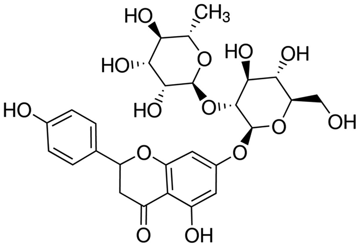
Naringin is (full name, naringenin-7-O-neohesperidin
glycoside) is a dihydrogen flavonoid, and the primary active
ingredient of a number of traditional Chinese medicines, including
orange, Drynaria and Citrus aurantium. Naringin
exhibits a wide range of biological activities, including
anti-oxidation (10), bone growth
promotion (11), plasma cholesterol
reduction (12),
anti-atherosclerotic (13), sedative
(14), anti-tumor (15) and anti-fungal (16) properties. However, the protective
effect of naringin against AS and the underlying molecular
mechanism, including the signal pathways affecting ossification,
inflammation and oxidative stress, remain unclear. The present
study aimed to evaluate the protective effect of naringin against
ossification, inflammation and oxidative stress, and to further
investigate the possible underlying mechanisms, including
anti-inflammatory signaling pathways.
Materials and methods
Materials and chemicals
Osteocalcin (OC; ml026391), alkaline phosphatase
(ALP; E-CL-M0075c) and triglyceride (TG; E-EL-M2603c), nuclear
factor (NF)-κB p65 unit (ml026326), tumor necrosis factor-α (TNF-α;
ml022566), interleukin-1β (IL-1β; ml028611), IL-6 (ml028608),
malonaldehyde (MDA; ml027131), SOD (ml026976), CAT (E-EL-H5408c)
and GSH-PX (E-EL-H5410c) enzyme-linked immunosorbent assay (ELISA)
determination kits were obtained from the Beyotime Institute of
Biotechnology (Nanjing, China). A bicinchoninic acid (BCA; 5000001)
assay kit was obtained from Bio-Rad Laboratories, Inc. (Hercules,
CA, USA). Naringin (purity, >95%) was purchased from
Sigma-Aldrich (Sigma-Aldrich). The chemical structure of naringin
is shown in Fig. 1.
Construction of AS mouse model
All animal studies were performed in accordance with
the regulations of the Zhejiang University Hospital for the care
and use of laboratory animals (Shandong, China). Kunming mice were
obtained from the Laboratory Animal Institute of The Second
Hospital Affiliated to Zhejiang University of Chinese Medicine
(Hangzhou, China). The AS model was established in the mice as
described previously (17). A total
of 50 mice were injected with 30 UI human chorionic gonadotropin
hormone (HCG; Sigma-Aldrich) to induce superovulation. Then,
zygotes were gathered and a HLAB2704 gene fragment was injected
using microinjection into the pronucleus. Surviving zygotes were
transferred into pseudocyesis mice for generation. In brief, AS
model mice were prepared by Biocytogen Biological Technology Co.
Ltd. (eijing, China). A total of 50 mice were injected with HCG
hormone to induce superovulation. Next, ovum and sperm were
integrated, zygotes were gathered and HLAB2704 gene fragments were
injected using a microinjection into the pronucleus. Surviving
zygotes were transferred into pseudocyesis mice for generation. The
mice were sacrificed by incision under 50 mg/kg of sodium
pentobarbital.
Grouping and treatment
Five treatment groups were established for the study
(n=10 per group), as follows: i) Control group, which included
normal mice that received 0.1 ml/100 g sodium pentobarbital
(Sigma-Aldrich) injected intraperitoneally (i.p.); ii) AS group,
which included AS mice that received 0.1 ml/100 g sodium
pentobarbital (i.p.); iii) Nar (20)
group, which included AS mice that received 20 mg/kg naringin
(i.p.) for 8 days (18); iv) Nar
(40) group, which included AS mice
that received 40 mg/kg naringin (i.p.) for 8 days; and v) Nar (80),
which included AS mice that received 80 mg/kg naringin (i.p.) for 8
days.
ELISA of OC, ALP and TG activity
Following treatment with naringin for 3 days, OC,
ALP and TG, NF-κB p65 unit, TNF-α, IL-1β and IL-6, (inflammatory
factors); MDA, SOD, CAT and activity of GSH-PX (oxidative stress)
activity were determined using ELISA determination kits, according
to the standard curve (Beyotime Institute of Biotechnology, China
(Liu, Fan). The optical density was read at 405 nm using a Bio-Rad
microplate reader (Bio-Rad Laboratories, Inc., Hercules, CA,
USA).
Western blot analysis of signal
transducer and activator of transcription 3 (STAT3) and Janus
kinase 2 (JAK2) protein expressions in AS mouse model
After the treatment with naringin for 3 days, 10 mg
AS tissue samples were removed, and incubated with 100 µl tissue
lysis buffer (Thomas Scientific, Swedesboro, NJ, USA) for 30 min on
ice. The protein concentration was measured using a BCA kit
(Bio-Rad Laboratories, Inc.). Equal quantities of protein (50 µg)
were resolved using 12% SDS-polyacrylamide gel and transferred onto
a polyvinylidene fluoride membrane (Bio-Rad Laboratories, Inc).
Next, the membrane was blocked with 5% non-fat milk and incubated
with anti-p-STAT3 (sc-135649; 1:2,000), anti-p-JAK2 (sc-16566-R; 1:
3,000) and β-actin (sc-130657; 1:5,000; all purchased from Santa
Cruz Biotechnology, Inc., Dallas, TX, USA) and incubated overnight
at 4°C followed by incubation with an anti-rabbit secondary
antibody (sc-2795; 1:5,000, Santa Cruz Biotechnology Inc.) for 30
min at 37°C. The signal was developed using an EasyBlot ECL kit
(Shanghai Sangon Biological Engineering Technology & Services
Co., Ltd., Shanghai, China) according to the manufacturer's
instructions. Densitometric measurement of the band intensity was
performed with Quantity One software, version 4.4.0 (Bio-Rad
Laboratories, Inc.).
Statistical analysis
Values were expressed as the mean ± standard
deviation and analysed using SPSS version 19.0, IBM SPSS, Inc.,
Chicago, IL, USA. One-way analysis of variance was performed for
the statistical analysis of data using GraphPad Prism 5 software
(GraphPad Software, San Diego, CA, USA). The Student's t-test was
used to analyze statistical significance. P<0.05 was considered
to indicate a statistically significant difference.
Results
Effect of naringin on OC, ALP and TG
activity in AS mouse model
To determine the protective effect of naringin on
ossification in the AS mouse model, the activity OC, ALP and TG
were evaluated. OC and ALP activity were found to be decreased,
while the TG activity was enhanced in the AS mouse compared with
the control (Fig. 2). Pretreatment
with naringin (20, 40 and 80 mg/kg) markedly altered the OC and ALP
activity, and reduced the TG activity in the AS mice (Fig. 2).
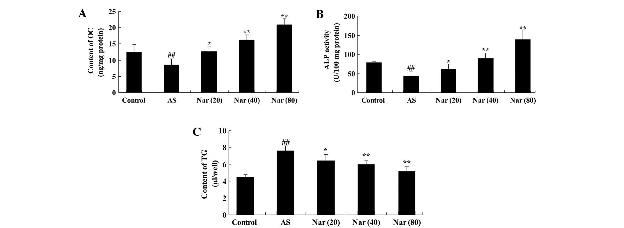 | Figure 2.Effect of naringin on OC, ALP and TG
activity values in AS mouse model. The protective effect of
naringin on (A) OC, (B) ALP and (C) TG activity values in the AS
mouse model. ##P<0.01 vs. control group; *P<0.05
vs. AS group; **P<0.01 vs. AS group. OC, osteocalcin; AS,
ankylosing spondylitis group; Nar (20), naringin (20 mg/kg)-treated; Nar
(40), naringin (40 mg/kg)-treated;
Nar (80), naringin (80 mg/kg)-treated; One-way analysis of variance
was performed for the statistical analysis of data, the Student's
t-test was used to analyze statistical significance. ALP, alkaline
phosphatase; TG, triglyceride. |
Effect of naringin on inflammatory
factor expression in AS model mice
To determined the protective effect of naringin
against the expression of inflammatory factors in the AS mouse
model, the activity NF-κB p65 unit, TNF-α, IL-1β and IL-6 values
were measured. As shown in Fig. 3,
NF-κB p65 unit, TNF-α, IL-1β and IL-6 activity values were
increased in the AS mouse group compared with the control. When
pretreated with naringin (20, 40 and 80 mg/kg) for 8 days, NF-κB
p65 unit, TNF-α, IL-1β and IL-6 activity values were notably
attenuated in the AS mice (Fig.
3).
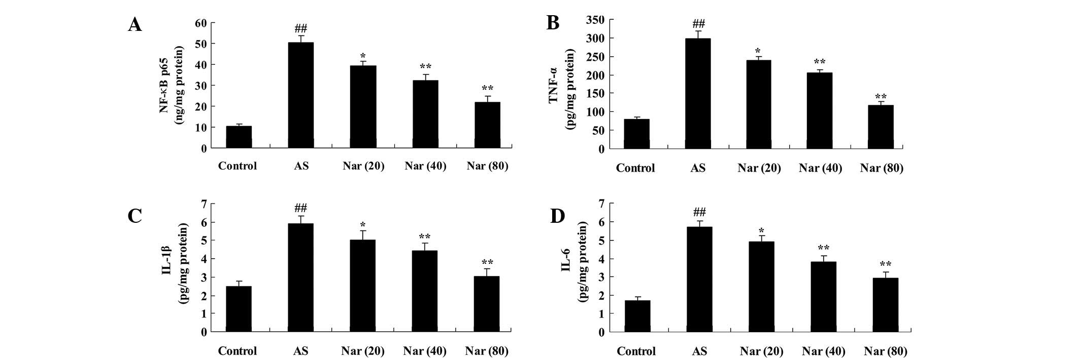 | Figure 3.Effect of naringin on inflammatory
factors in AS mouse model. The protective effect of naringin on the
activity (A) NF-κB p65 unit, (B) TNF-α, (C) IL-1β and (D) IL-6
values in the AS mouse model. ##P<0.01 vs. control
group; *P<0.05 vs. AS group; **P<0.01 vs. AS group. NF-κB
p65, nuclear factor-κB p65; AS, ankylosing spondylitis group; Nar
(20), naringin (20 mg/kg)-treated;
Nar (40), naringin (40
mg/kg)-treated; Nar (80), naringin (80 mg/kg)-treated; One way
analysis of variance was performed for the statistical analysis of
data, the Student's t-test was used to analyze statistical
significance. TNF-α, tumor necrosis factor-α; IL, interleukin. |
Effect of naringin on markers of
oxidative stress in AS mouse model
To infer the protective effect of naringin on
oxidative stress of AS mouse model, the activity of MDA, SOD, CAT
and GSH-PX were evaluated. In the AS group, the MDA activity was
elevated, while the SOD, CAT and GSH-PX activities were reduced
compared with the control (Fig. 4).
The MDA, SOD, CAT and GSH-PX activities were improved by treatment
with naringin (20, 40 and 80 mg/kg) (Fig. 4).
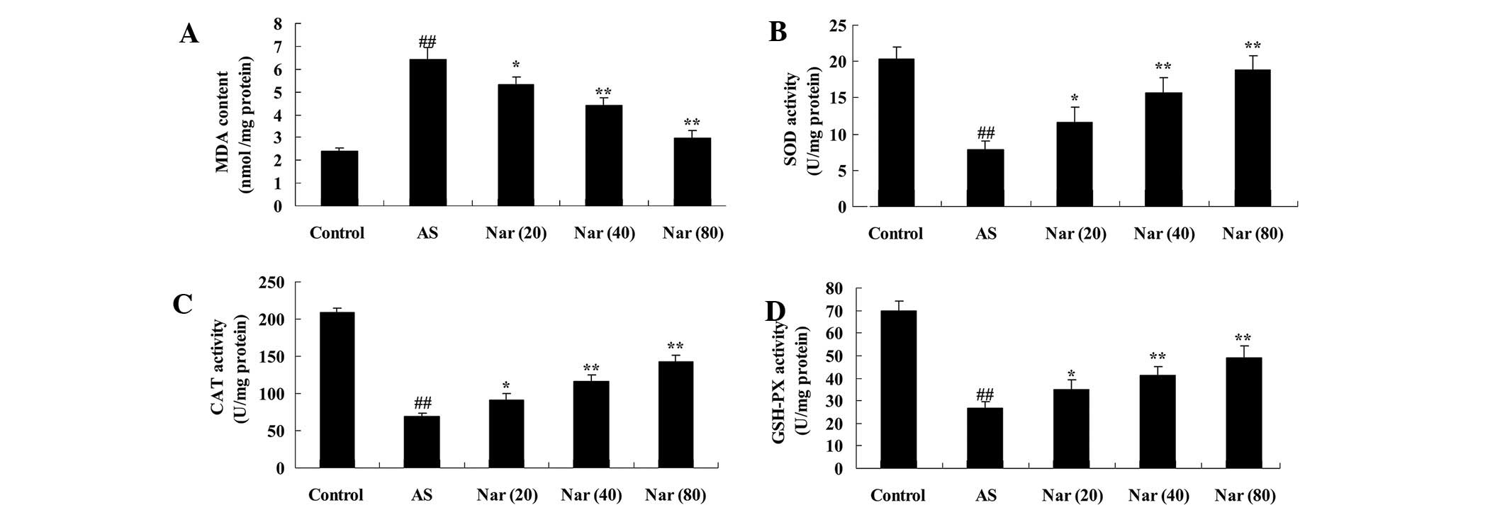 | Figure 4.Effect of naringin on oxidative stress
markers in AS mouse model. The protective effect of naringin on the
concentrations of (A) MDA, (B) SOD, (C) CAT and (D) GSH-PX in the
AS mouse model. ##P<0.01 vs. control group;
*P<0.05 vs. AS group; **P<0.01 vs. AS group. MDA,
malondialdehyde; AS, ankylosing spondylitis group; Nar (20), naringin (20 mg/kg)-treated; Nar
(40), naringin (40 mg/kg)-treated;
Nar (80), naringin (80 mg/kg)-treated; One way analysis of variance
was performed for the statistical analysis of data, the Student's
t-test was used to analyze statistical significance. SOD,
superoxide dismutase; CAT, catalase; GSH-PX, glutathione
peroxidase. |
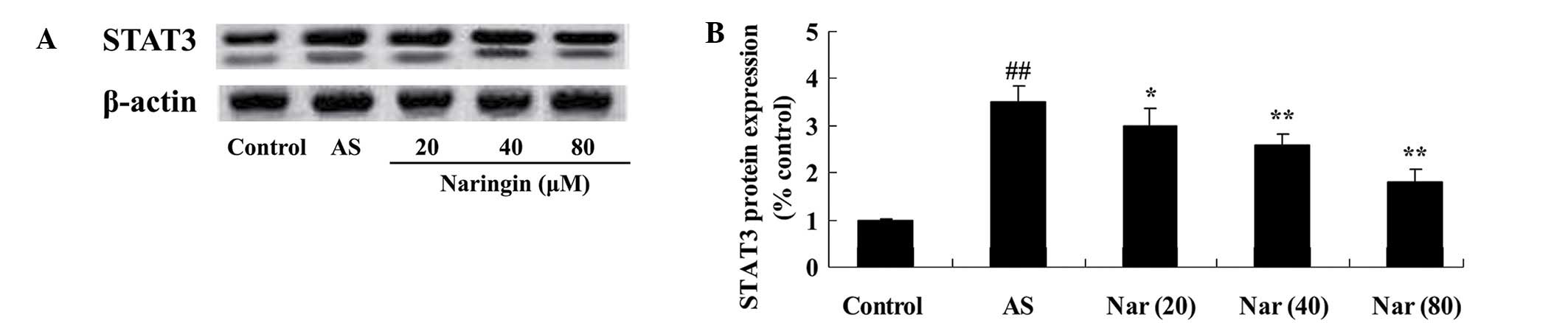
Effect of naringin on STAT3 protein
expression in AS mouse model
To elucidate the protective effect of naringin on
STAT3, STAT3 protein expression of AS mouse was tested using a
western blot assay. The STAT3 protein expression was induced in AS
mouse (Fig. 5). However, treatment
with naringin (20, 40 and 80 mg/kg) mitigated this increased
expression of STAT3 protein in AS mouse (Fig. 5).
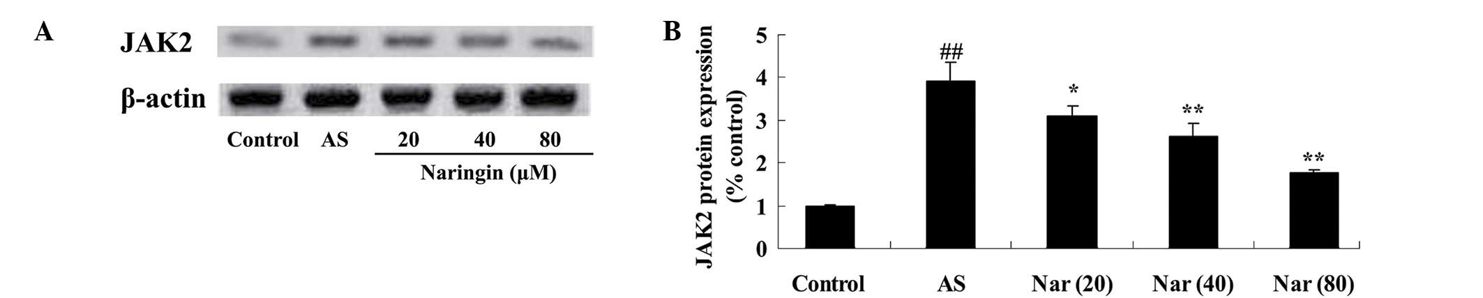
Protective effect of naringin on JAK2
protein expression in AS mouse model
To clarify the involvement the protective effect of
naringin on JAK2, JAK2 protein expression of AS mouse was
evaluated. The JAK2 protein expression was induced in AS mice;
however, treatment with naringin (20, 40 and 80 mg/kg) decreased
this enhanced JAK2 expression (Fig.
6).
Discussion
AS is a chronic inflammatory disease, in which the
rheumatoid factor is important, and it involves axial bone joints
and tendon ligament attachment points, and the lesion is finally
developed into the fibrosis of central joint and ankylosis, forming
a typical ‘bamboo vertebrae’ (19).
The pathogenesis of AS is currently unclear; however, its
occurrence is associated with heredity, chronic infections,
autoimmune disorders and endocrine disorders. The joint changes of
AS primarily include synovial thickening and infiltration of
macrophages, lymphocytes and plasma cells, accompanied by joint
fibrosis and bone ankylosis (20).
The primary site of the lesion is the region where the ligaments
and joint capsule are attached, in which the inflammation leads to
bone destruction, defects and replacement by connective tissue
containing lymph and plasma cells; the filled and repaired
cancellous bone develops ligament ossification in eroded bone
surface (21).
The present study showed that administration of
naringin increased OC and ALP activity, while reducing TG activity
in AS model mice. These results are consistent with those of
previous studies; for example, Liu et al suggested that the
effects of naringin upregulates osteogenesis in human amniotic
fluid-derived stem cells (22).
Furthermore, Li et al reported that naringin is able to
significantly improved ALP activity as well as upregulate the
expression of type I collagen in the osteoblastic cell line
MC3T3-E1 (23).
AS is a type of rheumatism characterized by chronic
inflammation of the axial joint, and may involve the internal
organs and other tissues (24). Most
scholars believe that this disease is an autoimmune inflammation
reaction caused predominantly by genetic factors, in addition to
being stimulated by trauma, infection, fatigue and other
environmental factors (25–27). The present results showed that
naringin attenuates NF-κB p65 unit, TNF-α, IL-1β and IL-6 activity
in AS mice. These results are consistent with a previous study in
which naringin was shown to ameliorate oxidative stress and
inflammation in mice (28). In
addition, it has been suggested that naringin may exert
anti-inflammatory effects in the adult brain (29). However, the detailed mechanisms
underlying the anti-inflammatory effects of naringin in AS mouse
remain unclear, and further clarification is required in
future.
In patients with AS, neutrophils are activated so
that reactive oxygen species are generated, leading to oxidative
stress (9). As a result of the
increase in myeloperoxidase activity and advanced oxidation protein
products in patients, the sulfhydryl level is decreased, from which
it may be inferred that activated neutrophils serve a crucial
function in the pathogenesis of AS (30). In the blood of patients with active
AS, MDA levels and catalase activity are increased compared with
those in control group (9). This
catalase activity is positively correlated with erythrocyte
sedimentation rate and C-reactive protein levels, and it is
believed that the increase of catalase activity is a response to
increased superoxide anion (31).
The present results indicated that naringin reduced the MDA
activity and increased the SOD, CAT and GSH-PX activities of the AS
mice. Chen et al indicated that naringin had effective
protection against paraquat-induced acute lung injury and pulmonary
fibrosis through increasing activities of SOD, GSH-PX in mice
(32). Cui et al suggested
that naringin benefited the recovery of traumatic brain injury by
reducing oxidative and inflammatory alterations in mice (33).
STATs are a group of cytoplasmic protein
transcription factors, mediating the cytoplasm, which play a key
role in the signaling of the nucleus (34). It has been reported that mutant mice
lacking STAT3 are highly sensitive to AS, and the concentrations of
serum inflammatory cytokines such as TNF-α, IL-1β and IL-6 are
increased (35). Furthermore,
macrophages lacking in STAT3 show abnormal activation phenotypes,
such as increased production of inflammatory cytokines in response
to endotoxin (36). STAT3 activation
is crucial for the prevention of chronic inflammation in mice
(37). The present results showed
that naringin inhibited the STAT3 protein expression in AS rat. A
previous study showed that naringin inhibited the development of
carrageenan-induced acute lung inflammation via suppression of
STAT3 (38).
AK2 belongs to Janus kinase family, and the gene is
located in the short arm of chromosome 9 (9p24), belonging to JAK
family together with JAK1, JAK3 and TYK2 as intracellular protein
tyrosine kinase (39). Under normal
physiological conditions, JAK2 mediates the signal transduction of
a variety of cytokines, including erythropoietin, thrombopoietin,
granulocyte-macrophage colony stimulating factor and IL-3, thus
regulating and promoting cell proliferation (40). The present study showed that naringin
also inhibited JAK2 protein expression in AS rat. In addition,
naringin appeared to exert an anti-inflammatory effect via the
suppression of the JAK2/STAT3 signaling pathway.
In summary, naringin exerted notable osteogenic,
anti-inflammatory and anti-oxidative effects, and the mechanism was
mediated by the downregulation of the JAK2/STAT3 signaling pathways
in AS mice. Future studies are required to investigate the
protective effect of naringin against AS.
References
|
1
|
Liu YF, Dong H, Tu SH, Zheng CH, Liu PL
and Hu YH: Etanercept in the treatment of ankylosing spondylitis: A
systematic review and meta-analysis. Exp Ther Med. 8:1585–1592.
2014.PubMed/NCBI
|
|
2
|
Chou CT, Huo AP, Chang HN, Tsai CY, Chen
WS and Wang HP: Cytokine production from peripheral blood
mononuclear cells in patients with ankylosing spondylitis and their
first-degree relatives. Arch Med Res. 38:190–195. 2007. View Article : Google Scholar : PubMed/NCBI
|
|
3
|
Zhou L, Zhang Y, Xu H, et al: Decreased
programmed death-1 expression on the T cells of patients with
ankylosing spondylitis. Am J Med Sci. 349:488–492. 2015. View Article : Google Scholar : PubMed/NCBI
|
|
4
|
Rednic S, Marinescu C, Chira R, Rogojan L
and Rednic N: Treatment with infliximab in a patient with
ankylosing spondylitis and Crohn's disease. J Gastrointestin Liver
Dis. 15:379–382. 2006.PubMed/NCBI
|
|
5
|
Mielants H, Veys EM, Cuvelier C, et al:
Gut inflammation in children with late onset pauciarticular
juvenile chronic arthritis and evolution to adult
spondyloarthropathy––a prospective study. J Rheumatol.
20:1567–1572. 1993.PubMed/NCBI
|
|
6
|
Weber U, Hodler J, Kubik RA, Rufibach K,
Lambert RG, Kissling RO, Pfirrmann CW and Maksymowych WP:
Sensitivity and specificity of spinal inflammatory lesions assessed
by whole-body magnetic resonance imaging in patients with
ankylosing spondylitis or recent-onset inflammatory back pain.
Arthritis Rheum. 61:900–908. 2009. View Article : Google Scholar : PubMed/NCBI
|
|
7
|
Maksymowych WP, Chiowchanwisawakit P,
Clare T, Pedersen SJ, Østergaard M and Lambert RG: Inflammatory
lesions of the spine on magnetic resonance imaging predict the
development of new syndesmophytes in ankylosing spondylitis:
Evidence of a relationship between inflammation and new bone
formation. Arthritis Rheum. 60:93–102. 2009. View Article : Google Scholar : PubMed/NCBI
|
|
8
|
Okano K, Kimura K, Tanaka Y, Tsuchiya K,
Akiba T and Nitta K: Direct measurement of reactive oxygen species
in leukocytes during hemodialysis therapy. Int J Clin Exp Med.
8:20959–20964. 2015.PubMed/NCBI
|
|
9
|
Kozaci LD, Sari I, Alacacioglu A, Akar S
and Akkoc N: Evaluation of inflammation and oxidative stress in
ankylosing spondylitis: A role for macrophage migration inhibitory
factor. Mod Rheumatol. 20:34–39. 2010. View Article : Google Scholar : PubMed/NCBI
|
|
10
|
Jagetia GC, Reddy TK, Venkatesha VA and
Kedlaya R: Influence of naringin on ferric iron induced oxidative
damage in vitro. Clin Chim Acta. 347:189–197. 2004. View Article : Google Scholar : PubMed/NCBI
|
|
11
|
Zhou X, Zhang P, Zhang C and Zhu Z:
Promotion of bone formation by naringin in a titanium
particle-induced diabetic murine calvarial osteolysis model. J
Orthop Res. 28:451–456. 2010.PubMed/NCBI
|
|
12
|
Kim SY, Kim HJ, Lee MK, Jeon SM, Do GM,
Kwon EY, Cho YY, Kim DJ, Jeong KS, Park YB, et al: Naringin
time-dependently lowers hepatic cholesterol biosynthesis and plasma
cholesterol in rats fed high-fat and high-cholesterol diet. J Med
Food. 9:582–586. 2006. View Article : Google Scholar : PubMed/NCBI
|
|
13
|
Choe SC, Kim HS, Jeong TS, Bok SH and Park
YB: Naringin has an antiatherogenic effect with the inhibition of
intercellular adhesion molecule-1 in hypercholesterolemic rabbits.
J Cardiovasc Pharmacol. 38:947–955. 2001. View Article : Google Scholar : PubMed/NCBI
|
|
14
|
Fernandez SP, Nguyen M, Yow TT, Chu C,
Johnston GA, Hanrahan JR and Chebib M: The flavonoid glycosides,
myricitrin, gossypin and naringin exert anxiolytic action in mice.
Neurochem Res. 34:1867–1875. 2009. View Article : Google Scholar : PubMed/NCBI
|
|
15
|
Camargo CA, Gomes-Marcondes MC, Wutzki NC
and Aoyama H: Naringin inhibits tumor growth and reduces
interleukin-6 and tumor necrosis factor alpha levels in rats with
Walker 256 carcinosarcoma. Anticancer Res. 32:129–133.
2012.PubMed/NCBI
|
|
16
|
Liu Q, Lu L and Xiao M: Cell surface
engineering of α-l-rhamnosidase for naringin hydrolysis. Bioresour
Technol. 123:144–149. 2012. View Article : Google Scholar : PubMed/NCBI
|
|
17
|
Gu X, Wu H and Fu P: Allicin attenuates
inflammation and suppresses HLA-B27 protein expression in
ankylosing spondylitis mice. Biomed Res Int. 2013:1715732013.
View Article : Google Scholar : PubMed/NCBI
|
|
18
|
Kumar VS, Rajmane AR, Adil M, Kandhare AD,
Ghosh P and Bodhankar SL: Naringin ameliorates acetic acid induced
colitis through modulation of endogenous oxido-nitrosative balance
and DNA damage in rats. J Biomed Res. 28:132–145. 2014.PubMed/NCBI
|
|
19
|
Shiau MY, Lo MK, Chang CP, Yang TP, Ho KT
and Chang YH: Association of tumour necrosis factor alpha promoter
polymorphisms with ankylosing spondylitis in Taiwan. Ann Rheum Dis.
66:562–563. 2007. View Article : Google Scholar : PubMed/NCBI
|
|
20
|
Tam LS, Chan KY and Li EK: The influence
of illness and variables associated with functional limitations in
Chinese patients with ankylosing spondylitis. J Rheumatol.
34:1032–1039. 2007.PubMed/NCBI
|
|
21
|
Gu X, Wu H and Fu P: Allicin attenuates
inflammation and suppresses HLA-B27 protein expression in
ankylosing spondylitis mice. Biomed Res Int. 2013:1715732013.
View Article : Google Scholar : PubMed/NCBI
|
|
22
|
Liu M, Li Y and Yang ST: Effects of
naringin on the proliferation and osteogenic differentiation of
human amniotic fluid-derived stem cells. J Tissue Eng Regen Med.
2014.(Epub ahead of print). View Article : Google Scholar
|
|
23
|
Li L, Zeng Z and Cai G: Comparison of
neoeriocitrin and naringin on proliferation and osteogenic
differentiation in MC3T3-E1. Phytomedicine. 18:985–989. 2011.
View Article : Google Scholar : PubMed/NCBI
|
|
24
|
Lange U, Teichmann J and Stracke H:
Correlation between plasma TNF-alpha, IGF-1, biochemical markers of
bone metabolism, markers of inflammation/disease activity and
clinical manifestations in ankylosing spondylitis. Eur J Med Res.
5:507–511. 2000.PubMed/NCBI
|
|
25
|
Schulz M, Dotzlaw H and Neeck G:
Ankylosing spondylitis and rheumatoid arthritis: serum levels of
TNF-alpha and Its soluble receptors during the course of therapy
with etanercept and infliximab. Biomed Res Int. 2014:6751082014.
View Article : Google Scholar : PubMed/NCBI
|
|
26
|
Huang WN, Tso TK, Kuo YC and Tsay GJ:
Distinct impacts of syndesmophyte formation on male and female
patients with ankylosing spondylitis. Int J Rheum Dis. 15:163–168.
2012. View Article : Google Scholar : PubMed/NCBI
|
|
27
|
Liu C, Hong T, Shao M, Chen Z and Wang C:
Melatonin synergized with cyclosporine A improves cardiac allograft
survival by suppressing inflammation and apoptosis. Mol Med Rep.
10:1323–1328. 2014.PubMed/NCBI
|
|
28
|
Golechha M, Sarangal V, Bhatia J, Chaudhry
U, Saluja D and Arya DS: Naringin ameliorates
pentylenetetrazol-induced seizures and associated oxidative stress,
inflammation, and cognitive impairment in rats: Possible mechanisms
of neuroprotection. Epilepsy Behav. 41:98–102. 2014. View Article : Google Scholar : PubMed/NCBI
|
|
29
|
Jung UJ and Kim SR: Effects of naringin, a
flavanone glycoside in grapefruits and citrus fruits, on the
nigrostriatal dopaminergic projection in the adult brain. Neural
Regen Res. 9:1514–1517. 2014. View Article : Google Scholar : PubMed/NCBI
|
|
30
|
Feijoo M, Tunez I, Tasset I, Montilla P,
Ruiz A and Collantes E: Infliximab reduces oxidative stress in
ankylosing spondylitis. Clin Exp Rheumatol. 27:167–168.
2009.PubMed/NCBI
|
|
31
|
Karakoc M, Altindag O, Keles H, Soran N
and Selek S: Serum oxidative-antioxidative status in patients with
ankylosing spondilitis. Rheumatol Int. 27:1131–1134. 2007.
View Article : Google Scholar : PubMed/NCBI
|
|
32
|
Chen Y, Nie YC, Luo YL, Lin F, Zheng YF,
Cheng GH, Wu H, Zhang KJ, Su WW, Shen JG and Li PB: Protective
effects of naringin against paraquat-induced acute lung injury and
pulmonary fibrosis in mice. Food Chem Toxicol. 58:133–140. 2013.
View Article : Google Scholar : PubMed/NCBI
|
|
33
|
Cui QJ, Wang LY, Wei ZX and Qu WS:
Continual naringin treatment benefits the recovery of traumatic
brain injury in rats through reducing oxidative and inflammatory
alterations. Neurochem Res. 39:1254–1262. 2014. View Article : Google Scholar : PubMed/NCBI
|
|
34
|
Kicinska A, Leluk J and Jarmuszkiewicz W:
Acanthamoeba castellanii STAT protein. PLoS One. 9:e1113452014.
View Article : Google Scholar : PubMed/NCBI
|
|
35
|
Danoy P, Pryce K, Hadler J, Bradbury LA,
Farrar C and Pointon J: Australo-Anglo-American Spondyloarthritis
Consortium, Ward M, Weisman M, Reveille JD, et al: Association of
variants at 1q32 and STAT3 with ankylosing spondylitis suggests
genetic overlap with Crohn's disease. PLoS Genet. 6:e10011952010.
View Article : Google Scholar : PubMed/NCBI
|
|
36
|
Ahmad SF, Attia SM, Bakheet SA, Zoheir KM,
Ansari MA, Korashy HM, Abdel-Hamied HE, Ashour AE and Abd-Allah AR:
Naringin attenuates the development of carrageenan-induced acute
lung inflammation through inhibition of NF-κb, STAT3 and
pro-inflammatory mediators and enhancement of IκBα and
anti-inflammatory cytokines. Inflammation. 38:846–857. 2015.
View Article : Google Scholar : PubMed/NCBI
|
|
37
|
Davidson SI, Liu Y, Danoy PA, Wu X, Thomas
GP, Jiang L, Sun L, Wang N, Han J, Han H, et al: Association of
STAT3 and TNFRSF1A with ankylosing spondylitis in Han Chinese. Ann
Rheum Dis. 70:289–292. 2011. View Article : Google Scholar : PubMed/NCBI
|
|
38
|
Zimmers TA, Fishel ML and Bonetto A: STAT3
in the Systemic Inflammation of Cancer Cachexia. Semin Cell Dev
Biol. 2016. View Article : Google Scholar : PubMed/NCBI
|
|
39
|
Chen C, Zhang X and Wang Y: Analysis of
JAK2 and STAT3 polymorphisms in patients with ankylosing
spondylitis in Chinese Han population. Clin Immunol. 136:442–446.
2010. View Article : Google Scholar : PubMed/NCBI
|
|
40
|
Liu H, Yang Z, Hu J, et al: Improvement of
thoracic aortic vasoreactivity by continuous and intermittent
exercise in high-fat diet-induced obese rats. Biomed Rep.
3:527–532. 2015.PubMed/NCBI
|