Open Access

Mechanism of intestinal mucosal barrier dysfunction in a rat model of chronic obstructive pulmonary disease: An observational study

  • Authors:
    • Xiaofeng Xin
    • Wei Dai
    • Jie Wu
    • Liping Fang
    • Ming Zhao
    • Pengpeng Zhang
    • Min Chen
  • View Affiliations

  • Published online on: June 30, 2016     https://doi.org/10.3892/etm.2016.3493
  • Pages: 1331-1336
  • Copyright: © Xin et al. This is an open access article distributed under the terms of Creative Commons Attribution License.

Metrics: Total Views: 0 (Spandidos Publications: | PMC Statistics: )
Total PDF Downloads: 0 (Spandidos Publications: | PMC Statistics: )


Abstract

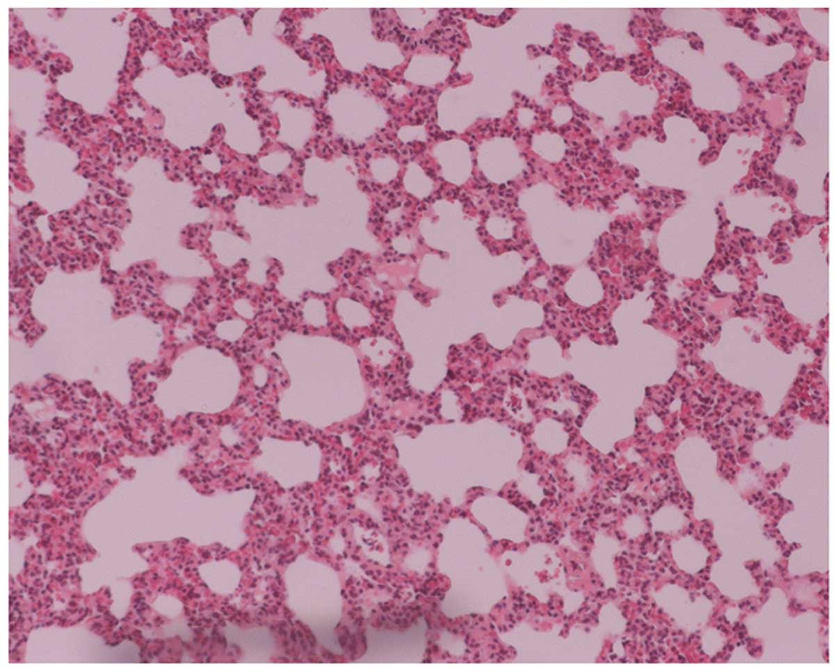
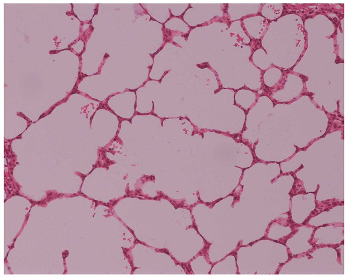
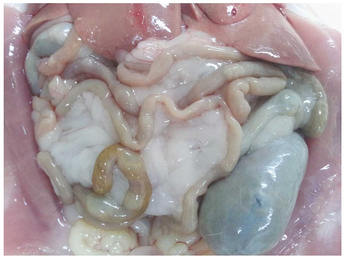
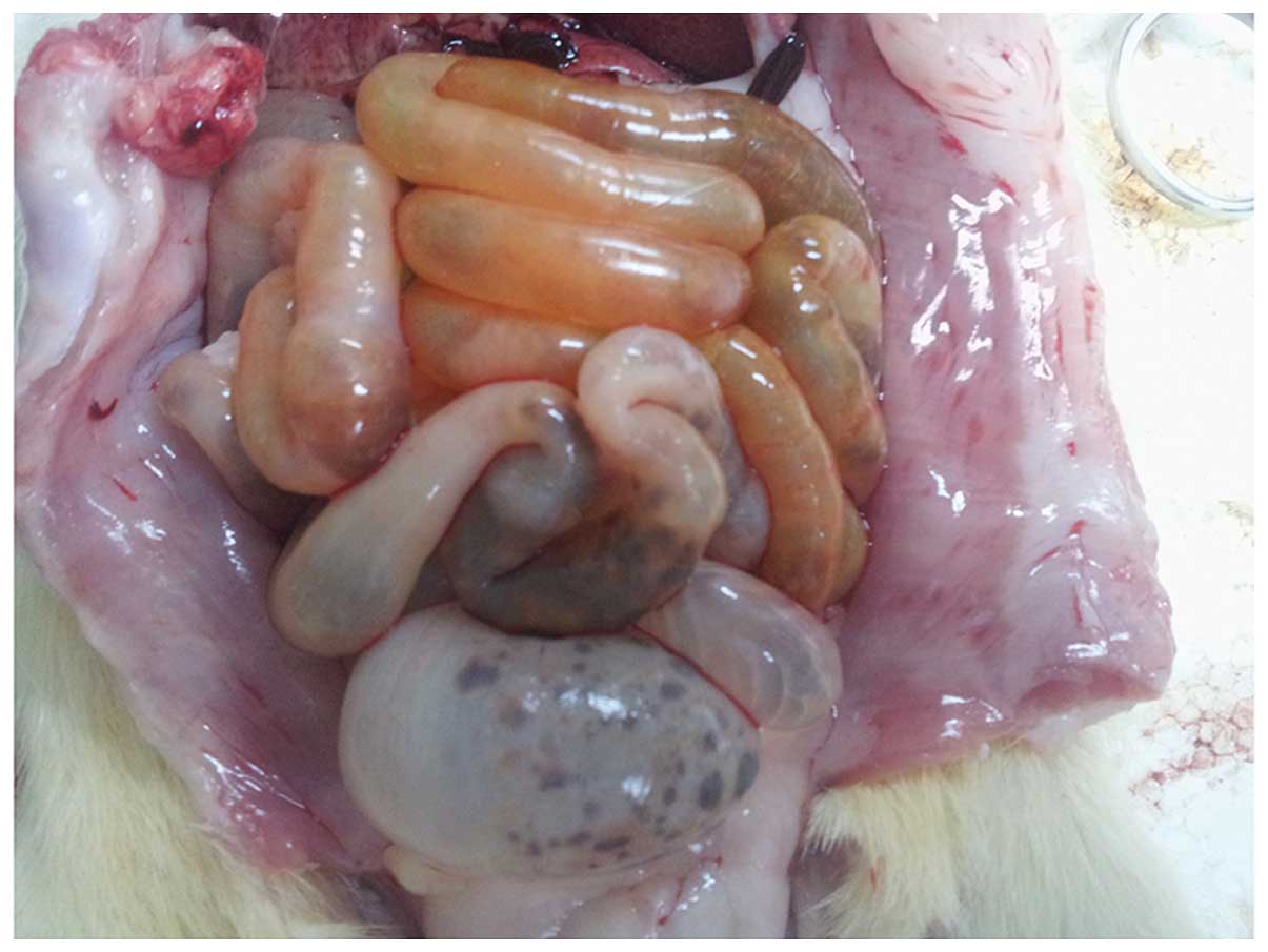
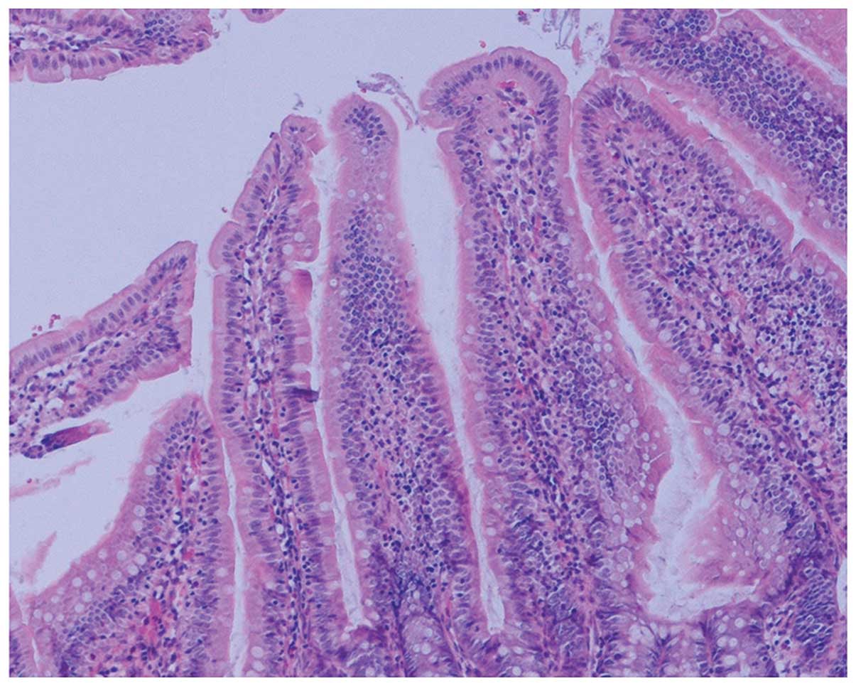
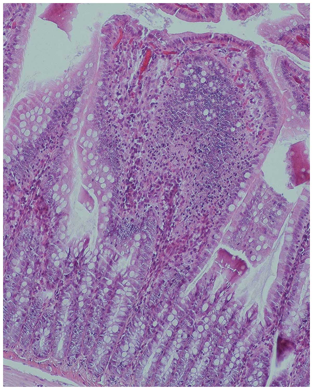
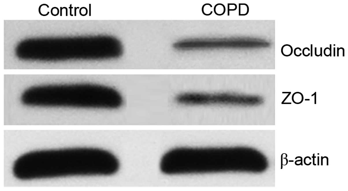
The aim of the present study was to investigate intestinal mucosal barrier dysfunction in a rat model of chronic obstructive pulmonary disease (COPD). Male Sprague Dawley rats (n=40) were evenly randomized into control and COPD groups and the COPD model was established by regulated exposure to cigarette smoke for 6 months. Histopathological changes of the lung and intestinal tissues were detected by hematoxylin and eosin staining. Expression of the tight junction proteins occludin and zona occludens‑1 (ZO‑1) in the intestinal tissues were analyzed by western blotting, serum diamine oxidase (DAO) activity was detected by spectrophotometry, the urinary lactulose to mannitol ratio (L/M) was evaluated by high performance liquid chromatography, and intestinal tissue secretion of tumor necrosis factor (TNF)‑α, interferon (IFN)‑γ and interleukin (IL)‑8 were detected by ELISA. Lung histopathology revealed thinned alveolar walls, ruptured alveolar septa, enlarged and deformed alveoli, and the formation of bullae and emphysema due to alveolar fusion in the COPD group, while intestinal histopathology indicated clearly swollen intestines with darkened and gray mucosa, neutrophil infiltration of the intestinal mucosal and regional epithelial shedding. The occludin and ZO‑1 expression levels were significantly lower in the COPD group compared with those in the corresponding control group (P<0.05), while the urinary L/M ratio was significantly higher (P<0.05). Furthermore, the serum DAO activity and secretion of TNF‑α, IFN‑γ and IL‑8 in the intestinal tissues were significantly higher in the COPD group than in the control group (each P<0.05). Dysfunctional and structural changes were observed in the intestinal mucosal barrier in COPD model rats, which may be associated with the increased intestinal inflammatory responses.
View Figures
View References

Related Articles

Journal Cover

September-2016
Volume 12 Issue 3

Print ISSN: 1792-0981
Online ISSN:1792-1015

Sign up for eToc alerts

Recommend to Library

Copy and paste a formatted citation
x
Spandidos Publications style
Xin X, Dai W, Wu J, Fang L, Zhao M, Zhang P and Chen M: Mechanism of intestinal mucosal barrier dysfunction in a rat model of chronic obstructive pulmonary disease: An observational study. Exp Ther Med 12: 1331-1336, 2016
APA
Xin, X., Dai, W., Wu, J., Fang, L., Zhao, M., Zhang, P., & Chen, M. (2016). Mechanism of intestinal mucosal barrier dysfunction in a rat model of chronic obstructive pulmonary disease: An observational study. Experimental and Therapeutic Medicine, 12, 1331-1336. https://doi.org/10.3892/etm.2016.3493
MLA
Xin, X., Dai, W., Wu, J., Fang, L., Zhao, M., Zhang, P., Chen, M."Mechanism of intestinal mucosal barrier dysfunction in a rat model of chronic obstructive pulmonary disease: An observational study". Experimental and Therapeutic Medicine 12.3 (2016): 1331-1336.
Chicago
Xin, X., Dai, W., Wu, J., Fang, L., Zhao, M., Zhang, P., Chen, M."Mechanism of intestinal mucosal barrier dysfunction in a rat model of chronic obstructive pulmonary disease: An observational study". Experimental and Therapeutic Medicine 12, no. 3 (2016): 1331-1336. https://doi.org/10.3892/etm.2016.3493