Introduction
Deep vein thrombosis (DVT) is a severe postoperative
complication, which commonly occurs in 40–80% of patients
undergoing orthopedic surgeries, such as total hip replacement,
total knee replacement, and hip fractures surgery (1–3).
Pulmonary embolism, a potentially life-threatening disease with a
high mortality rate in China, may result from a detached thrombus
that travels to the lungs (4).
Formation of thrombus is closely related to the
activation of platelets (5,6). Under the action of coagulation factor,
platelets are activated and aggregated, eventually forming a
thrombus (7). Vesicle-associated
membrane protein 8 (VAMP8) is a vesicle-associated major membrane
protein of soluble N-ethylmaleimide-sensitive factor attachment
protein receptor (8). VAMP8 is
necessary for platelet activation, which is involved in the process
of platelet degranulation (9,10). A
previous study showed that increased expression of microRNA-96
(miR-96) lead to a reduction in platelet activation by inhibiting
the expression of VAMP8 protein (11). It has been suggest that the
occurrence of DVT may be predicted by detection of miR-96
expression in platelets (12).
D-dimer is a fibrin degradation product in blood after a thrombus
is degraded by thrombin, coagulation factor XIIIa and fibrinolysin
(13–15). Levels of D-dimer may reflect the
activity of coagulation and the fibrinolytic system, which may be
used for diagnosis of DVT (16).
However, the correlation between miR-96 expression and D-dimer in
the development of DVT is unknown.
In the present study, levels of miR-96 expression in
platelets were detected in patients with orthopedic conditions
following surgery. The association between miR-96 expression and
occurrence of DVT was thus determined. Furthermore, the correlation
between miR-96 expression in platelets and of D-dimer levels in
plasma was investigated.
Materials and methods
Patients
A total of consecutive 8 patients with DVT after
orthopedic surgery (DVT group; 3 males and 5 females) were enrolled
in this study in Laiwu People's Hospital (Laiwu, China). The age of
these patients ranged between 51 and 68 years, with an average age
of 63.4 years. The prognosis of DVT referred to the clinical
manifestations, imaging and laboratory test results, including
whether deep vein thrombosis, limb swelling, thickened
circumference, pain or tenderness, superficial vein dilation, skin
pigmentation and/or abnormal D-dimer values were detected. In the
corresponding period, a total of consecutive 61 orthopedic patients
without DVT following orthopedic surgery (non-DVT group) were
enrolled in this study. These patients included 35 males and 26
females, with ages ranging between 31 and 72 years, and an average
age of 58.3 years. As a control, a total of consecutive 30 healthy
individuals (control group) were enrolled in this study, which
included 15 males and 15 females. The age of these healthy
individuals ranged between 30 and 88 years, with an average age of
56.5 years.
The exclusion criteria of DVT group were as follows:
i) Patients with DVT before orthopedic surgeries; ii) patients with
active bleeding and spontaneous intracranial hemorrhage recently;
iii) patients with bleeding disorders and undergoing anticoagulant
treatment; iv) patients with severe liver and kidney disease; v)
patients had not undergone surgery and trauma; vi) patients had
history of thrombosis; and vii) patients with malignancies,
inflammatory diseases. Prior written and informed consent were
obtained from every patient. The study was approved by the ethics
review board of Laiwu People's Hospital.
Color Doppler ultrasonography
The lower limb veins were detected in patients
before orthopedic surgery and at days 3 and 7 after orthopedic
surgery. Venous diameter, blood flow, intraluminal pressure, deep
venous valve function and thrombosis formation were determined
using a color Doppler ultrasonography imaging system (ACUSON
Antares Color Doppler Ultrasound System; Siemens AG, Berlin,
German).
Purification of platelets
Peripheral blood was extracted from patients after
orthopedic surgery and from healthy individuals. The samples were
mixed with 85 mM trisodium citrate, 78 mM citric acid and 111 mM
glucose (all from Sigma-Aldrich, St. Louis, MO. USA). Following
centrifugation at 80 × g for 10 min, the mixtures were mixed with 2
mM ethylenediaminetetraacetic acid (EDTA; pH 8.0; Sigma-Aldrich)
and centrifuged at 1,000 × g for 10 min. The pellets of platelets
were resuspended in 3 ml Beads Buffer [0.8% NaCl, 0.02% KCl, 0.144%
Na2HPO4, 0.024% KH2PO4,
0.5% bovine serum albumin and 2 mM EDTA (pH 8.0)], then 40 µl human
CD45 MicroBeads reagent (Miltenyi Biotec GmbH, Bergisch Gladbach,
German) was added and mixed gently. The platelets were separated
using a MACS® magnetic cell sorting system (Miltenyi
Biotec GmbH). The purity of platelets was >99.99%.
Quantitative polymerase chain reaction
(qPCR) assay
Total RNA of platelets were extracted using TRIzol
reagent (Invitrogen; Thermo Fisher Scientific, Inc., Carlsbad, CA,
USA). RNA quality was detected by RNA electrophoresis and
spectrophotometer at an optical density of 260/280 nm. All RNA was
reverse transcribed into cDNA using a PrimeScript RT Regent kits
(Takara Bio, Inc., Otsu, Japan) according to the manufacturer's
protocol. Quantitative PCR assay was conducted using a SYBR
PrimeScript RT-PCR kit II (Takara Bio, Inc.) in accordance with the
manufacturer's protocol. β-actin was used as an internal control
for miR-96. The primers for miR-96 were as follows:
5′-TTTGGCACTAGCACATT-3′ and 5′-GAGCAGGCTGGAGAA-3′. The primers for
β-actin were: 5′-CATGTACGTTGCTATCCAGGC-3′ and
5′-CTCCTTAATGTCACGCACGAT-3′. Oligonucleotide primers of miR-96 and
β-actin were synthesized by Invitrogen (Thermo Fisher Scientific,
Inc.). The 20 µl reaction system consisted of 0.5 µl cDNA, 10 µl
SYBR Ex Taq, 0.5 µl forward and reverse primer, and 8.5 µl
ddH2O. The PCR thermocycling conditions were as follows:
Denaturation at 95°C for 10 sec, 95°C for 5 sec, and 60°C for 20
sec for a total of 40 cycles. For each sample, PCR reaction was
repeated for at least 3 times. The gene expression levels were
calculated with the 2−ΔΔCq method.
Western blot analysis
Total proteins of platelets were extracted using a
total protein extraction reagent (BioTeke Corporation, Beijing,
China). Total proteins (100 µl) were separated on 12% SDS-PAGE gel,
and transferred onto a polyvinylidene fluoride membrane (KLH
Bioscience, Shanghai, China). After blocking with 5% non-fat milk
in Tris-buffered saline with Tween-20 at 37°C for 2 h, the membrane
was incubated at 4°C for 24 h with rabbit anti-human VAMP8
polyclonal primary antibodies (1:800; cat. no. ab76021; Abcam,
Cambridge, MA, USA). The membrane was subsequently incubated at
37°C for 1 h with horse radish peroxidase-conjugated goat
anti-rabbit secondary antibodies (1:2,000; cat. no. ab181658;
Abcam). β-tubulin was used as an internal control. Blots were
detected using ECL Plus enhanced chemiluminescence reagent (EMD
Millipore, Billerica, MA, USA). Image quantifications were
performed using ImageLab software version 4.1 (Bio-Rad
Laboratories, Inc., Hercules, CA, USA). Experiments were repeated
at least 3 times.
Plasma D-dimer detection
Peripheral venous blood samples were collected from
patients at days 1, 3 and 7 after orthopedic surgery. For control,
peripheral venous blood samples were collected from healthy
individuals. Subsequently, ~1.8-ml samples were transferred into
anticoagulant tube with 0.2 ml citric acid (109 mM) and
mixed. The mixtures were centrifuged at 400 × g for 15 min. The
plasma supernatants were finally transferred to a new tube for the
analysis of D-dimer. D-dimer was detected by enzyme-linked
fluorescent assay using a VIDAS instrument and VIDAS D-dimer
Exclusion II kit (VIDAS S60; BioMerieux, Inc., Grenoble, France),
in accordance with the manufacturer's instructions.
Statistical analysis
All results are expressed as the mean ± standard
deviation. All statistical analyses were performed with SPSS
software, version 13.0 for Windows (SPSS, Inc., Chicago, IL, USA).
Paired t-test was used to analyze comparisons between groups
and analysis of paired data. Pearson correlation was used to
analyze the association between miR-96 expression and plasma
D-dimer levels for the prediction of DVT. P<0.05 was considered
to indicate a statistically significant difference.
Results
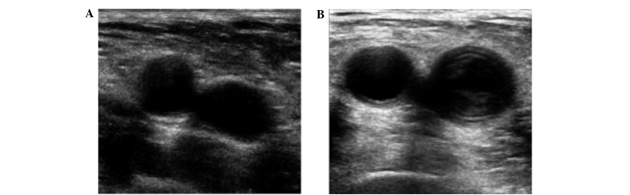
Diagnosis of patients with DVT by
color Doppler ultrasonography
To determine the occurrence of DVT, ultrasonic color
Doppler imaging was performed on the lower limb veins of patients
and healthy individuals. As shown in Fig. 1A, for the non-DVT group, there was no
sign of thrombus in the femoral vein. However, for the DVT group,
coronal imaging showed significant vein stenosis and several
thrombi in the femoral vein (Fig.
1B). These results suggest that occurrence of DVT can be
clearly diagnosed using color Doppler ultrasonography.
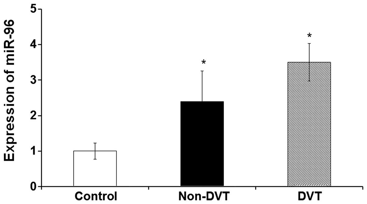
Expression levels of platelet miR-96
are significantly increased in patients following orthopedic
surgery
To determine the expression levels of miR-96 between
patients with orthopedic disease and healthy individuals, a qPCR
assay was performed. As shown in Fig.
2, expression levels of platelet miR-96 in the DVT and non-DVT
group were significantly increased compared with the control group
(P<0.05). The expression levels of miR-96 in the DVT group were
higher than those in the non-DVT group. However, there was no
significant difference between the DVT and the non-DVT group. These
results indicate that the expression levels of platelet miR-96 are
significantly increased in orthopedic patients after orthopedic
surgery. High expression of miR-96 may be associated with the
occurrence of DVT.
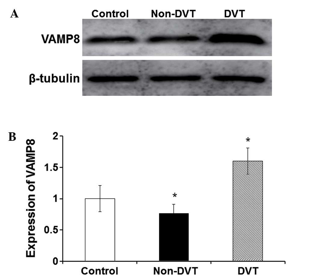
Expression levels of VAMP8 are
increased in the DVT group and reduced in the non-DVT group
To investigate the expression levels of the
platelet-related protein VAMP8 between orthopedic patients and
healthy individuals, western blot analysis was performed. As shown
in Fig. 3, the expression levels of
VAMP8 in the non-DVT group were significantly decreased when
compared with those in the control group (P<0.05). However, the
expression levels of VAMP8 in the DVT group were significantly
increased when compared with those in the control group
(P<0.05). These results indicate that the expression levels of
VAMP8 are increased in the DVT group while decreased in the non-DVT
group. Expression of VAMP8 in platelets may be inhibited by the
high expression of miR-96 in the non-DVT group.
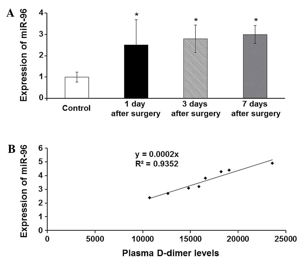
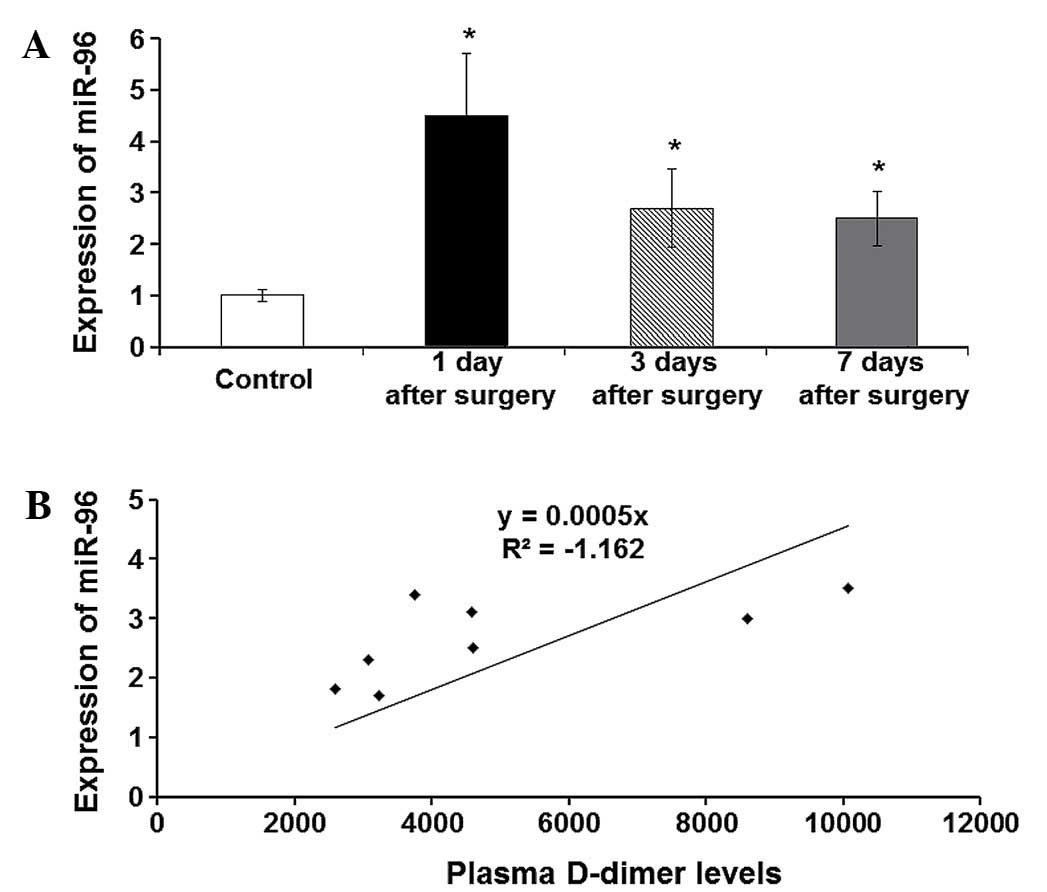
miR-96 expression levels are increased
in the non-DVT group, but are not correlated with plasma D-dimer
levels
To investigate the association between miR-96
expression levels and plasma D-dimer levels in the non-DVT group,
Pearson correlation analysis was performed. The expression levels
of miR-96 were detected using a qPCR assay in the non-DVT group at
days 1, 3 and 7 after orthopedic surgery. For control, the
expression levels of miR-96 in the control group were detected. As
shown in Fig. 4A, miR-96 expression
levels in the non-DVT group at days 1, 3 and 7 after orthopedic
surgery were significantly increased when compared with those in
the control group (P<0.05). Pearson correlation analysis results
showed that the gradually increased miR-96 expression levels were
not linearly correlated with plasma D-dimer levels in the non-DVT
group (Fig. 4B). Levels of plasma
D-dimer are commonly used for the prediction of DVT (17). These results suggest although miR-96
expression levels are increased in the non-DVT group, miR-96
expression levels are not correlated with plasma D-dimer
levels.
Increased miR-96 expression levels are
correlated with plasma D-dimer levels in the DVT group
To investigate whether miR-96 expression levels were
correlated with plasma D-dimer levels in the DVT group, Pearson
correlation analysis were performed. In the DVT group at days 1, 3
and 7 after orthopedic surgery, the expression levels of miR-96
were detected using a qPCR assay. As shown in Fig. 5A, miR-96 expression levels in the DVT
group at days 1, 3 and 7 after orthopedic surgery were
significantly increased when compared with those in the control
group (P<0.05). Pearson correlation analysis results showed that
miR-96 expression levels were correlated with plasma D-dimer levels
in the DVT group (Fig. 5B). These
results indicate that the occurrence of DVT may be predicted by
comprehensive analysis of the levels of miR-96 and plasma
D-dimer.
Discussion
DVT is a common postoperative complication in
orthopedic patients, accounting for ~50% of the total patients with
embolization (18). Pulmonary
embolism caused by DVT is a potentially life-threatening disease
with a high mortality rate of 0.5% (4). Therefore, early prevention and accurate
diagnosis are key to reducing morbidity and mortality.
As an important miRNA in platelets, miR-96 inhibits
the expression of VAMP8, which serves a crucial function in
reducing the activation of platelets (11). In the present study, the expression
levels of miR-96 and VAMP8 were determined in the DVT, non-DVT and
control groups. The results showed that in the non-DVT group,
miR-96 expression levels were increased while VAMP8 expression
levels were decreased when compared with those in the control
group. For the DVT group, miR-96 expression levels were also
significantly increased in platelets. The expression levels of
miR-96 in the DVT group were higher than those in the non-DVT
group. However, VAMP8 expression levels were not decreased in the
DVT group, as compared with the non-DVT group. These results
indicate that the expression of VAMP8 after orthopedic surgery may
be regulated by miR-96, in addition to other signaling pathways.
Plasma D-dimer levels indicate the activity of the
coagulation/fibrinolytic system in vivo, which can be used
as a marker for the prediction of DVT.
The present results showed that miR-96 expression
levels in the DVT group at days 1, 3 and 7 after orthopedic surgery
were significantly increased when compared with those in the
control group. The increased miR-96 expression levels were
correlated with plasma D-dimer levels in the DVT group. However,
although miR-96 expression levels were increased in the non-DVT
group, miR-96 expression levels were not correlated with plasma
D-dimer levels. Typically, due to the stress responses, compared
with normal subjects the levels of various proteins and factors
would be elevated in the patients following surgery. However, the
single factor detection may not be sufficient for disease
prediction. The results of the present study demonstrated that the
expression levels of miR-96 in the patients following surgery were
elevated in both the DVT and non-DVT groups. Compared with the DVT
group, the D-dimer value was also increased in the DVT group, in
addition to elevated miR-96 expression levels, indicating an
association between the two events in these patients. Based on
these findings, the simultaneous detection of elevated miR-96
expression levels and D-dimer values could contribute to a more
accurate prediction of the incidence of DVT.
In conclusion, the expression levels of platelets
miR-96 are significantly increased in patients after orthopedic
surgery. The expression levels of platelets miR-96 are correlated
with the levels of plasma D-dimer in the DVT group. These results
suggest that the occurrence of DVT may be accurately predicted by
comprehensive analysis with levels of miR-96 and plasma
D-dimer.
Acknowledgements
The authors thank Professor Xuesheng Sun, Mr. Yichen
Dong and Ms. Huiyun Zhang from Department of Orthopedics of Laiwu
People's Hospital (Laiwu, China).
References
|
1
|
Dua A, Neiva S and Sutherland A: Does
previous varicose vein surgery alter deep vein thrombosis risk
after lower limb arthroplasty? Orthop Surg. 4:222–2226. 2012.
View Article : Google Scholar : PubMed/NCBI
|
|
2
|
Shah K, Thevendran G, Younger A and Pinney
SJ: Deep-vein thrombosis prophylaxis in foot and ankle surgery:
What is the current state of practice? Foot Ankle Spec. 8:101–116.
2015. View Article : Google Scholar : PubMed/NCBI
|
|
3
|
Rahman JR, Magan A and Tytherleigh-Strong
GM: Subclavian vein thrombosis following acute internal fixation of
a clavicular fracture. Shoulder Elb. 5:108–110. 2013. View Article : Google Scholar
|
|
4
|
Wu O, Clark P, Lowe GD, Walker ID and
Greer IA: Thrombosis: Risk and Economic Assessment of Thrombophilia
Screening (TREATS) Study Group: Thrombophilia and venous
thromboembolism after total hip or knee replacement surgery: A
systematic review. J Thromb Haemost. 3:811–813. 2005. View Article : Google Scholar : PubMed/NCBI
|
|
5
|
Silverstein RL: Platelet CD36 links not
only α-granule-derived proteins to thrombus stability but also
metabolic and oxidant stress to a prothrombotic phenotype.
Arterioscler Thromb Vasc Biol. 34:1120–1121. 2014. View Article : Google Scholar : PubMed/NCBI
|
|
6
|
Kuijpers MJ, de Witt S, Nergiz-Unal R, van
Kruchten R, Korporaal SJ, Verhamme P, Febbraio M, Tjwa M, Voshol
PJ, Hoylaerts MF, et al: Supporting roles of platelet
thrombospondin-1 and CD36 in thrombus formation on collagen.
Arterioscler Thromb Vasc Biol. 34:1187–1192. 2014. View Article : Google Scholar : PubMed/NCBI
|
|
7
|
Heestermans M, Salloum-Asfar S, Salvatori
D, Laghmani EH, Luken BM, Zeerleder SS, Spronk HM, Korporaal SJ,
Wagenaar GT, Reitsma PH and van Vlijmen BJ: Role of platelets,
neutrophils, and factor XII in spontaneous venous thrombosis in
mice. Blood. [Epub ahead of print]. 2016. View Article : Google Scholar : PubMed/NCBI
|
|
8
|
Marshall MR, Pattu V, Halimani M,
Maier-Peuschel M, Müller ML, Becherer U, Hong W, Hoth M, Tschernig
T, Bryceson YT and Rettig J: VAMP8-dependent fusion of recycling
endosomes with the plasma membrane facilitates T lymphocyte
cytotoxicity. J Cell Biol. 210:135–151. 2015. View Article : Google Scholar : PubMed/NCBI
|
|
9
|
Polgár J, Chung SH and Reed GL:
Vesicle-associated membrane protein 3 (VAMP-3) and VAMP-8 are
present in human platelets and are required for granule secretion.
Blood. 100:1081–1083. 2002. View Article : Google Scholar : PubMed/NCBI
|
|
10
|
Ren Q, Barber HK, Crawford GL, Karim ZA,
Zhao C, Choi W, Wang CC, Hong W and Whiteheart SW:
Endobrevin/VAMP-8 is the primary v-SNARE for the platelet release
reaction. Mol Biol Cell. 18:24–33. 2007. View Article : Google Scholar : PubMed/NCBI
|
|
11
|
Kondkar AA, Bray MS, Leal SM, Nagalla S,
Liu DJ, Jin Y, Dong JF, Ren Q, Whiteheart SW, Shaw C and Bray PF:
VAMP8/endobrevin is overexpressed in hyperreactive human platelets:
Suggested role for platelet microRNA. J Thromb Haemost. 8:369–378.
2010. View Article : Google Scholar : PubMed/NCBI
|
|
12
|
Zhu HQ: Differential expression of miR-223
and miR-96 in platelets and its association with clopidogrel
reactivity in patients with coronary heart disease. Cardiovascular
Disease Journal Of integrated traditional Chinese and Western
Medicine. 1:7–8. 2015.(In Chinese).
|
|
13
|
Molugu C, Fisher G, Hirons B, Hughes D and
Raftery S: P151 V-DimERS study-value of D-Dimers in estimating risk
of significant pulmonary embolism and deep vein thrombosis. Thorax.
68(Suppl 3): A144. 2013. View Article : Google Scholar
|
|
14
|
Wells P: Overview and comparison of
D-dimer assay kits for DVT and PE. Clin Adv Hematol Oncol.
2:1602004.PubMed/NCBI
|
|
15
|
Wedlund A and Voslar A: Is the D-dimer
test a viable option for the detection of PE? J Nucl Med. 55(Suppl
1): 27082014.
|
|
16
|
Lees D, Griffiths P, Paxton C and Wahbi Z:
Can D-dimer assay, together with clinical probability predict
computed tomography pulmonary angiogram (CTPA) outcomes for
pulmonary embolism (PE)? Eur Respir J. 38(Suppl 55): 5822011.
|
|
17
|
Hodgkiss-Harlow KD and Betti K:
Institutional quality outcome measures: Investigating the impact of
raising the positive D-dimer threshold for DVT as confirmed by
duplex ultrasound. J Vasc Surg. 62:5272015. View Article : Google Scholar
|
|
18
|
Shulman RM, Buchan C, Bleakney RR and
White LM: Low prevalence of unexpected popliteal DVT detected on
routine MRI assessment of the knee. Clin Imaging. 40:79–85. 2016.
View Article : Google Scholar : PubMed/NCBI
|