Introduction
Hydrocephalus, which is characterized by
cerebrospinal fluid circulation disorder, pathological expansion of
the ventricle and/or subarachnoid space, and cerebral shrinkage, is
a common complication occurring after intraventricular hemorrhage
(IVH) (1,2). Hydrocephalus leads to decreased
cognitive function and neurological damage, seriously affecting the
prognosis of patients (3). Previous
studies have suggested that the key causes of hydrocephalus
subsequent to IVH may be the apoptosis or death of stem cells as a
result of the presence of various inflammatory and pro-apoptotic
factors in the micro-environment, which are released after IVH
(2,4). However, the underlying mechanism of
hydrocephalus occurring after IVH is not fully understood.
Although surgery has been used as the main treatment
for hydrocephalus, it commonly leads to certain complications and
has a poor success rate. Currently, studies focus on the
reconstruction of the body's resources in order to repair
neurologic function (5,6). In addition, the mobilization of
endogenous neural stem cells has been investigated as an
alternative and less invasive approach for brain injury (7). Granulocyte-colony stimulating factor
(G-CSF) is a well-established stem-cell mobilizer for endogenous
neural stem cell transplantation, in which CD34 is used as a marker
of hemopoietic stem cells (8). G-CSF
stimulates the proliferation, survival and maturation of cells
committed to the neutrophilic granulocyte lineage through binding
to the specific G-CSF receptor (9).
In brain injury, G-CSF has been shown to be upregulated and to
present strong nerve regeneration ability in vivo and in
vitro (10). Lithium chloride is
the most commonly used drug for the treatment of manic depressive
illness and is reported to be a neuroprotective agent (11), exerting this effect through the
inhibition of cell apoptosis and inflammation (12,13). Wnt
pathway serves a key role in the proliferation and differentiation
of neural stem cells (14). Lithium
chloride may regulate the Wnt signaling pathway in order to reduce
the apoptosis of nerve cells and promote the differentiation of
neural stem cells (15,16).
As there are multiple factors causing hydrocephalus
subsequent to IVH, it is difficult to achieve the best treatment
effect using a single drug alone. Therefore, an increasing number
of studies have researched the effect of treatment using a
combination of drugs on hemorrhage (17,18).
Vose et al reported that treatment with TH accelerated the
proliferation and maturation of oligodendrocytes and restored
neurological function in pups with IVH (19). In the present study, the protective
effect of the combination of endogenous neural stem cell
mobilization and lithium chloride treatment on hydrocephalus
following IVH was investigated. The study examined the
inflammation, apoptosis and neuranagenesis in the brain following
the combination treatment. The current findings provide evidence on
the effect of this noninvasive treatment for hydrocephalus
subsequent to IVH.
Materials and methods
Animal groups
Animal use protocols were approved by People's
Hospital of Zhengzhou University Committee on the Use and Care of
Animals (Zhengzhou, China). Animals were provided by Henan
Experimental Animal Center of Zhengzhou. A total of 130 newborn
Sprague-Dawley (SD) rats at postnatal day 4 (weight, 5–10 g) were
used in the present study and were reared with their dams. The dams
and the newborn rats were housed at 18–25°C in an atmosphere of
50–70% relative humidity, with a 12-h light/dark cycle. Rats were
randomly divided into five groups: IVH control group (n=30; Group
1); G-CSF treatment group (n=30; Group 2); lithium chloride
treatment group (n=30; Group 3); combination treatment group (n=30;
Group 4); and sham surgery group (n=10; Group 5). Rats in Group 1
received hypodermic injection of saline on days 2, 4, 6, and 8 and
intraperitoneal injection after 2 days of IVH. Rats in Group 2
received a hypodermic injection of 50 µg/kg/day G-CSF
(Sigma-Aldrich, St. Louis, MO, USA) on days 2, 4, 6 and 8, and
intraperitoneal injection of saline with equal amounts with lithium
chloride after 2 days of IVH. Rats in Group 3 were given a
hypodermic injection of saline with equal amounts with G-CSF and
intraperitoneal injection of 3 mmol/kg/day lithium chloride after 2
days of IVH. Group 4 rats received combination treatment with a
hypodermic injection of 50 µg/kg/day G-CSF on days 2, 4, 6 and 8,
as well as intraperitoneal injection of 3 mmol/kg/day lithium
chloride after 2 days of IVH. Rats in Group 5 were subjected to
sham surgery and received no treatment.
Animal model
The IVH rat model was established as previously
described by Ahn et al (20)
and Lodhia et al (21).
Briefly, SD rats were anesthetized using 1.5–2% isoflurane
(Sigma-Aldrich) in an oxygen-enriched atmosphere (20,21). A
total of 100 µl fresh whole blood from the pregnant rats was slowly
infused into the right ventricles over 5 min at a rate of 10
µl/min, 1.7 mm lateral to the sagittal suture, 3.8 mm posterior to
the coronal suture and at a depth of 3.5 mm. After 10 min, equal
amount of fresh maternal whole blood was infused into the left
ventricles. For rats in Group 1, the same amount of saline was
infused into the right and left ventricles rather than fresh
maternal whole blood, according to the aforementioned procedure.
The animals were then allowed to recover from anesthesia. Rats
received intraperitoneal injection of 10 mg/kg/day
bromodeoxyuridine (BrdU; Sigma-Aldrich; one day after surgery) to
mark neural stem cells under proliferation. Rats in group 5
underwent a sham operation without blood injection.
Tissue collection
Rats in each group were divided into the day 11, 18
and 32 time points. At each time point, rats were fixed on the
experimental platform under deep anesthesia with 400 mg/kg chloral
hydrate (Sigma-Aldrich). Next, the hair on the chest and abdomen
was removed and the heart was exposed. The rats were decapitated
and the brain was placed in paraformaldehyde solution. Brain tissue
was collected in the periventricular cerebral mass, corpus callosum
region, subventricular zone (SVZ) and hippocampal dentate gyrus and
cut into sections of 2 mm. Subsequently, the obtained tissue was
moved into sucrose solution treated with diethyl pyrocarbonate
(Sigma-Aldrich) for 48 h. The tissue was then sliced into 5 µm
using a freezing microtome and loaded onto glass slides. The
samples were wrapped up with aluminized paper and stored at
−70°C.
Cell apoptosis assay
Cell death of nerve cells in the periventricular
cerebral mass, corpus callosum region, SVZ and hippocampal dentate
gyrus brain tissue was assessed using the immunofluorescent
terminal deoxynucleotidyl transferase-mediated deoxyuridine
triphosphate nick-end labeling (TUNEL) in the ApopTag Fluorescein
In Situ Apoptosis Detection kit (cat. no. S7110; Chemicon;
Merck Millipore, Temecula, CA, USA) according to the manufacturer's
instructions. The nucleus presenting yellow or light yellow bulk or
appearing granular under the light microscope was considered as
TUNEL-positive. A blinded evaluator counted the number of
TUNEL-positive nuclei in the brain tissue samples from the
periventricular cerebral mass, corpus callosum region, SVZ and
hippocampal dentate gyrus. A total of 10 random non-overlapping
fields of view from each brain were counted.
Western blot analysis
In order to perform immunoblotting analysis, brain
tissue obtained from rats in each group was lysed by
radioimmunoprecipitation assay lysis buffer (Beyotime Institute of
Biotechnology, Inc., Shanghai, China) and homogenates were
centrifuged at 9,200 × g for 15 min at 4°C. Then the
supernatant was collected and used for protein concentration
determination using a Bradford protein assay kit (Beyotime
Institute of Biotechnology). A total of 15 mg protein was loaded
onto each lane of an 8% sodium dodecyl sulfate-polyacrylamide gel
containing 4 mol/l urea, and electrophoresis was performed under
standard conditions. Next, the protein sample was
electrophoretically transferred to Immobilon-P polyvinylidene
difluoride membranes (Merck Millipore) by semidry blotting.
Subsequent to blocking in 3% nonfat dry milk in Tris-buffered
saline/Tween 20 (TBST; containing 20 mmol/l Tris base, pH 7.6, 137
mmol/l NaCl and 0.05% Tween 20) for 1 h at room temperature, the
membranes were incubated with antibodies against rabbit polyclonal
anti-B cell lymphoma-2 (Bcl-2; 1:500; cat. no. ab59348; Abcam,
Cambridge, UK), rabbit polyclonal anti-Bcl-2-associated X protein
(Bax; 1:500; cat. no. ab69643; Abcam), or rabbit polyclonal
anti-GAPDH (1:2,500; cat. no. ab9485; Abcam) overnight at 4°C. The
membranes were washed in TBST and then incubated for 1 h at room
temperature with horseradish peroxidase-conjugated goat anti-rabbit
IgG H&L (1:10,000; cat. no. ab97051; Abcam). Immunoreactive
bands were visualized in the linear range with enhanced
chemoluminescence (Sigma-Aldrich). The scanned images were
semi-quantitated using Quantity One software (Bio-Rad Laboratories,
Inc., Hercules, CA, USA). The relative integrated density values
were calculated using FluorChem 2.0 software (Alpha Innotech
Corporation, San Leandro, CA, USA) and normalized with GAPDH.
Double immunofluorescence
For the double immunofluorescence detection of BrdU
along with glial fibrillary acidic protein (GFAP), neuronal nuclei
(NeuN) or polysialylated-neural cell adhesion molecule (PSA-NCAM).
Brain sections were washed in phosphate-buffered saline (PBS) and
pre-incubated for 60 min in blocking buffer (5% normal donkey serum
in PBS with 0.3% Triton X-100; both obtained from Sigma-Aldrich).
Next, the brain tissue sections were incubated with the sheep
polyclonal anti-BrdU antibody (1:1,000; cat. no. M20105S; Biodesign
Inc., Saco, ME, USA), in addition to the goat polyclonal anti-GFAP
(1:400; cat. no. sc-9065; Santa Cruz Biotechnology Inc., Santa
Cruz, CA, USA), the mouse monoclonal anti-NeuN (1:500; cat. no.
MAB377; Chemicon; Merck Millipore) or the mouse monoclonal
anti-PSA-NCAM antibody (1:1,000; cat. no. MAB5324; Chemicon; Merck
Millipore) for 12 h at 4°C. Subsequently, the sections were
incubated with the fluorescein isothiocyanate (FITC)-labeled donkey
anti-sheep IgG (1:500; Molecular Probes; Thermo Fisher Scientific
Inc., Eugene, OR, USA) and rhodamine-labeled horse anti-mouse IgG
(1:500; Chemicon; Merck Millipore) antibodies for 1 h. The sections
were then rinsed in the dark, mounted on polylysine-coated slides
and coverslipped using anti-fadent solution (Biomart, Shanghai,
China). The tissue sections were viewed and photographed using
epifluorescence on an Axiophot epifluorescence microscope with
appropriate filter sets for fluorescein (excitation, 450–490 nm;
emission, 514–565 nm) and Texas red (excitation, 530–585 nm;
emission, >615 nm).
Statistics
Data are presented in the present study as the mean
± standard deviation. Statistical differences among groups were
determined using a Student's t-test. Analysis of variance with a
Newman-Keuls post-hoc test was used for multiple comparisons. SPSS
version 13.0 software (SPSS, Inc., Chicago, IL, USA) was used to
analyze the data. A value of P<0.05 was considered to indicate
statistically significant differences.
Results
Effect of combination treatment with
G-CSF and lithium chloride on hydrocephalus incidence following
IVH
To investigate the effect of combination treatment
of G-CSF and lithium chloride on hydrocephalus following IVH, rats
were divided into five groups (Group 1, IVH control; Group 2, G-CSF
treatment; Group 3, lithium chloride treatment; Group 4,
combination treatment; and Group 5, sham surgery). G-CSF was used
to mobilize endogenous neural stem cells (10). In addition, lithium chloride was
observed to have neuroprotective effects by promoting neural stem
cell differentiation (11). As shown
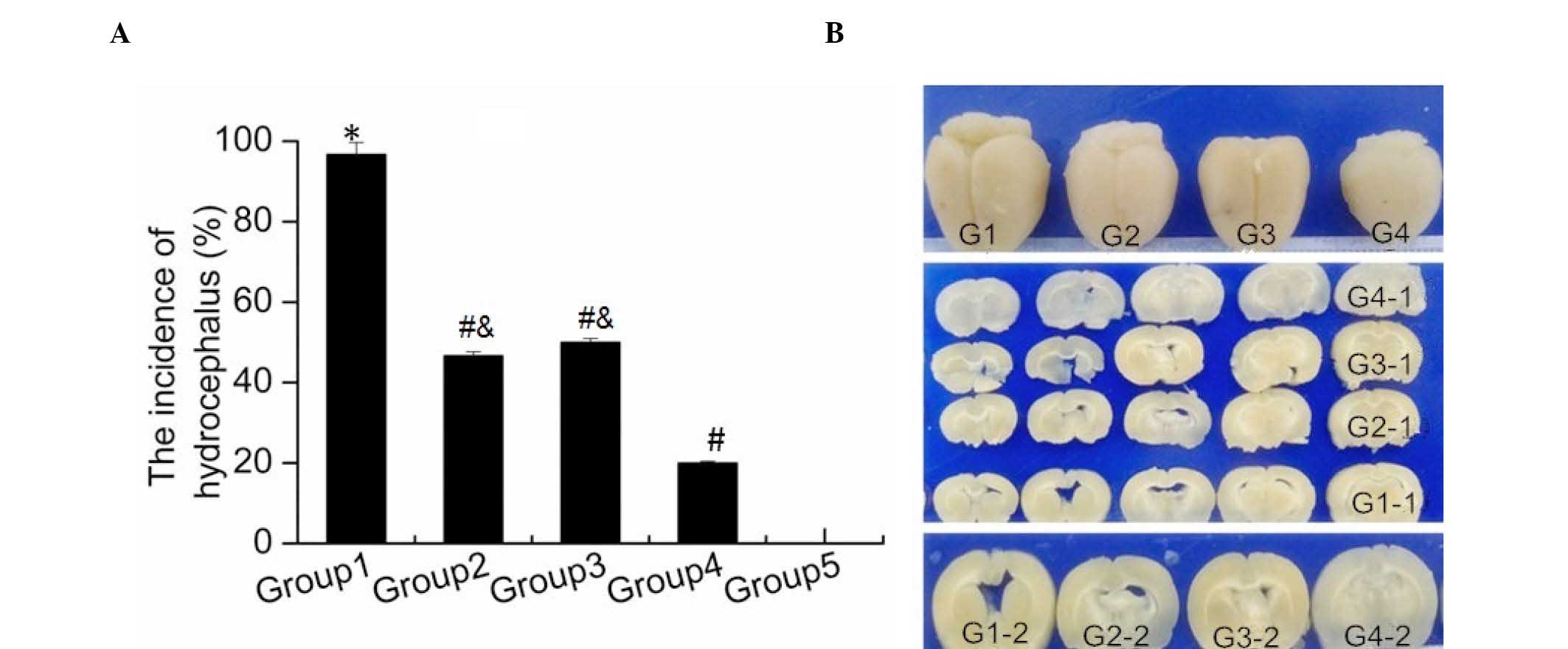
in Fig. 1A, severe hydrocephalus
occurred in the majority of rats in Group 1 after IVH, compared
with the rats only subjected to sham surgery (Group 5; P<0.001).
In addition, the hydrocephalus incidence after IVH in Group 2 and
Group 3 rats was significantly decreased compared with that in
Group 1 rats (both P<0.001). Furthermore, a significant reduced
hydrocephalus incidence was observed in Group 4 when compared with
that in Groups 2 and 3 (P=0.028; P=0.015). Images of brain tissue
obtained from rats in the different groups were captured, and are
presented in Fig. 1B. The brain of
rats in Group 1 appeared to be larger in size when compared with
that in other groups. The brains of rats in Groups 2 and 3 were
smaller than those in Group 1, however they were still larger in
size compared with those in Group 4, which appeared to be closer to
the normal brain size in Group 5 (Fig.
1B). These data indicated that combination treatment with G-CSF
and lithium chloride was more effective against hydrocephalus after
IVH compared with single drug administration, and significantly
attenuated the development of hydrocephalus.
Effect of combination treatment with
G-CSF and lithium chloride on nerve cell apoptosis in brain
tissue
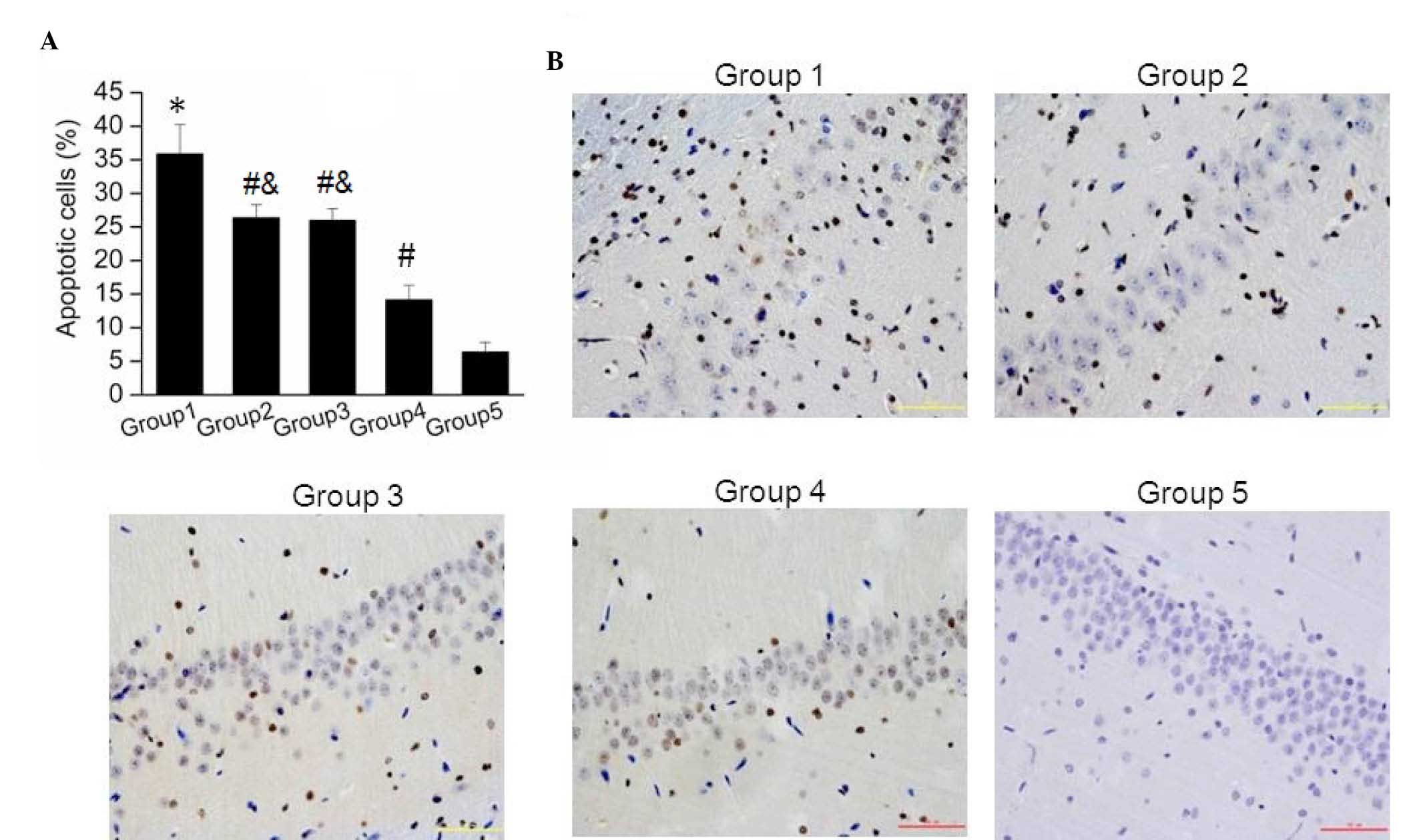
The current study further detected the nerve cell
apoptosis in brain tissue samples obtained from the five groups
using TUNEL assay. It was observed that the apoptotic cell number
in Group 1 was significantly higher than that in Group 5 (P=0.002).
As shown in Fig. 2A, the amount of
apoptotic cells in Groups 2, 3 and 4 were significantly reduced
compared with that in Group 1. Furthermore, apoptosis of nerve
cells was significantly reduced in Group 4 when compared with that
in Groups 2 and 3 (Fig. 2A; both
P=0.001). Images of the apoptosis assay in brain tissue samples
from the five groups are shown in Fig.
2B, further verifying these results. These findings suggested
that combination treatment with G-CSF and lithium chloride
significantly reduced the nerve cell apoptosis induced by
hydrocephalus subsequent to IVH.
Protein expression levels of Bcl-2 and
Bax in nerve cells in brain tissue
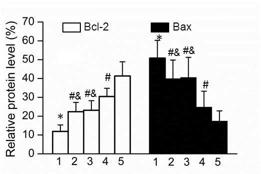
As Bcl-2 and Bax are known to be involved in cell
apoptosis, their protein expression levels were examined in nerve
cells of brain tissue samples obtained from the various rat groups.
As showed in Fig. 3, the protein
expression of Bcl-2 in Groups 2 and 3 was significantly higher
compared with that in Group 1; however, the expression in Groups 2
and 3 was significantly lower compared with that in Group 4
(P=0.0015; P=0.001). By contrast, the expression of Bax was
significantly upregulated in Group 1, while a lower protein level
was observed in Group 4, when compared with Bax expression in
Groups 2 and 3. In addition, the expression levels of Bcl-2 and Bax
were significantly decreased or increased in Group 1 compared with
that in Group 5, respectively (P=0.003; P=0.0025).
Expression of nerve cell
differentiation markers in brain tissue
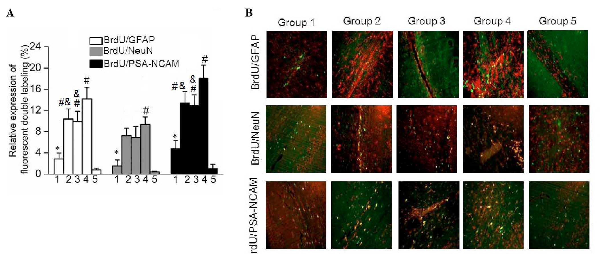
In order to detect the differentiation of nerve
cells in brain tissue samples obtained from the five rat groups,
the expression levels of GFAP, NeuN and PSA-NCAM were examined
using a double immunofluorescence labeling method with BrdU to
evaluate cell proliferation intensity. As shown in Fig. 4, the results demonstrated that the
expression levels of BrdU/GFAP, BrdU/NeuN and BrdU/PSA-NCAM were
significantly upregulated in Group 1 compared with those in Group 5
(P=0.03). In addition, the expression levels of BrdU/GFAP,
BrdU/NeuN and BrdU/PSA-NCAM in Groups 2 and 3 were significantly
increased when compared with that in Group 1 (Fig. 4A). In addition, highest expression
levels of BrdU/GFAP, BrdU/NeuN and BrdU/PSA-NCAM were observed in
Group 4. The fluorescence distribution images shown in Fig. 4B further verify this differential
expression. These data indicated that combination treatment with
G-CSF and lithium chloride was more effective at promoting nerve
cell differentiation compared with single drug administration.
Discussion
IVH is the most common severe disease in the area of
neurosurgery and results in several complications, including
hydrocephalus, high intracranial pressure and the injury of
ventricular tissue in the brain (1).
Among these, hydrocephalus is the most common complication, which
aggravates the damage in brain tissue. It is widely recognized that
hydrocephalus subsequent to IVH results from the increased
secretion of blood stimulated subarachnoid granules and blood
coagulation (22). A series of
pathophysiological changes occur during the formation of
hydrocephalus, including apoptosis of nerve cells and glial
hyperplasia (23). It has been
reported that neuronal apoptosis and proliferation of white matter
mainly results in irreversible damage of hydrocephalus (5).
Mobilization of bone marrow stem cells is a novel
method for cell transplantation, and contributes to the
regeneration or repair of nerve cells (24). A specific type of bone marrow stem
cells have been shown to positively express C-X-C chemokine
receptor 4 (CXCR4) and possess the function of directional
differentiation and migration (25).
In addition, CXCR4 was found to be the unique receptor of stromal
cell-derived factor-1 (SDF-1) (26).
When pathological changes occurred in the central nervous system,
SDF-1 was demonstrated to be upregulated and cause the migration of
monocytes/macrophages in the blood system into the injury zone,
exerting chemotactic effects (27).
Reported evidence has indicated that G-CSF may mobilize bone marrow
stem cells to stimulate proliferation and differentiation of stem
cells (28). G-CSF may exert an
anti-apoptotic function by regulating the expression of Bcl-2 and
Bax (29). In addition, it is widely
known as a growth factor for hematopoietic cells that promotes
survival, proliferation and differentiation of cells of the
neutrophil lineage (30). Kawabe
et al have also suggested that G-CSF exerts neuroprotective
effects via the promotion of angiogenesis subsequent to spinal cord
injury (31). Furthermore, lithium
chloride has been previously reported to reduce the apoptosis of
nerve cells by suppressing the activity of glycogen synthase kinase
3β and enhancing the absorption of BrdU (15). In the current study, the effect of
combination treatment with G-CSF and lithium chloride on
hydrocephalus following IVH was investigated. The results
demonstrated that single use of G-CSF or lithium chloride only
partly relieved the incidence of hydrocephalus following IVH. By
contrast, combination treatment with G-CSF and lithium chloride
significantly attenuated the development of hydrocephalus following
IVH in rats.
The present study further examined the neuronal
apoptosis using TUNEL assay in brain tissues. The results showed
that neuronal apoptosis was significantly reduced by the
combination treatment with G-CSF and lithium chloride. Furthermore,
the expression of Bcl-2 was upregulated and Bax was downregulated
in the combination treatment group. Bcl-2 has been reported to
inhibit cell apoptosis, while Bax may stimulate the expression of
apoptotic genes (32). These data
observed in the present study indicated that combination treatment
of G-CSF and lithium chloride was more effective in treating
hydrocephalus after IVH compared with treatment with a single
agent. In addition, combination treatment exerted a neuroprotective
function by reducing the apoptotic cells.
The expression levels of neural stem cell marker
NeuN, mature neuron marker PSA-NCAM and mature astrocyte marker
GFAP commonly characterize the differentiation of nerve cells
(33). The present study observed
that the expression levels of BrdU/GFAP, BrdU/NeuN and
BrdU/PSA-NCAM in the G-CSF alone or lithium chloride alone
treatment groups were significantly increased when compared with
those in the IVH control group (Fig.
4A). In addition, the highest levels of BrdU/GFAP, BrdU/NeuN
and BrdU/PSA-NCAM were expressed in the combination treatment
group. These data indicated that combination treatment with G-CSF
and lithium chloride may be able to mobilize bone marrow stromal
cells to assemble into the hemorrhage region of brain tissue.
Regenerative nerve cells may replace the local necrosis and loss
nerve cells. A synaptic linkage between the nerve cells and the
surrounding neurons may then be established and the function of
neuron cells may be recovered.
In conclusion, the present study demonstrated that
the combination of endogenous neural stem cell mobilization using
G-CSF and lithium chloride treatment highly reduced the incidence
of hydrocephalus following IVH by inhibiting neuronal apoptosis.
The findings of the current study provide evidence on the
effectiveness of this noninvasive treatment for hydrocephalus
occurring subsequent to IVH.
Acknowledgements
The study was supported by a grant from the
Foundation and Advanced Technology of Henan Province (no.
142300410267).
References
|
1
|
Vassilyadi M, Tataryn Z, Shamji MF and
Ventureyra EC: Functional outcomes among premature infants with
intraventricular hemorrhage. Pediatr Neurosurg. 45:247–255. 2009.
View Article : Google Scholar : PubMed/NCBI
|
|
2
|
Cherian SS, Love S, Silver IA, Porter HJ,
Whitelaw AG and Thoresen M: Posthemorrhagic ventricular dilation in
the neonate: Development and characterization of a rat model. J
Neuropathol Exp Neurol. 62:292–303. 2003. View Article : Google Scholar : PubMed/NCBI
|
|
3
|
Zacharia BE, Vaughan KA, Hickman ZL, Bruce
SS, Carpenter AM, Petersen NH, Deiner S, Badjatia N and Connolly ES
Jr: Predictors of long-term shunt-dependent hydrocephalus in
patients with intracerebral hemorrhage requiring emergency
cerebrospinal fluid diversion. Neurosurg Focus. 32:E52012.
View Article : Google Scholar : PubMed/NCBI
|
|
4
|
Simard PF, Tosun C, Melnichenko L, Ivanova
S, Gerzanich V and Simard JM: Inflammation of the choroid plexus
and ependymal layer of the ventricle following intraventricular
hemorrhage. Transl Stroke Res. 2:227–231. 2011. View Article : Google Scholar : PubMed/NCBI
|
|
5
|
Chen FM, Wu LA, Zhang M, Zhang R and Sun
H: Homing of endogenous stem/progenitor cells for in situ tissue
regeneration: Promises, strategies, and translational perspectives.
Biomaterials. 32:3189–3209. 2011. View Article : Google Scholar : PubMed/NCBI
|
|
6
|
Bellenchi GC, Volpicelli F, Piscopo V,
Perrone-Capano C and di Porzio U: Adult neural stem cells: An
endogenous tool to repair brain injury? J Neurochem. 124:159–167.
2013. View Article : Google Scholar : PubMed/NCBI
|
|
7
|
Lindvall O, Kokaia Z and Martinez-Serrano
A: Stem cell therapy for human neurodegenerative disorders-how to
make it work. Nat Med Suppl. S42–S50. 2004. View Article : Google Scholar
|
|
8
|
Yanqing Z, Yu-Min L, Jian Q, Bao-Guo X and
Chuan-Zhen L: Fibronectin and neuroprotective effect of granulocyte
colony-stimulating factor in focal cerebral ischemia. Brain Res.
1098:161–169. 2006. View Article : Google Scholar : PubMed/NCBI
|
|
9
|
Hartung T: Anti-inflammatory effects of
granulocyte colony-stimulating factor. Curr Opin Hematol.
5:221–225. 1998. View Article : Google Scholar : PubMed/NCBI
|
|
10
|
Schäbitz WR, Kollmar R, Schwaninger M,
Juettler E, Bardutzky J, Schölzke MN, Sommer C and Schwab S:
Neuroprotective effect of granulocyte colony-stimulating factor
after focal cerebral ischemia. Stroke. 34:745–751. 2003. View Article : Google Scholar : PubMed/NCBI
|
|
11
|
Chuang DM, Chen RW, Chalecka-Franaszek E,
Ren M, Hashimoto R, Senatorov V, Kanai H, Hough C, Hiroi T and
Leeds P: Neuroprotective effects of lithium in cultured cells and
animal models of diseases. Bipolar Disord. 4:129–136. 2002.
View Article : Google Scholar : PubMed/NCBI
|
|
12
|
Chen RW and Chuang DM: Long term lithium
treatment suppresses p53 and Bax expression but increases Bcl-2
expression. A prominent role in neuroprotection against
excitotoxicity. J Biol Chem. 274:6039–6042. 1999. View Article : Google Scholar : PubMed/NCBI
|
|
13
|
Chen G, Zeng WZ, Yuan PX, Huang LD, Jiang
YM, Zhao ZH and Manji HK: The mood-stabilizing agents lithium and
valproate robustly increase the levels of the neuroprotective
protein bcl-2 in the CNS. J Neurochem. 72:879–882. 1999. View Article : Google Scholar : PubMed/NCBI
|
|
14
|
Katoh M: WNT signaling in stem cell
biology and regenerative medicine. Curr Drug Targets. 9:565–570.
2008. View Article : Google Scholar : PubMed/NCBI
|
|
15
|
Hirsch C, Campano LM, Wöhrle S and Hecht
A: Canonical Wnt signaling transiently stimulates proliferation and
enhances neurogenesis in neonatal neural progenitor cultures. Exp
Cell Res. 313:572–587. 2007. View Article : Google Scholar : PubMed/NCBI
|
|
16
|
Chamnanvanakij S, Margraf LR, Burns D and
Perlman JM: Apoptosis and white matter injury in preterm infants.
Pediatr Dev Pathol. 5:184–189. 2002. View Article : Google Scholar : PubMed/NCBI
|
|
17
|
Di Renzo GC, Mignosa M, Gerli S, Burnelli
L, Luzi G, Clerici G, Taddei F, Marinelli D, Bragetti P, Mezzetti
D, et al: The combined maternal administration of magnesium sulfate
and aminophylline reduces intraventricular hemorrhage in very
preterm neonates. Am J Obstet Gynecol. 192:433–438. 2005.
View Article : Google Scholar : PubMed/NCBI
|
|
18
|
Chemtob S, Laudignon N and Aranda JV: Drug
therapy in hypoxic-ischemic cerebral insults and intraventricular
hemorrhage of the newborn. Clin Perinatol. 14:817–842.
1987.PubMed/NCBI
|
|
19
|
Vose LR, Vinukonda G, Jo S, Miry O,
Diamond D, Korumilli R, Arshad A, Zia MT, Hu F, Kayton RJ, et al:
Treatment with thyroxine restores myelination and clinical recovery
after intraventricular hemorrhage. J Neurosci. 33:17232–17246.
2013. View Article : Google Scholar : PubMed/NCBI
|
|
20
|
Ahn SY, Chang YS, Sung DK, Sung SI, Yoo
HS, Lee JH, Oh WI and Park WS: Mesenchymal stem cells prevent
hydrocephalus after severe intraventricular hemorrhage. Stroke.
44:497–504. 2013. View Article : Google Scholar : PubMed/NCBI
|
|
21
|
Lodhia KR, Shakui P and Keep RF:
Hydrocephalus in a rat model of intraventricular hemorrhage. Acta
Neurochir Suppl. 96:207–211. 2006. View Article : Google Scholar : PubMed/NCBI
|
|
22
|
Paczkowska E, Dabkowska E, Nowacki P and
Machaliński B: Stem cell-based therapy in central nervous system
diseases. Neurol Neurochir Pol. 43:550–558. 2009.(In Polish).
PubMed/NCBI
|
|
23
|
Rodríguez EM, Guerra MM, Vío K, González
C, Ortloff A, Bátiz LF, Rodríguez S, Jara MC, Muñoz RI, Ortega E,
et al: A cell junction pathology of neural stem cells leads to
abnormal neurogenesis and hydrocephalus. Biol Res. 45:231–242.
2012. View Article : Google Scholar : PubMed/NCBI
|
|
24
|
Urdzíková L, Jendelová P, Glogarová K,
Burian M, Hájek M and Syková E: Transplantation of bone marrow stem
cells as well as mobilization by granulocyte-colony stimulating
factor promotes recovery after spinal cord injury in rats. J
Neurotrauma. 23:1379–1391. 2006. View Article : Google Scholar : PubMed/NCBI
|
|
25
|
Stumm RK, Rummel J, Junker V, Culmsee C,
Pfeiffer M, Krieglstein J, Höllt V and Schulz S: A dual role for
the SDF-1/CXCR4 chemokine receptor system in adult brain:
isoform-selective regulation of SDF-1 expression modulates
CXCR4-dependent neuronal plasticity and cerebral leukocyte
recruitment after focal ischemia. J Neurosci. 22:5865–5878.
2002.PubMed/NCBI
|
|
26
|
Rath D, Chatterjee M, Borst O, Müller K,
Langer H, Mack AF, Schwab M, Winter S, Gawaz M and Geisler T:
Platelet surface expression of stromal cell-derived factor-1
receptors CXCR4 and CXCR7 is associated with clinical outcomes in
patients with coronary artery disease. J Thromb Haemost.
13:719–728. 2015. View Article : Google Scholar : PubMed/NCBI
|
|
27
|
Blum A, Childs RW, Smith A, Patibandla S,
Zalos G, Samsel L, McCoy JP, Calandra G, Csako G and Cannon RO III:
Targeted antagonism of CXCR4 mobilizes progenitor cells under
investigation for cardiovascular disease. Cytotherapy.
11:1016–1019. 2009. View Article : Google Scholar : PubMed/NCBI
|
|
28
|
Shyu WC, Lin SZ, Yang HI, Tzeng YS, Pang
CY, Yen PS and Li H: Functional recovery of stroke rats induced by
granulocyte colony-stimulating factor-stimulated stem cells.
Circulation. 110:1847–1854. 2004. View Article : Google Scholar : PubMed/NCBI
|
|
29
|
Yata K, Matchett GA, Tsubokawa T, Tang J,
Kanamaru K and Zhang JH: Granulocyte-colony stimulating factor
inhibits apoptotic neuron loss after neonatal hypoxia-ischemia in
rats. Brain Res. 1145:227–238. 2007. View Article : Google Scholar : PubMed/NCBI
|
|
30
|
Roberts AW: G-CSF: A key regulator of
neutrophil production, but that's not all! Growth Factors.
23:33–41. 2005. View Article : Google Scholar : PubMed/NCBI
|
|
31
|
Kawabe J, Koda M, Hashimoto M, Fujiyoshi
T, Furuya T, Endo T, Okawa A and Yamazaki M: Neuroprotective
effects of granulocyte colony-stimulating factor and relationship
to promotion of angiogenesis after spinal cord injury in rats:
Laboratory investigation. J Neurosurg Spine. 15:414–421. 2011.
View Article : Google Scholar : PubMed/NCBI
|
|
32
|
Shen ZJ, Esnault S, Schinzel A, Borner C
and Malter JS: The peptidyl-prolyl isomerase Pin1 facilitates
cytokine-induced survival of eosinophils by suppressing Bax
activation. Nat Immunol. 10:257–265. 2009. View Article : Google Scholar : PubMed/NCBI
|
|
33
|
Garcia AD, Doan NB, Imura T, Bush TG and
Sofroniew MV: GFAP-expressing progenitors are the principal source
of constitutive neurogenesis in adult mouse forebrain. Nat
Neurosci. 7:1233–1241. 2004. View
Article : Google Scholar : PubMed/NCBI
|