Introduction
Focal cortical dysplasia is a type of intractable
epilepsy-related congenital malformation of cortical development
(MCDs) in children and adults (1).
The statistical results of epilepsy in focal cortical dysplasia
from Sloan and Barres showed that the incidence of intractable
epilepsy in MCDs was as high as 32.5% (2). Study results from Canpolat et al
(3) showed that, MCDs was most
common in children with epilepsy and physical retardation or
neurological deficits limitations, thus, MCDs may be considered a
predisposing factor for adults and children with intractable
epilepsy. Zhang suggested that epilepsy is a type of chronic
disease featured by transient brain dysfunction caused by sudden
abnormal discharge from brain neurons, with an incidence of
approximately 7% in China and nearly 40 million new cases are
reported each year in China (4).
Myeloid-related protein 8 (MRP8), an endogenous ligand of low
molecular weight calcium-binding protein, has been demonstrated to
be associated with various autoimmune diseases such as asthma
(5), arthritis and inflammatory
bowel disease and other autoimmune deficiencies (6). The study results from Pellegrini et
al have shown that the extremely important cytokine in human
body, interleukin-7 (IL-7) was closely associated with many
immunoreactions in the body (7).
According to Xu et al (8), it
has been shown that IL-7 plays important roles in the aspects of
messaging, activation and regulation of immune cells, and
intracellular and intercellular immune response in the body,
mediated activation of T and B cells, proliferation and
differentiation and inflammation treatment. Although MRP8 and IL-7
have certain therapeutic effects on the associated immune
overreaction in the body, the studies on the effect of these
proteins on treating focal cortical dysplasia with secondary
intractable epilepsy are rarely reported.
In the present study, the effect of lamotrigine in
treating focal cortical dysplasia with secondary intractable
epilepsy is expected to provide a theoretical and experimental
basis for the sequential treatment of the disease cortical
dysplasia with secondary intractable epilepsy.
Materials and methods
Animal treatment
The rat model of focal cortical dysplasia with
secondary intractable epilepsy used in this experiment was
constructed and preserved in our laboratory. In the present study,
21 healthy Sprague-Dawley rats were selected as the subjects and
randomly divided into the control group normal mice (n=38),
observation group I mice having the disease and treated (n=39), and
observation group II mice with the disease but not treated (n=38).
The observation group I was given intraperitoneal injection of 0.02
mg/kg lamotrigine (Sigma-Aldrich, St. Louis, MO, USA) each day, and
the observation II and the control group were given intraperitoneal
injection of the same amount of normal saline each day.
RNA extraction
RNA extraction was conducted in accordance with the
Axygen kit instructions (Axygen Biosciences, Union City, CA, USA)
with modifications (9).
Fluorescence quantitative polymerase
chain reaction (qPCR)
In this study, fluorescence qPCR kit was purchased
from Takara Bio (Dalian, China). The experiment was carried out in
three steps, and the specific scheme was carried out in accordance
with the instructions and improved. The primers used are shown in
Table I.
 | Table I.Primers used for the PCR reaction. |
Table I.
Primers used for the PCR reaction.
| Gene | Primer sequence |
|---|
| MRP8 | F:
5′-CGACATGGCAACTGAACTGGA-3′ |
|
| R:
5′-ACGCCCACCCTTATCACCAAC-3′ |
| IL-7 | F:
5′-CGTCGGGTTAGCTAGCATAGC-3′ |
|
| R:
5′-TGCTGACGCCTAGCATCGATAC-3′ |
| GAPDH | F:
5′-TCATGGGTGTGAACCATGAGAA-3′ |
|
| R:
5′-GGCAGGACTGTGGTCATGAG-3′ |
Expression of MRP8 and IL-7 in the
hippocampus by enzyme-linked immunosorbent assay (ELISA)
The double antibody sabdwich method was employed to
detect the expression of TAG1/APP gene. The specific methods
were:
i) Coating: phosphate-buffered saline (PBS), pH 9.0
buffer was used to appropriately dilute the antibody protein, and
the concentration was 1–10 µg/ml. Then 0.1 ml was added into the
96-well plate, leaving it overnight at the temperature of 4°C, then
the liquid was discarded the following day and the plate was washed
5 times, each time 2 min, with washing liquid.
ii) The sample: The treated serum samples of 0.1 ml
were added into the 96-well plate, leaving it for 1 h at the
temperature of 37°C. Then the plate was washed 5 times, each time 2
min, with washing buffer (the blank well, the negative control, and
the positive control were prepared).
iii) Secondary antibody: The secondary antibody of
0.1 ml was added into the 96-well plate after washing, leaving it
for 0.5–1.2 h at the temperature of 37°C, then washing 5 times,
each time 2 min, with washing buffer after dyeing red.
iv) Chromogenic substrate: Newly prepared
chromogenic substrate was added into the 96-well plate after
washing, and then incubated for 30 min at the temperature of
37°C.
v) Terminating solution: 0.2 M of sulfuric acid
termination solution of 0.005 ml was added into the plate on
termination.
vi) Qualitative detection: The 96-well plate was
quantitatively observed for the depth of color, and the deeper the
color, the stronger the positive degree was; and a higher content
of GAG1/APP indicated the negative control well was colorless.
Quantitative detection involved placing a 96-well plate on the
enzyme standard instrument for quantitative detection with a
wavelength of 450 nm. Zero adjustment was conducted by the blank
well. If the level of OD was 1.2-fold greater than the level of
negative control, it was recorded as positive.
Expression quantity of MRP8 and IL-7
in the hippocampus by western blot analysis
The experimental operation of western blot analysis
was conducted according to the Molecular Cloning Manual with
modifications. Primary rabbit monoclonal MRP8 antibody (dilution,
1/500; cat. no. ab92331), rabbit polyclonal IL-7 antibody
(dilution, 1/500; cat. no. ab9732), and secondary goat anti-rabbit
(HRP) IgG antibody (dilution, 1/2,000; cat. no. ab6721) were all
purchased from Abcam (Cambridge, MA, USA).
Expression quantity of MRP8 and IL-7
in the hippocampus by the immune group
According to the experimental methods of Xiang et
al (10), the experimental rats
were treated and the tissue samples were obtained. The samples were
soaked for 10 min with 3% hydrogen peroxide after the conventional
dewaxing hydration treatment, and then placed in the microwave oven
with gentle heat for 1 min. The experimental samples were removed,
the first antibody was added when cooled (MRP8, 1:400 and 1:350,
both from Roche Diagnostics), and the samples were incubated for 4
h at 20°C. Subsequently, the samples were washed 5 times with 0.1
mol/l PBS, each time for 5 min, the antibodies were detected in
accordance with the two steps of the instructions of ELISA kit
[Tiangen Biotech (Beijing) Co., Ltd., Beijing, China], and then a
polymer assistant was added and incubated for 30 min at 37°C.
Treatment samples were washed 5 times with PBS, each time for 5
min, and then goat anti-rabbit IgG [Tiangen Biotech (Beijing) Co.,
Ltd.] with horseradish peroxidase was added, incubated for 2 h at
room temperature, and washed 5 times with 0.1 ml PBS, each time for
5 min. The samples were stained with DAB for 10 min, and washed
fully with running water. Subsequently, the nucleus was stained
with hematoxylin, and conventionally mounted. The above experiments
were repeated 3 times.
Data analysis
Data were presented as mean ± SD, the mean among
various samples were analyzed by one-way ANOVA, the mean comparison
between two groups was tested using a t-test, and the inter-group
comparison was tested using a q test. SPSS 2.0 software (Chicago,
IL, USA) was used to conduct statistical analysis, P<0.05 was
set as the statistically significant difference.
Results
Expression quantity of MRP8 and IL-7
in the control group, the observation group I and the observation
group II measured by RT-PCR
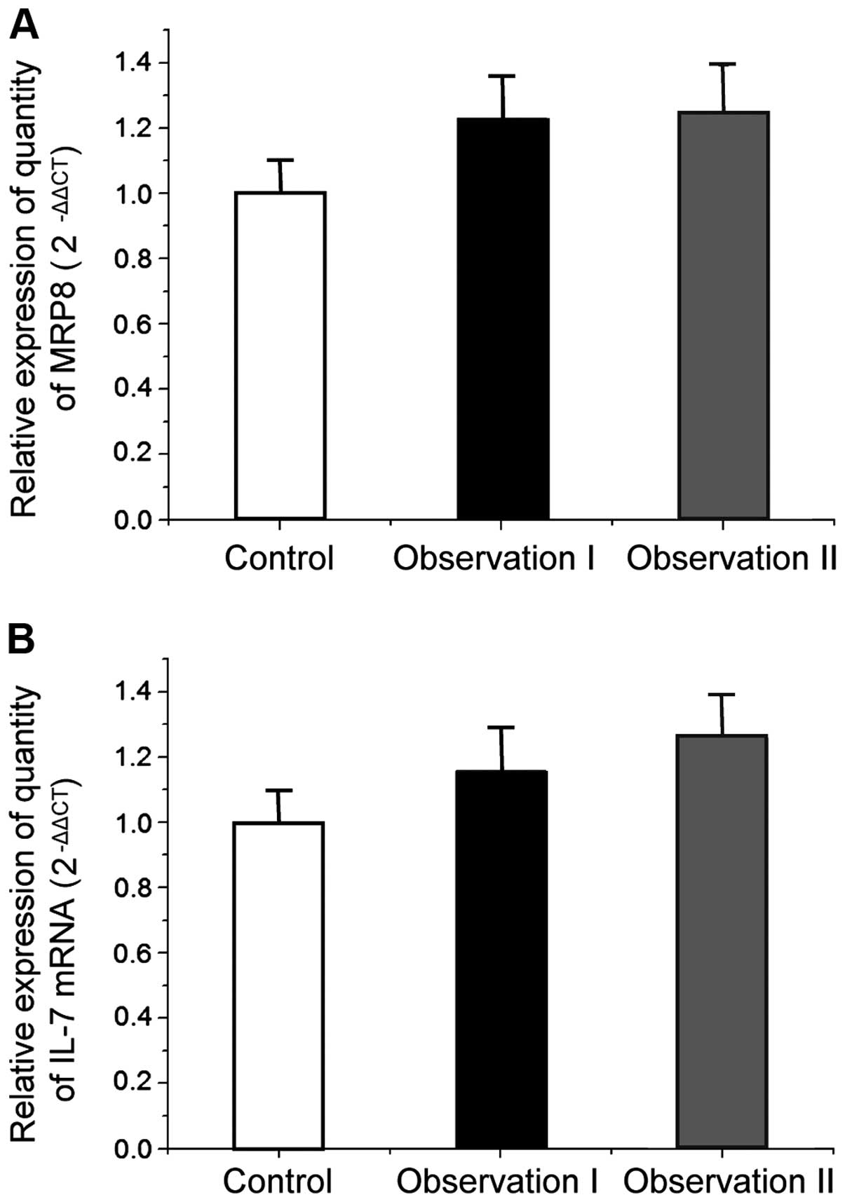
According to the study results from Schroten-Loef
et al (12), taking
lamotrigine can treat focal cortical dysplasia with secondary
intractable epilepsy to a certain extent, but the involved
mechanism was not clear. By quantitative PCR method on gene mRNA
relative expression quantity of MRP8 1 and IL-7 in the samples of
rat hippocampus with different treatments in the control group, in
the observation group I and the observation group II, it was found
that MRP8 and IL-7 gene mRNA had no significant difference between
the observation group I and the control group (P<0.05) (Fig. 1); and there were no significant
differences in MRP8 1 and IL-7 gene mRNA expression
between the observation group I treated by lamotrigine and the
expression in the observation group I (P<0.05), which showed
that lamotrigine did not affect the transcription of MRP8
and IL-7 genes.
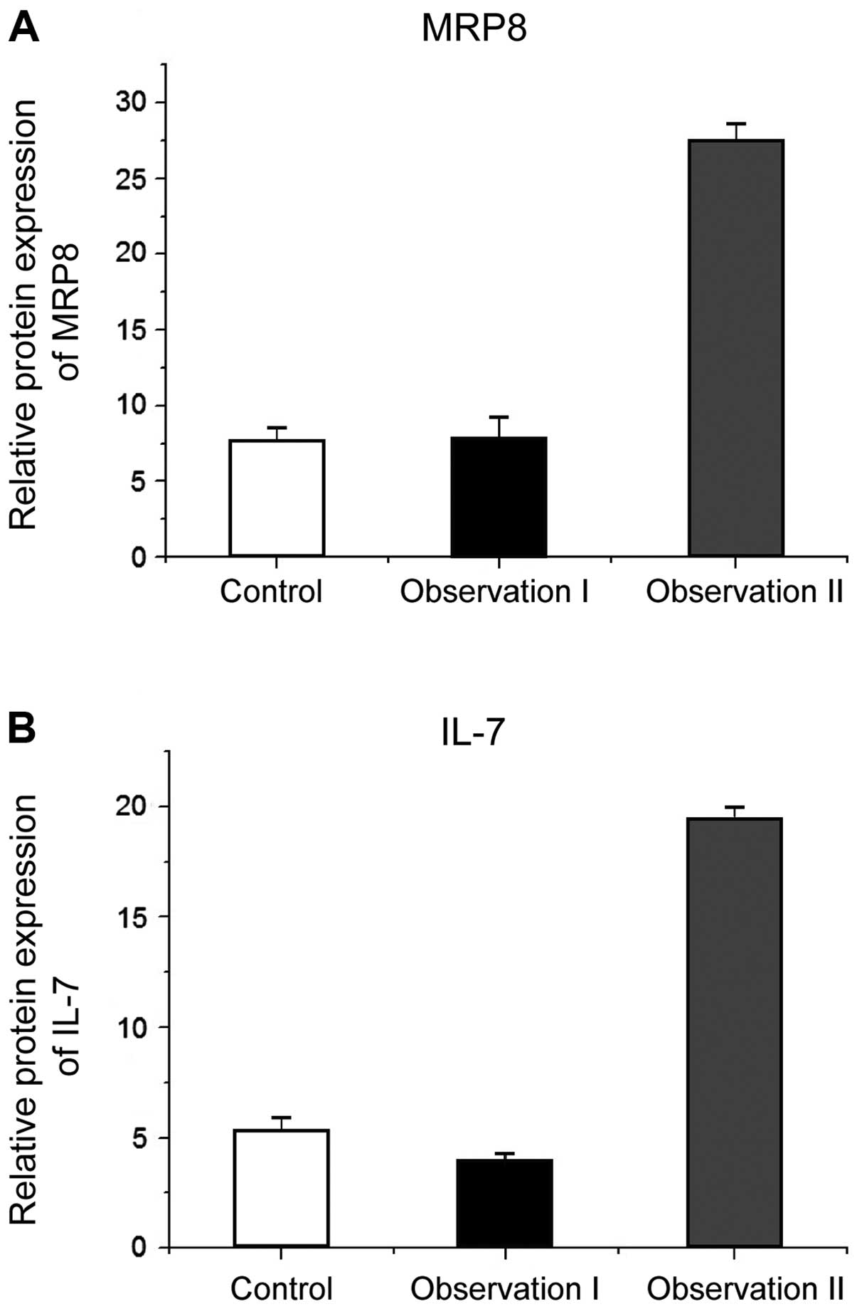
Expression quantity of MRP8 and IL-7
in the control group, the observation group I and the observation
group II by ELISA
There was no significant difference (P<0.05) of
MRP8 and IL-7 in the control group (7.52±1.03, 3.62±0.29) and the
observation group I (7.91±1.3, 3.86±0.38) treated with lamotrigine,
whereas MRP8 and IL-7 content had a significant difference
(P<0.05) between the observation group II (27.47±1.13,
19.45±0.48) and the observation group I (7.91±1.3, 3.86±0.38)
treated with lamotrigine. This indicated that taking lamotrigine
can treat focal cortical dysplasia with secondary intractable
epilepsy by lowering MRP8 and IL-7 content in rats to some extent
(Fig. 2).
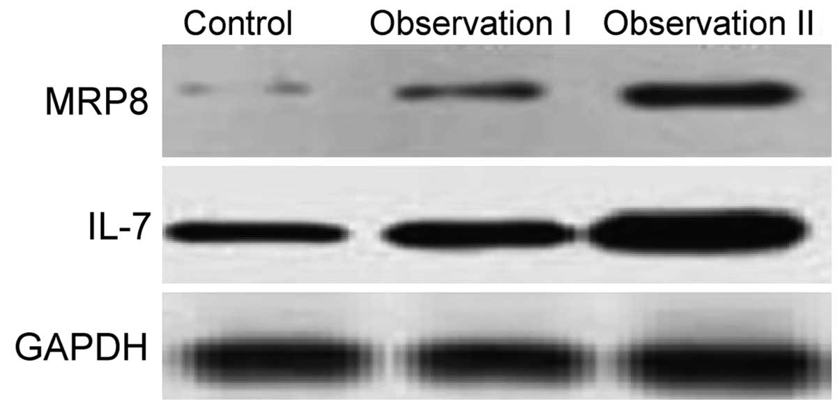
Expression quantity of MRP8 and IL-7
in the control group, the observation group I and the observation
group II by western blotting
Results of western blotting (Fig. 3) mirrored the above results that the
protein contents of MPR8 and IL-7 did not show differences, but
those in the observation group I with lamotrigine injection were
significantly lower than those in the observation group II without
amotrigine injection. This result was consistent with the results
of ELISA, which indicated that the treatment of lamotrigine for
focal cortical dysplasia with secondary intractable epilepsy
treatment was mainly due to reducing MRP8 and IL-7 protein content
in the body.
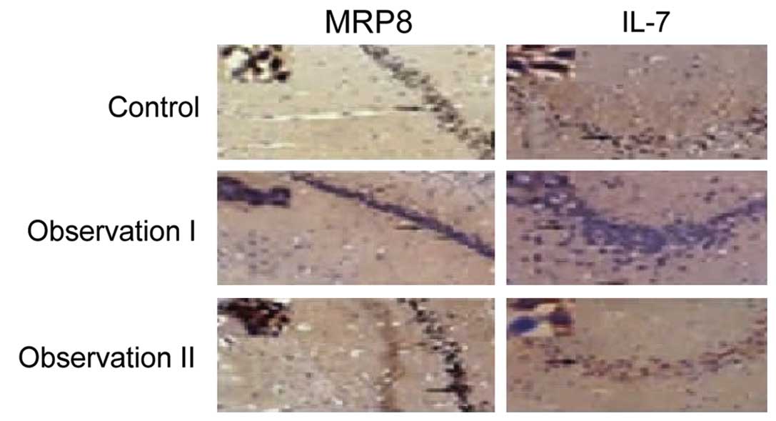
Expression quantity of MRP8 and IL-7
in the control group, the observation group I and the observation
group II by immunohistochemistry
From the immunohistochemical staining on hippocampal
tissue of the samples in the control group and in the observation
group I and group II, it was found that the stained positive cells
of MRP8 and IL-7 were less in the sample tissues of the control
group and the observation group I than those in the observation
group. In addition, from the observation of the immunohistochemical
staining image, it was found that the cells presenting positive
after staining were mostly irregular in shape, with larger cell
size and with cytoplasmic vacuoles and in disorder, in addition,
from the statistical results of the quantities of positive cells
and invisible cells in the samples of the control group, the
observation group I and II, it was found that the positive cell
quantity in the observation group II was significantly higher than
that in the observation group I, and there was a significant
difference (P<0.05) (Fig. 4;
Tables II and III).
| Table II.MRP8 expression result in
immunohistochemical hippocampus tissue of rats. |
Table II.
MRP8 expression result in
immunohistochemical hippocampus tissue of rats.
| Groups | Paraffin section
no. | Positive cell
no. | Negative cell
no. | P-value |
|---|
| Control | 38 | 9 | 29 | <0.05 |
| Observation I | 39 | 11 | 28 |
|
| Observation II | 38 | 29 | 9 |
|
| Table III.IL-7 expression results in
immunohistochemical hippocampus tissue of rats. |
Table III.
IL-7 expression results in
immunohistochemical hippocampus tissue of rats.
| Groups | Paraffin section
no. | Positive cell
no. | Negative cell
no. | P-value |
|---|
| Control | 38 | 10 | 28 | <0.05 |
| Observation I | 39 | 9 | 30 |
|
| Observation II | 38 | 32 | 6 |
|
Discussion
As a spontaneous severe convulsion behavior caused
by the internal neuron erethism of the body, the incidence and
disability rate of epilepsy increased significantly (11–13).
Related study results showed that the main pathogens of epilepsy
are the anaphylactic reactions of the nervous system and dominated
relevant tissues, which were caused by neurons and associated nerve
tissue structural abnormalities or functional abnormalities, thus
far the main effect target of the treatment on epilepsy is
associated with neuron cells (14,15).
However, statistical data indicate that in the current treatment of
epilepsy, nearly one-third of patients with epilepsy have related
drug resistance (16). In recent
years, the relevant study results indicate that MRP8 protein can
mediate the inflammatory response of endothelial cells and
inflammation of neurons in the body, and as one of internal nervous
system diseases of the body, epilepsy has been proven to be related
closely with neuron structural abnormalities and function disorders
(17,18). As a class of cytokines, IL-7 plays an
essential role in regulating activation of immune system in the
body itself, proliferation of relevant immune cells and function
(19). For example, the previous
findings showed that IL-7 can stimulate myeloid precursor cells and
megakaryocytes to produce colonies and form units and platelets to
aid the body recover from the immunosuppressive effect of cyclic
amide phospholipids (20).
In this study, fluorescence quantitative PCR, enzyme
immunoassay, western blotting and immunohistochemical staining
methods were used to explore the effect of lamotrigine on
expression of MRP8 and IL-7 in rat models of focal cortical
dysplasia with secondary refractory epilepsy. Results indicated
that despite significant effects on treating focal cortical
dysplasia with secondary refractory epilepsy, the MRP8 and IL-7
protein levels of the observation group II after lamotrigine
treatment showed no significant differences with normal mice in the
control group, but its content was much lower than that in the
observation group II (rats without the lamotrigine treatment),
which indicates that lamotrigine may be used to treat focal
cortical dysplasia with secondary intractable epilepsy by reducing
MRP8 and IL-7 protein levels in the body. However, since we found
that lamotrigine does not reduce mRNA content of MRP8 and IL-7
within the tissues, it can be considered that lamotrigine
potentially acts on the protein translation process rather than the
transcription process to achieve the regulation of MRP8 and IL-7
content in a different manner, which requires further
investigation.
References
|
1
|
Ashhab MU, Omran A, Kong H, Gan N, He F,
Peng J and Yin F: Expressions of tumor necrosis factor alpha and
microRNA-155 in immature rat model of status epilepticus and
children with mesial temporal lobe epilepsy. J Mol Neurosci.
51:950–958. 2013. View Article : Google Scholar : PubMed/NCBI
|
|
2
|
Sloan SA and Barres BA: Mechanisms of
astrocyte development and their contributions to neurodevelopmental
disorders. Curr Opin Neurobiol. 27:75–81. 2014. View Article : Google Scholar : PubMed/NCBI
|
|
3
|
Canpolat M, Per H, Gumus H, Yikilmaz A,
Unal E, Patiroglu T, Cinar L, Kurtsoy A and Kumandas S: Rapamycin
has a beneficial effect on controlling epilepsy in children with
tuberous sclerosis complex: results of 7 children from a cohort of
86. Childs Nerv Syst. 30:227–240. 2014. View Article : Google Scholar : PubMed/NCBI
|
|
4
|
Zhang CL: Study on the relationship and
mechanism between dynamin-1 and mesial temporal lobe epilepsy
(unpublished PhD thesis)Central South University; China: 2014
|
|
5
|
Kong H, Omran A, Ashhab MU, Gan N, Peng J,
He F, Wu L, Deng X and Yin F: Changes in microglial
inflammation-related and brain-enriched MicroRNAs expressions in
response to in vitro oxygen-glucose deprivation. Neurochem Res.
39:233–243. 2014. View Article : Google Scholar : PubMed/NCBI
|
|
6
|
Kwon CH, Moon HJ, Park HJ, Choi JH and
Park Y: S100A8 and S100A9 promotes invasion and migration through
p38 mitogen-activated protein kinase-dependent NF-κB activation in
gastric cancer cells. Mol Cells. 35:226–234. 2013. View Article : Google Scholar : PubMed/NCBI
|
|
7
|
Pellegrini M, Calzascia T, Toe JG, Preston
SP, Lin AE, Elford AR, Shahinian A, Lang PA, Lang KS, Morre M, et
al: IL-7 engages multiple mechanisms to overcome chronic viral
infection and limit organ pathology. Cell. 144:601–613. 2011.
View Article : Google Scholar : PubMed/NCBI
|
|
8
|
Xu YY, Wang Y, Li YP, Xu XX, Zhu SB and
Zhang XG: Interleukin-7 influences the development of thymic T
lymphocytes and thymic dendritic cells. Chinese J Immunol.
26:108–112. 2010.(In Chinese).
|
|
9
|
Holtman L, van Vliet EA, Aronica E,
Wouters D, Wadman WJ and Gorter JA: Blood plasma inflammation
markers during epileptogenesis in post-status epilepticus rat model
for temporal lobe epilepsy. Epilepsia. 54:589–595. 2013. View Article : Google Scholar : PubMed/NCBI
|
|
10
|
Xiang QL, Zhang CL, Peng J, He F, Wu LW,
Ou M and Yin F: Expression of TLR4 and MRP8 in the developing
immature rats with mesial temporal lobe epilepsy. J Apoplexy
Nervous Diseases. 7:580–584. 2012.(In Chinese). http://www.cnki.com.cn/Article/CJFDTotal-ZFSJ201207002.htm
|
|
11
|
Mohammad G, Siddiquei MM, Othman A,
Al-Shabrawey M and Abu El-Asrar AM: High-mobility group box-1
protein activates inflammatory signaling pathway components and
disrupts retinal vascular-barrier in the diabetic retina. Exp Eye
Res. 107:101–109. 2013. View Article : Google Scholar : PubMed/NCBI
|
|
12
|
Schroten-Loef C, de Ridder CM, Reneman S,
Crezee M, Dalgleish A, Todryk SM, Bangma CH and Kraaij R: A
prostate cancer vaccine comprising whole cells secreting IL-7,
effective against subcutaneous challenge, requires local GM-CSF for
intra-prostatic efficacy. Cancer Immunol Immunother. 58:373–381.
2009. View Article : Google Scholar : PubMed/NCBI
|
|
13
|
Buchler T, Bortlicek Z, Poprach A,
Kubackova K, Kiss I, Zemanova M, Fiala O, Dusek L, Vyzula R and
Melichar B: Czech Renal Cancer Cooperative Group: Efficacy of
everolimus in second- and third-line therapy for metastatic renal
cell carcinoma: a registry-based analysis. Urol Oncol. 32:569–575.
2014. View Article : Google Scholar : PubMed/NCBI
|
|
14
|
Hille A, Grüger S, Christiansen H, Wolff
HA, Volkmer B, Lehmann J, Dörr W and Rave-Fränk M: Effect of
tumour-cell-derived or recombinant keratinocyte growth factor (KGF)
on proliferation and radioresponse of human epithelial tumour cells
(HNSCC) and normal keratinocytes in vitro. Radiat Environ Biophys.
49:261–270. 2010. View Article : Google Scholar : PubMed/NCBI
|
|
15
|
Yucel AF, Kanter M, Pergel A, Erboga M and
Guzel A: The role of curcumin on intestinal oxidative stress, cell
proliferation and apoptosis after ischemia/reperfusion injury in
rats. J Mol Histol. 42:579–587. 2011. View Article : Google Scholar : PubMed/NCBI
|
|
16
|
Wu W, Huang Q, He F, Xiao M, Pang S, Guo
X, Brunk UT, Zhao K and Zhao M: Roles of mitogen-activated protein
kinases in the modulation of endothelial cell function following
thermal injury. Shock. 35:618–625. 2011. View Article : Google Scholar : PubMed/NCBI
|
|
17
|
Lan L, Tao J, Chen A, Xie G, Huang J, Lin
J, Peng J and Chen L: Electroacupuncture exerts anti-inflammatory
effects in cerebral ischemia-reperfusion injured rats via
suppression of the TLR4/NF-κB pathway. Int J Mol Med. 31:75–80.
2013.PubMed/NCBI
|
|
18
|
Schelbergen RF, Blom AB, van den Bosch MH,
Slöetjes A, Abdollahi-Roodsaz S, Schreurs BW, Mort JS, Vogl T, Roth
J, van den Berg WB, et al: Alarmins S100A8 and S100A9 elicit a
catabolic effect in human osteoarthritic chondrocytes that is
dependent on Toll-like receptor 4. Arthritis Rheum. 64:1477–1487.
2012. View Article : Google Scholar : PubMed/NCBI
|
|
19
|
Wang FL, Jiang JM, Wang F, Fu ZZ and Zhang
ZF: Expressions of interleukin 18 and prostaglandin E2 and their
correlation in the synoviocytes of patients with osteoarthritis. J
Southern Med University. 30:731–733. 2010.(In Chinese).
|
|
20
|
Peng J, Omran A, Ashhab MU, Kong H, Gan N,
He F and Yin F: Expression patterns of miR-124, miR-134, miR-132,
and miR-21 in an immature rat model and children with mesial
temporal lobe epilepsy. J Mol Neurosci. 50:291–297. 2013.
View Article : Google Scholar : PubMed/NCBI
|