Introduction
Initially extracted from the pheochromocytoma tissue
excised in a surgery (1),
adrenomedullin (ADM) is a polypeptide sharing homology with
calcitonin gene-related peptide and has functions of vascular
dilation and diuretic activity (2).
In a healthy individual, a considerable plasmatic concentration of
ADM is involved in the regulation of plasma (3). In addition, as a bioactive peptide, ADM
has extensive biological functions and is distributed in almost all
tissues (4), particularly in normal
adrenal medulla, heart, lung, kidney, blood plasma, and urine and
is able to reduce high blood pressure, dilate blood vessels,
pulmonary and renal arteries, and relieve bronchospasms (5).
ADM has multiple biological functions, including the
regulation of internal secretion in organisms and inhibition of
hyperplasia of vascular smooth muscles. Furthermore, ADM
participates in multiple physiological accommodations in an
autocrine and a paracrine manner as a circulating hormone.
Currently, the pursuit of regulating measures for immune
dysfunctions after trauma is becoming a field of interest. In the
central nervous system, ADM and its mRNA are also widely
distributed and located in neurons (6). Research has shown an increased
expression of ADM after spinal cord injury, but there are few
reports on its mechanism of expression, role and regulation
(7).
For this reason, we aimed to detect the expression
of ADM in a rat model of the spinal cord injury and attempted to
discuss the mechanism of expression, effectiveness and regulation
availability. The aim was to provide theoretical bases for clinical
treatment of spinal cord injury by establishing a model of
Sprague-Dawley (SD) rat for spinal cord injury and intervention of
recombinant human erythropoietin.
Materials and methods
Animals
The experiment was completed in the Central
Laboratory of the Department of Orthopaedics, The First Affiliated
Hospital of Liaoning Medical University (Liaoning, China) from
October, 2012 to March, 2013. A total of 45 Sprague-Dawley (SD)
healthy rats aged 60 days and weighing 180–200 g were selected. The
rats were provided by the Shanghai Laboratory Animal Center (SLAC)
Co., Ltd. [animal license no. SCXK (Shanghai) 2012–0138; Shanghai,
China]. The rats were divided into 3 groups randomly:
Sham-operation group (n=15), group of spinal cord injury model
(n=15), and group of recombinant human erythropoietin (n=15).
Establishment of animal models
Pentobarbital sodium (5 g/l) was intraperitoneally
injected as per 10 mg/kg. Surgery was performed aseptically after
anesthesia. An incision, ~3 cm long, was made in the middle of the
dorsal side to expose or injure the spinal cord section
T10. Excision of vertebral plates was performed in the
sham-operation group to expose the spinal cord. For the spinal cord
injury model, a model of dorsal side injury of rat spinal cord was
prepared using the Allen's WD method. The vertebral plate was
excised to expose the spinal cord. A concave-type plastic sheet
with the diameter of 2 mm was horizontally placed at the site of
the spinal cord to be injured. A cylindrical metal bar was made and
dropped freely and vertically in a thin glass delivery tube to
establish an animal model of spinal cord injury. The metal bar
weighed 10 g and was 10 cm high with injury energy of 10×10 (g·cm).
A model of dorsal side injury of rat spinal cord was prepared with
the same method in the group of recombinant human erythropoietin.
Caudal vein injection of 300 U/kg recombinant human erythropoietin
was administered to the group of recombinant human erythropoietin
at 1, 3, 5, 7 and 9 days after injury, respectively. An equivalent
volume of normal saline was administered to the group of spinal
cord injury after injury at the same time. The completeness of
spinal dura mater was maintained and each layer was sutured in all
the groups. The success marks of the injury animal model were tail
wagging reflex and symmetric convulsion of both lower extremities
(8).
The study was approved by the Animal Ethics
Committee of Jinzhou Medical University.
Histopathological and
immunohistochemical examination
All the groups of animals were anesthetized again at
3, 6 and 9 days after operation, respectively. A piece of spinal
cord tissue with the approximate length of 1.0 cm was taken with
the injured spinal cord section as the center, fixed in
formaldehyde with the volume fraction of 0.3, embedded with
paraffin and serially sectioned to 5 µm sections with a microtome,
with 3 rats at each time. Regular hematoxylin and eosin staining
and ADM immunohistochemical staining were performed. Regular
hematoxylin and eosin staining was performed to observe edema,
degeneration and necrosis, inflammatory cell infiltration in
various groups of spinal cord tissues. The SABC method was used for
immunohistochemical staining of ADM. The rabbit anti-rat ADM
polyclonal antibody (1:100) and the SABC immunohistochemical
staining reagent kit were purchased from Beijing Ruixiang
Biotechnology Co., Ltd. (Beijing, China). Operations were in strict
accordance with the instructions in the reagent kit. The expression
of ADM was observed under a light microscope.
Image analysis
Five immunohistochemically-stained ADM sections were
selected randomly from each rat. Five high-power fields were
selected from the anterior angle to the relief angle of the grey
matter of each section. The HMIAS-2000 high-definition color
medical image-text analysis system was applied to determine the
average gray value of the positive reactant of the ADM and to
calculate the gray value.
Determination of motor function
The improved Tarlov scoring and the improved Rivlin
and Tator tilting plate scoring were used to observe the recovery
of the motor function of the lower limbs in rats at 3, 6 and 9 days
after operation, respectively (9).
Criteria for improved Tarlov scoring: 0 point:
Complete paralysis, there is no response to acupuncture in lower
limbs; 1 point: Complete paralysis, there is response to
acupuncture in lower limbs but the limbs are not able to move; 2
points: The limbs are able to move but the rat cannot stand or
stand steadily (<5 sec); 3 points: The rat can stand but cannot
walk; 4 points: The rat can walk several steps but not steadily; 5
points: The rat can walk slowly but not flexibly and the walking
has certain defects; 6 points: The rat can walk normally.
Improved Rivlin and Tator tilting plate method: The
rat was placed on the same smooth board with its body axis
perpendicular to the longitudinal axis of the tilting plate. The
tilting plate was raised by 5° each time and the maximum angle at
which the rat could stay for 5 sec was regarded as the functional
value.
Statistical analysis
SPSS 13.0 statistical software (SPSS, Inc., Chicago,
IL, USA) was used for statistical analysis. Data were expressed as
mean ± standard deviation. The F test was used for variance
analysis. P<0.05 was considered to indicate a statistically
significant difference.
Results
Quantitative analysis for experimental
animals
A result analysis was conducted for all of the 45
rats with attrition value.
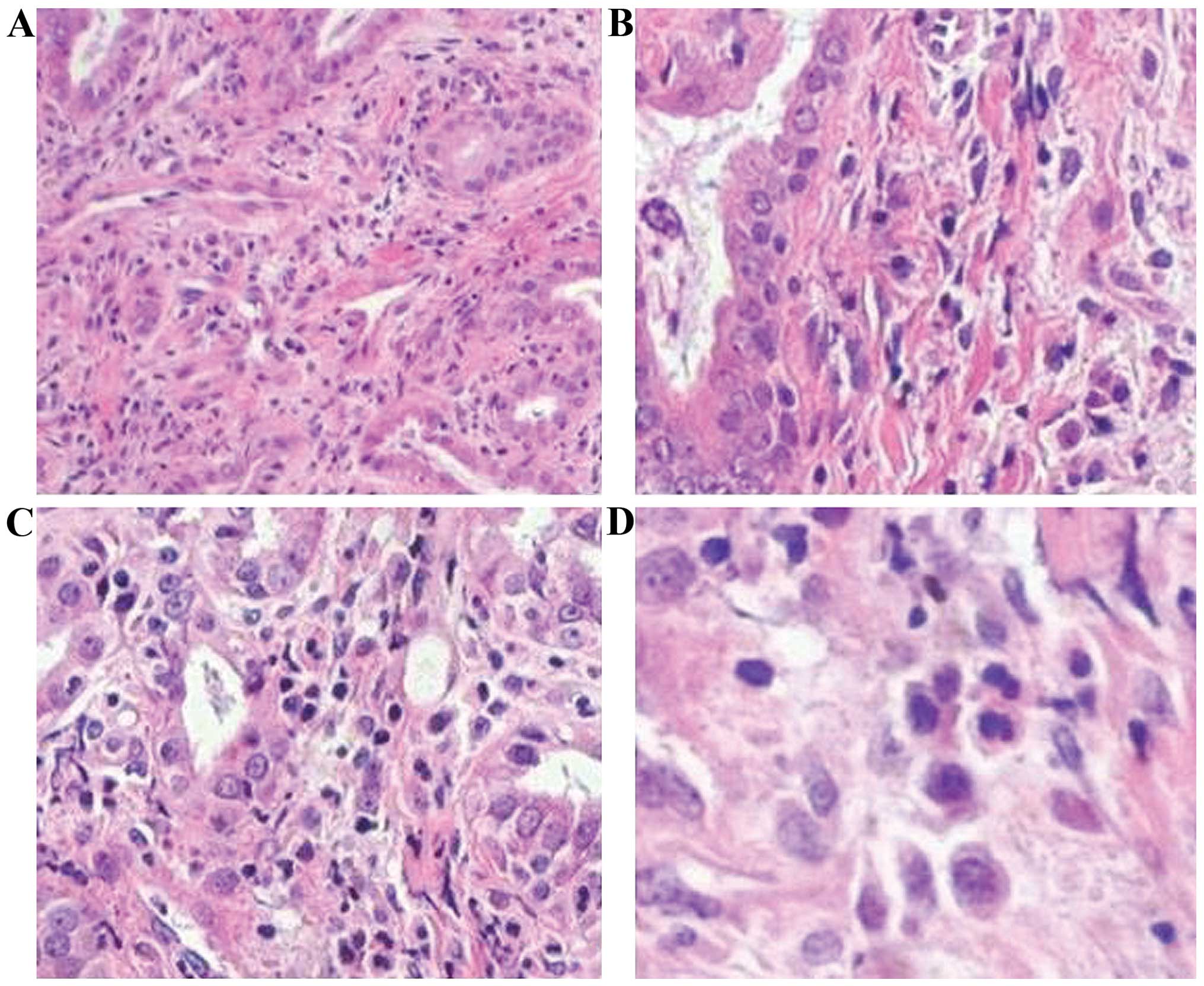
Result of hematoxylin and eosin
staining
No significant abnormality was present in material
sampled from the sham-operation group at various times. In the
group of spinal cord injury model, 3 days after injury, a large
number of erythrocytes were present within the grey matter; neural
cells were swollen and exhibited a round shape; substantial
karyopyknosis and karyorrhexis occurred; the border of grey matter
disappeared; voids formed in the grey matter; 6 days after injury,
erythrocytes fused into clusters; the border of grey matter was
clear; the residual neurons decreased significantly and were
slightly swollen; a small number of neutrophile granulocytes were
present; 11 days after injury, the morphology of the residual
neurons were basically normal. Pathological changes in the group of
recombinant human erythropoietin at various times were
significantly less severe than those in the group of spinal cord
injury model (Fig. 1).
Result of immunohistochemical staining
of ADM
The expression of ADM was primarily located in
neurons. The expression of ADM was low at various times in the
sham-operation group; compared with the sham-operation group, the
number of neurons decreased and the expression of ADM was increased
in the group of spinal cord injury model and group of recombinant
human erythropoietin. The expression of ADM was increased
particularly in the group of recombinant human erythropoietin. The
expression was restored to normal at 6 and 9 days after injury.
Expression of ADM in various groups of
rats at different time points
The difference in gray values of the sham-operation
group at various times was not significant indicating that an
uninjured spinal cord did not affect the expression of ADM; the
gray values of the group of spinal cord injury model and the group
of recombinant human erythropoietin were higher than that of the
sham-operation group at 3 days after injury and the gray value of
the group of recombinant human erythropoietin was higher in the
group of spinal cord injury model. Both differences were
significant (P<0.01), which indicated that the expression of ADM
after spinal cord injury was increased and the recombinant human
erythropoietin was able to promote the high expression of ADM
(Table I).
 | Table I.Comparison of gray values of positive
reactants of ADM in various groups of rats at different times. |
Table I.
Comparison of gray values of positive
reactants of ADM in various groups of rats at different times.
|
| t(injury)/days |
|---|
|
|
|
|---|
| Group | 3 | 6 | 9 |
|---|
| Sham-operation | 0.328±0.007 | 0.330±0.006 | 0.325±0.007 |
| Spinal cord injury
model |
0.376±0.008a | 0.332±0.008 | 0.331±0.009 |
| Recombinant human
erythropoietin |
0.412±0.010a,b | 0.332±0.009 | 0.334±0.007 |
Result of improved Tarlov scoring
The improved Tarlov scores of the group of spinal
cord injury model and the group of recombinant human erythropoietin
were significantly lower than those of the sham-operation group at
3, 6 and 9 days at the same time and the difference was significant
(P<0.01). The improved Tarlov scores of the group of recombinant
human erythropoietin were higher than those of the group of spinal
cord injury model at 3, 6 and 9 days at the same time and the
difference was significant (P<0.05; Table II).
| Table II.Result of improved Tarlov scoring. |
Table II.
Result of improved Tarlov scoring.
|
| t(injury)/days |
|---|
|
|
|
|---|
| Group | 3 | 6 | 9 |
|---|
| Sham-operation | 7.36±0.67 | 7.42±0.69 | 7.43±0.66 |
| Spinal cord injury
model | 3.36±0.42 |
2.97±0.38a |
3.64±0.47a |
| Recombinant human
erythropoietin |
4.78±0.48a,c |
4.83±0.51a.b |
5.31±0.48a,c |
Result of improved Rivlin and Tator
scoring
The improved Rivlin and Tator scores of the group of
spinal cord injury model and the group of recombinant human
erythropoietin were significantly lower than those of the
sham-operation group at 3, 6 and 9 days at the same time and the
difference was significant (P<0.01); the improved scores of the
group of recombinant human erythropoietin were higher than those of
the group of spinal cord injury model at 3, 6 and 9 days at the
same time and the difference was significant (P<0.01, Table III).
| Table III.Result of improved Rivlin and Tator
scoring. |
Table III.
Result of improved Rivlin and Tator
scoring.
|
| t(injury)/days |
|---|
|
|
|
|---|
| Group | 3 | 6 | 9 |
|---|
| Sham-operation | 67.16±8.13 | 69.34±8.20 | 70.28±8.17 |
| Spinal cord injury
model |
23.64±4.67a |
26.16±4.81a | 28.33±4.86 |
| Recombinant human
erythropoietin |
29.35±6.13a,b |
35.26±6.32a.b |
38.61±6.37a,b |
Discussion
ADM is an active peptide comprising 52 amino acids
with many important biological functions. ADM is distributed in
almost all tissues (10) and serves
as a circulating hormone with autocrine and paracrine functions, in
several tissues and organs such as blood plasma, adrenal gland,
heart, kidney, central nervous system, and blood vessels. ADM
participates in the regulation of blood vessel activities and
stress response in organisms and exerts the compensatory regulation
effect under many pathological conditions (11). Recently, the spinal cord injury has
been raising general concerns due to its high occurrence rate,
expenses and disability rate. In addition to primary mechanical
injury, the spinal cord injury may also cause a series of secondary
pathological changes. Currently, it is considered that the
secondary spinal cord injury is a process of mutual cascading of
many nerve biochemical and vascular mechanisms and continuous
exacerbation. An irreversible injury within 4 h is a primary injury
and various biochemical changes occurring after 4 h are secondary
injuries and have certain reversibility. It is crucial to maximize
recovery of spinal cord injury during the reversible period
(12).
The expression of ADM increases in a compensatory
manner after spinal cord injury and creates advantages for recovery
of neurological functions. It plays an important role in recovery
of an injured spinal cord (13). Its
increasing and action mechanisms are: Firstly, local ischemic and
hypoxia environment. The driving force for incremental regulation
of ADM is hypoxia and the local ischemic and hypoxia environment
after spinal cord injury provides conditions for incremental
regulation of ADM that exists as a cytoprotection factor in the
local ischemic and hypoxia environment and increases stability of
the cytomembrane (14). Secondly,
oxidative stress may increase the expression of ADM. Indeed, it has
been shown that oxidative stress is one of the causes of organ
damage under pathological conditions. Oxidative stress can increase
the expression of ADM, and ADM can inhibit the oxidative stress.
Therefore, ADM is determined as an endogenous antioxidant. The
therapeutic effect of endogenous antioxidant (e.g., vitamin E) on
spinal cord trauma has been demonstrated (15). Free radicals are cellular metabolites
with high activity and removed by antioxidants (e.g., vitamin C and
E) or converted into oxygen and water by some enzymes (e.g.,
superoxide dismutase). After secondary spinal cord injury,
depletion of endogenous antioxidant and decrease of superoxide
dismutase are not able to remove the increasing free radicals,
which can induce cell apoptosis and irreversible cell death
(16). The increase of ADM is of
great significance (17). The
expression of ADM increases after spinal cord injury, which
strengthens the oxidation resistance in spinal cord injury and
alleviates the spinal cord injury via antioxidation (18). In the experiment, the expression of
ADM is increased after spinal cord injury and further increased
after intervention of recombinant human erythropoietin, which
demonstrates that the ADM can be regulated. It is found through a
pathological examination that neuronal injuries are significantly
less severe than those in the group of spinal cord injury model at
various times and the scores of neurological functions at 3, 6 and
9 days after spinal cord injury are higher than those in the group
of spinal cord injury model, which indicates the correlation
between the increase of ADM and alleviation of the degree of spinal
cord injury and promotion of the recovery of neurological
functions.
Recombinant human erythropoietin is a member of the
cytokine superfamily. In addition to the function of promoting
proliferation, differentiation, and maturation of progenitor
erythrocytes, it also has anti-vasospasm, anti-apoptosis and
anti-inflammatory functions (19).
The recombinant human erythropoietin and its receptor exist in the
hemopoietin system, the central nervous system, and the peripheral
nervous system and can protect, not only cerebral injuries due to
various causes, but also the acute spinal cord injury and secondary
injury (19). The recombinant human
erythropoietin is capable of protecting injuries of neurocytes and
neural cells due to various causes such as excitotoxicity and lack
of neurotrophic factors (20).
Hypoxia can promote the expression of recombinant human
erythropoietin and it has been demonstrated that the recombinant
human erythropoietin can promote survival of neurocytes under
hypoxia (21). In the experiment,
the recombinant human erythropoietin was used to treat the spinal
cord injury of rats. It is observed through a pathological
examination that neuronal injuries are significantly less severe
than those in the group of recombinant human erythropoietin at
various times and the scores of neurological functions are higher
than those in the group of spinal cord injury model, which
indicates the therapeutical effect of recombinant human
erythropoietin.
In conclusion, the ADM increases compensatory
effects after spinal cord injury and the ADM promotes the recovery
of neurological functions of the spinal cord primarily by
antioxidation. The recombinant human erythropoietin can promote
further high expression of the ADM. The correlation between the
therapeutical effect of the recombinant human erythropoietin and
the high expression of ADM suggested a mechanisms of treating
spinal cord injury. Thus, treatment with recombinant human
erythropoietin may be expected as an effective method for treatment
of spinal cord injury.
References
|
1
|
Kitamura K, Kangawa K, Kawamoto M, Ichiki
Y, Nakamura S, Matsuo H and Eto T: Adrenomedullin: A novel
hypotensive peptide isolated from human pheochromocytoma. Biochem
Biophys Res Commun. 192:553–560. 1993. View Article : Google Scholar : PubMed/NCBI
|
|
2
|
Tazaki M, Endoh T, Kobayashi H, Nobushima
H, Shibukawa Y, Tsumura M, Sato M, Ubaidus S and Sueishi K:
Adrenomedullin facilitates calcium channel currents in osteoblasts.
Bull Tokyo Dent Coll. 53:203–206. 2012. View Article : Google Scholar : PubMed/NCBI
|
|
3
|
Yuan M, Wang Q, Li C, Tao L, Zhang H, Wang
H, Zhang Y and Ren J: Adrenomedullin in vascular endothelial injury
and combination therapy: Time for a new paradigm. Curr Vasc
Pharmacol. 13:459–466. 2015. View Article : Google Scholar : PubMed/NCBI
|
|
4
|
Yoshizawa T, Sakurai T, Kamiyoshi A,
Ichikawa-Shindo Y, Kawate H, Iesato Y, Koyama T, Uetake R, Yang L,
Yamauchi A, et al: Novel regulation of cardiac metabolism and
homeostasis by the adrenomedullin-receptor activity-modifying
protein 2 system. Hypertension. 61:341–351. 2013. View Article : Google Scholar : PubMed/NCBI
|
|
5
|
Sakimoto S, Kidoya H, Kamei M, Naito H,
Yamakawa D, Sakaguchi H, Wakabayashi T, Nishida K and Takakura N:
An angiogenic role for adrenomedullin in choroidal
neovascularization. PLoS One. 8:e580962013. View Article : Google Scholar : PubMed/NCBI
|
|
6
|
Dai X, Ma W, He XJ and Jha RK: Elevated
expression of adrenomedullin is correlated with prognosis and
disease severity in osteosarcoma. Med Oncol. 30:3472013. View Article : Google Scholar : PubMed/NCBI
|
|
7
|
Hong Y, Liu Y, Chabot J-G, Fournier A and
Quirion R: Upregulation of adrenomedullin in the spinal cord and
dorsal root ganglia in the early phase of CFA-induced inflammation
in rats. Pain. 146:105–113. 2009. View Article : Google Scholar : PubMed/NCBI
|
|
8
|
Saxena T, Deng B, Stelzner D, Hasenwinkel
J and Chaiken J: Raman spectroscopic investigation of spinal cord
injury in a rat model. J Biomed Opt. 16:0270032011. View Article : Google Scholar : PubMed/NCBI
|
|
9
|
Jiang Y, Lv H, Huang S, Tan H, Zhang Y and
Li H: Bone marrow mesenchymal stem cells can improve the motor
function of a Huntington's disease rat model. Neurol Res.
33:331–337. 2011. View Article : Google Scholar : PubMed/NCBI
|
|
10
|
Brouwers FP, de Boer RA, van der Harst P,
Struck J, de Jong PE, de Zeeuw D, Gans RO, Gansevoort RT, Hillege
HL, van Gilst WH, et al: Influence of age on the prognostic value
of mid-regional pro-adrenomedullin in the general population.
Heart. 98:1348–1353. 2012. View Article : Google Scholar : PubMed/NCBI
|
|
11
|
Tsuchiya K, Hida K, Hida Y, Muraki C, Ohga
N, Akino T, Kondo T, Miseki T, Nakagawa K, Shindoh M, et al:
Adrenomedullin antagonist suppresses tumor formation in renal cell
carcinoma through inhibitory effects on tumor endothelial cells and
endothelial progenitor mobilization. Int J Oncol. 36:1379–1386.
2010.PubMed/NCBI
|
|
12
|
Shibasaki I, Nishikimi T, Mochizuki Y,
Yamada Y, Yoshitatsu M, Inoue Y, Kuwata T, Ogawa H, Tsuchiya G,
Ishimitsu T, et al: Greater expression of inflammatory cytokines,
adrenomedullin, and natriuretic peptide receptor-C in epicardial
adipose tissue in coronary artery disease. Regul Pept. 165:210–217.
2010. View Article : Google Scholar : PubMed/NCBI
|
|
13
|
Marinoni E, Pacioni K, Sambuchini A,
Moscarini M, Letizia C and DI Iorio R: Regulation by hypoxia of
adrenomedullin output and expression in human trophoblast cells.
Eur J Obstet Gynecol Reprod Biol. 154:146–150. 2011. View Article : Google Scholar : PubMed/NCBI
|
|
14
|
Kim SM, Kim JY, Lee S and Park JH:
Adrenomedullin protects against hypoxia/reoxygenation-induced cell
death by suppression of reactive oxygen species via thiol redox
systems. FEBS Lett. 584:213–218. 2010. View Article : Google Scholar : PubMed/NCBI
|
|
15
|
Oz Oyar E, Korkmaz A, Kardesş O and
Omeroğlu S: Aortic cross-clamping-induced spinal cord oxidative
stress in rabbits: The role of a novel antioxidant adrenomedullin.
J Surg Res. 147:143–147. 2008. View Article : Google Scholar : PubMed/NCBI
|
|
16
|
Pfeil U, Aslam M, Paddenberg R, Quanz K,
Chang CL, Park JI, Gries B, Rafiq A, Faulhammer P, Goldenberg A, et
al: Intermedin/adrenomedullin-2 is a hypoxia-induced endothelial
peptide that stabilizes pulmonary microvascular permeability. Am J
Physiol Lung Cell Mol Physiol. 297:L837–L845. 2009. View Article : Google Scholar : PubMed/NCBI
|
|
17
|
Arduini A, Escobar J, Vento M, Escrig R,
Quintás G, Sastre J, Saugstad OD and Solberg R: Metabolic
adaptation and neuroprotection differ in the retina and choroid in
a piglet model of acute postnatal hypoxia. Pediatr Res. 76:127–134.
2014. View Article : Google Scholar : PubMed/NCBI
|
|
18
|
Yen DH, Chen LC, Shen YC, Chiu YC, Ho IC,
Lou YJ, Chen IC and Yen JC: Protein kinase A-dependent neuronal
nitric oxide synthase activation mediates the enhancement of
baroreflex response by adrenomedullin in the nucleus tractus
solitarii of rats. J Biomed Sci. 18:322011. View Article : Google Scholar : PubMed/NCBI
|
|
19
|
Ning B, Zhang A, Song H, Gong W, Ding Y,
Guo S, Zhao Y, Jiang J and Jia T: Recombinant human erythropoietin
prevents motor neuron apoptosis in a rat model of cervical
sub-acute spinal cord compression. Neurosci Lett. 490:57–62. 2011.
View Article : Google Scholar : PubMed/NCBI
|
|
20
|
Huang H, Fan S, Ji X, Zhang Y, Bao F and
Zhang G: Recombinant human erythropoietin protects against
experimental spinal cord trauma injury by regulating expression of
the proteins MKP-1 and p-ERK. J Int Med Res. 37:511–519. 2009.
View Article : Google Scholar : PubMed/NCBI
|
|
21
|
Wang H, Liu ZL, Zhuang XT, Wang MF and Xu
L: Neuroprotective effect of recombinant human erythropoietin on
optic nerve injury in rats. Chin Med J (Engl). 122:2008–2012.
2009.PubMed/NCBI
|