Introduction
Extramedullary hematopoiesis (EMH) refers to the
hematopoiesis that occurs in organs other than bone marrow
(1). It is a compensatory phenomenon
that results in the production of blood cell precursors outside the
marrow in patients with thalassemia intermedia. It has been
reported to occur in ~15% of cases of thalassemia, and it occurs in
myelofibrosis and in other anemic conditions (2). The process can involve virtually any
organ or tissue, such as the liver, spleen, abdominal viscera,
pleura, lymph nodes, adrenal glands, breast, thymus, kidneys,
gastrointestinal tract, intracranial structures and paraspinal
regions (3–6). EMH is a rare entity and often
asymptomatic but can sometimes lead to symptomatic tumor-like
masses. Treatment options are controversial and include
hypertransfusion, surgical excision, radiotherapy, and hydroxyurea
(7). Paraspinal involvement is the
associated with morbidity secondary to spinal cord compression
(8). Among the various body regions
reported, paraspinal involvement deserves special attention due to
the debilitating clinical consequences and challenges in its
diagnosis and management. The present study described two cases
with paraspinal medullary hematopoiesis in two thalassemic
patients. Verbal informed consent was obtained from both
patients.
Case report
Case 1
A 54-year-old male was admitted to the First
Affiliated Hospital of Guangzhou Medical University (Guangzhou,
China) in May 2012 with complaints of dizziness and hypodynamic
condition for one month. The patient was known to have
α-thalassemia. On the day of admission, the patient's hemoglobin
level was 71 g/l (normal range, 120–160 g/l), the red blood cell
count was 3.48×10¹² cells/l (normal range,
4.0–5.5×10¹² cells/l) and other routine hematological
examinations and biochemical tests were within normal limits.
Non-contrast and contrast-enhanced chest computed tomography (CT)
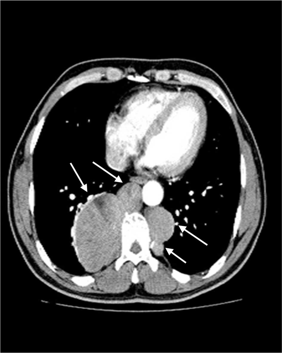
scan revealed multiple paraspinal masses in the rear mediastinum,
and the largest was measured 6.2×8.0 cm (Fig. 1). The tumors were well-demarcated,
and demonstrated heterogeneous density, ranging between 57 and 97
Hounsfield units. Considering the size of the mass and the
possibility of multiple neurofibroma, a surgical biopsy was
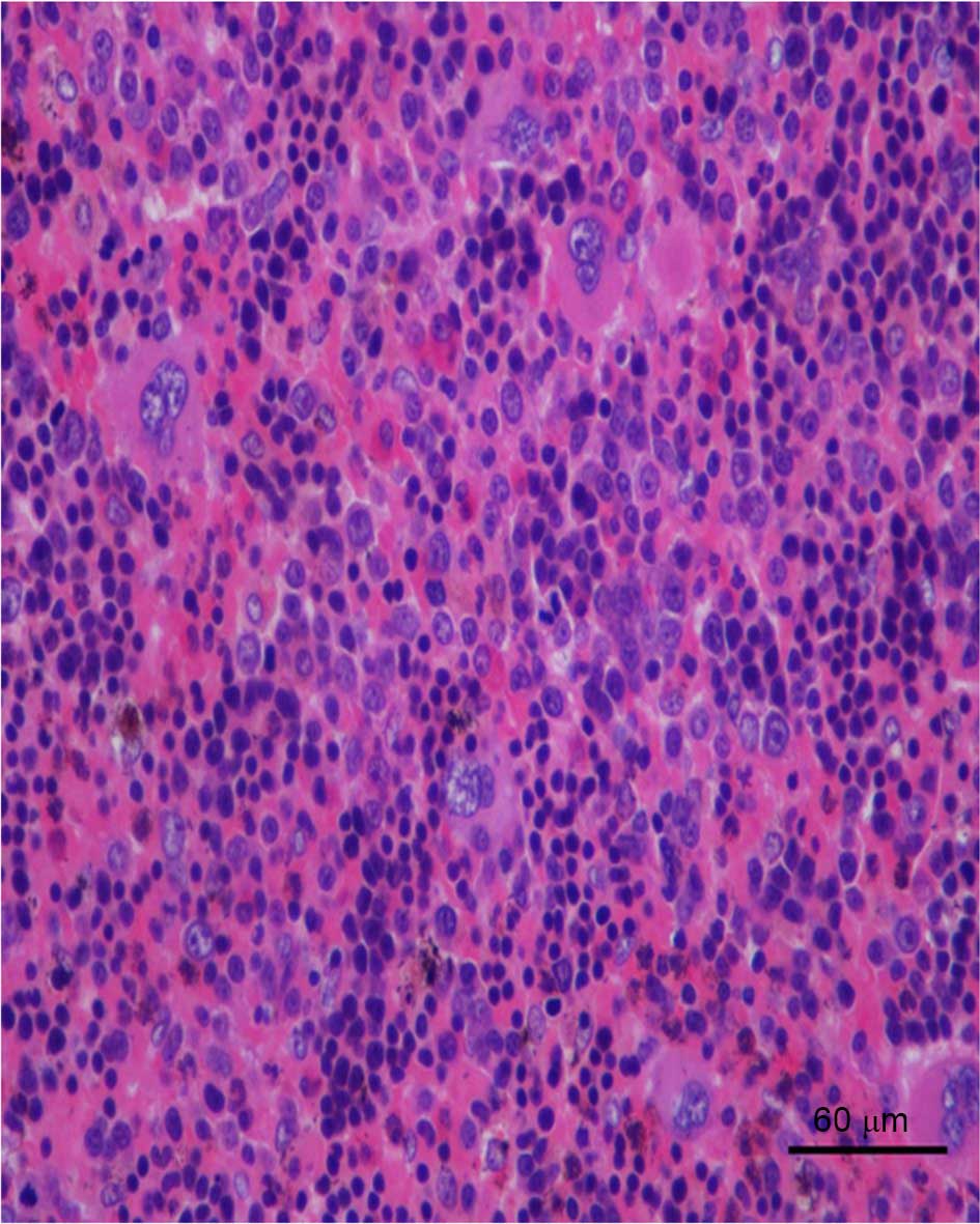
performed and showed infiltration by megakaryocytes, erythroblasts
and myeloid cells (Fig. 2).
Immunohistochemical staining was performed by obtaining paraspinal
mass from the rear mediastinum, fixing in 10% paraformaldehyde for
6 h at room temperature and embedding it in paraffin. Sections (4
µm) were then dewaxed in xylene, rehydrated and blocked using 3%
hydrogen peroxide in methanol for 30 min at room temperature with
10% bovine serum albumin/Tris-buffered saline for 30 min at room
temperature. Sections were then incubated with primary antibodies
against cluster of differentiation (CD)61 (1:100; cat. no.
MAB-0604; Maixin Biotech Co., Ltd., Fuzhou, China), MP0 (1:1,000;
cat. no. A0398; Dako Medical Device Technology Service Co., Ltd.,
Shanghai, China), CD15 (1:100; cat. no. M3631; Dako Medical Device
Technology Service Co., Ltd.), CD3 (1:50; cat. no. IR503; Dako
Medical Device Technology Service Co., Ltd.), CD5 (1:100; cat. no.
NCL-L-CD5-4CT; Quanhui Business Co., Ltd., Gaungzhou, China), CD20
(1:200; cat. no. IR604; Maixin Biotech Co., Ltd.), CD79a (1:200;
cat. no. IR621; Dako Medical Device Technology Service Co., Ltd.),
CD138 (operating fluid; cat. no. IR642; Dako Medical Device
Technology Service Co., Ltd.), LCA (1:100; cat. no. MAB-0037;
Maixin Biotech Co., Ltd.) and CD117 (1:500; cat. no. A4502; Dako
Medical Device Technology Service Co., Ltd.) at 37°C for 25–60 min.
Then, sections were then incubated with
horseradish-peroxidase-conjugated goat and rabbit secondary
antibodies. Slides were then counterstained with hematoxylin.
Negative controls were performed by replacing the primary
antibodies with phosphate-buffered saline.
Immunohistochemical analysis showed
CD3/CD20/CD5/CD79a/CD138/CD68/leukocyte common antigen diffuse
positive, CD117 negative, CD15 positive and myeloperoxidase
(MPO) positive corresponding to EMH. The patient experienced
ablation of the biggest mass, and was free from recurrence after 2
years of follow-up.
Case 2
The second patient was a 48-year-old male, who
presented with multiple masses in left thoracic cavity for 18 years
and shortness of breath for 3 months. The patient was admitted to
the First Affiliated Hospital of Guangzhou Medical University in
December 2013, and was known to have severe β-thalassemia. The
patient's hemoglobin level was 59 g/l and the red blood cell count
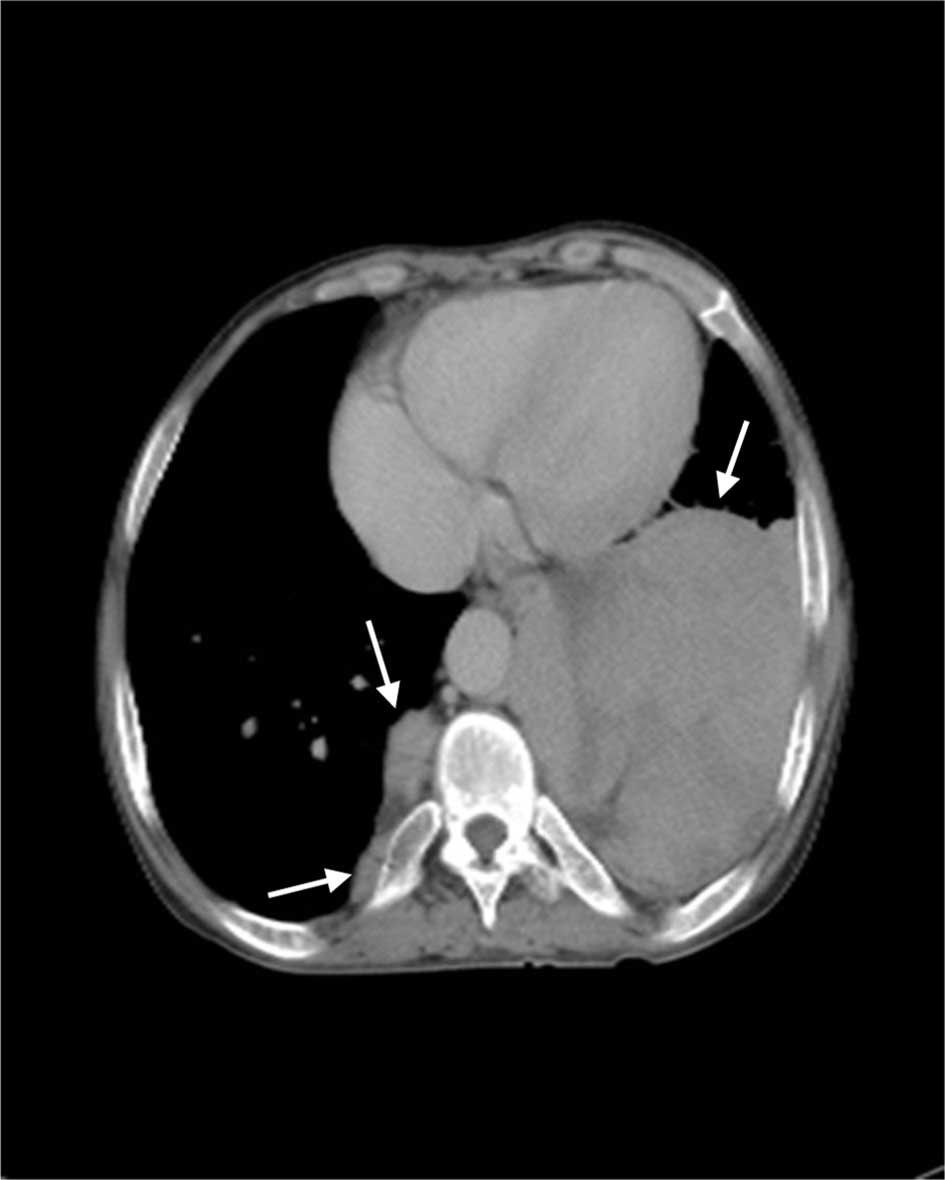
was 3.06×10¹²/l. CT scan revealed multiple paraspinal
masses in the bottom of the left thoracic cavity, and the largest
measured 10.1×10.5 cm (Fig. 3). The
tumors were well-demarcated and demonstrated heterogeneous density,
ranging between 51 and 89 Hounsfield units. Considering the size of
the mass and the possibility of multiple neurofibroma, a surgical
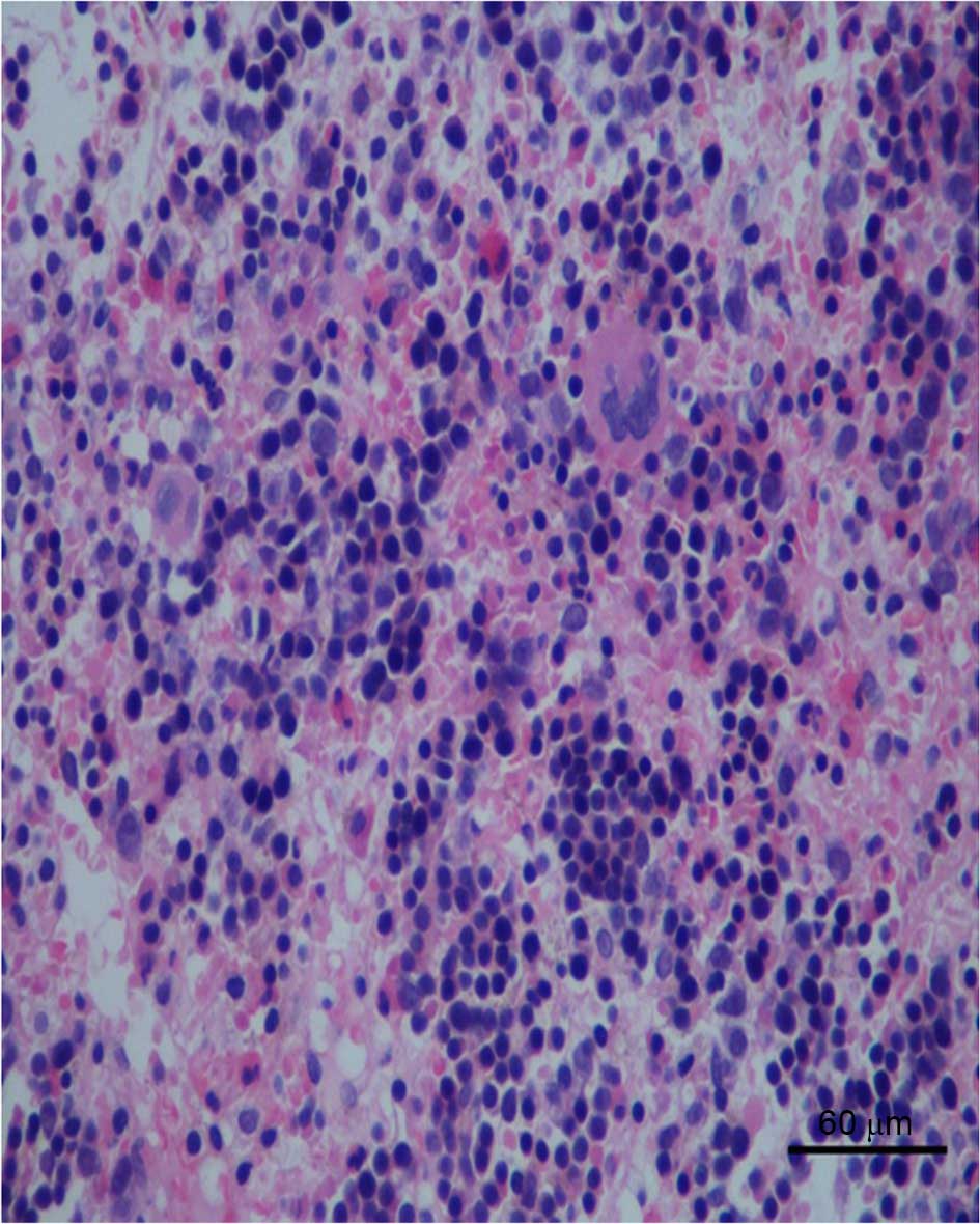
biopsy was performed. Microscopically, the excised tumor exhibited
characteristic megakaryocytes, erythroblasts and myeloid cells
(Fig. 4). Immunohistochemical
staining, performed as described above, showed that the tumor was
positive for MPO, glycophorin and CD61, and diffuse positive for
CD20/CD3. The findings were compatible with a diagnosis of EMH. The
patient was not provided any other treatment following a surgical
biopsy, and was free from recurrence after 2 years of
follow-up.
Discussion
EMH is defined as hematopoiesis occurring in organs
outside of the bone marrow. It occurs if the bone marrow is no
longer functional and is identified in a number of hematological
diseases; its occurrence in chronic hemolytic anemias remains
highest, particularly in transfusion-independent thalassemia
intermedia (9–12). Ineffective red cell production by the
bone marrow forces expansion of the hematopoietic tissue outside
the marrow medulla and results in hematopoietic compensatory
involvement, particularly in the form of masses, of other areas in
the body (13).
Among the various body regions described, paraspinal
involvement requires particular attention due to the debilitating
clinical consequences and challenges in management and diagnosis. A
paraspinal location in hematopoietic tissue occurs in 11–15% of
cases with EMH (8). There is some
predilection for the site of spinal cord involvement by the
hematopoietic tissue. The thoracic region and, to a lesser extent,
the lumbar region are the most frequently involved sites (14). The cause of this predilection is
uncertain; however, these sites are understood to normally engage
in active hematopoiesis in the fetus during gestation (14). This pathway typically stops at birth,
but the extramedullary hematopoietic vascular connective tissues
retain the ability to produce red cells under conditions of
long-standing ineffective erythropoiesis (14).
Paraspinal EMH typically presents as pseudotumors,
and can sometimes result in symptomatic tumor-like masses, which
may cause a variety of neurological symptoms due to spinal
compression. However, it is understood that >80% of cases do not
have signs and symptoms related directly to the disorder, and the
lesions are typically discovered incidentally by radiologic
techniques (15–17). The development of neurologic symptoms
is suggested to depend on the chronicity of the disease, with
neurologic symptoms most frequently being reported during the third
and fourth decades of life (18),
although few reports described presentation as early as the first
decade of life (19–21). The male to female ratio reaches 5:1
(18). Various clinical
presentations have been reported including back pain, lower
extremity pain, parasthesia, abnormal proprioception, exaggerated
or brisk deep tendon reflexes, Babinski response, Lasègue sign,
paraparesis, paraplegia, ankle clonus, spastic gate, urgency of
urination and bowel incontinence (22). The size and location of lesions and
the extent of spinal cord involvement determines the severity,
acuteness and multiplicity of signs and symptoms (22).
Although the history and physical examination may
help narrow the differential diagnosis, radiographic imaging
remains essential to confirm the existence of hematopoietic tissue.
Characteristic appearance has been observed primarily using
magnetic resonance imaging (MRI) or CT scan. MRI is the diagnostic
investigation of choice. Paraspinal EMH appears as unique, multiple
iso- or hyperintense masses, with homogeneous enhancement following
contrast administration (23). These
masses are usually lobular, well-circumscribed masses of
intermediate signal intensity on T1-weighted images and low signal
intensity on T2-weighted images (23,24). The
CT appearance is characterized by the heterogeneous soft tissue
density mass. The diagnosis is confirmed following surgical removal
of the mass. Biopsy remains the gold standard (25).
Because of its rarity, there is no standard
treatment approach in patients with symptomatic EMH, and no
evidence-based guidelines for the treatment of EMH. Therapy
typically depends on the severity of symptoms, size of the mass,
patient's clinical condition and previous treatment. Numerous
treatment options have been described, including transfusion
therapy, laminectomy, radiotherapy and the use of fetal hemoglobin,
inducing agents that decrease the hematopoietic drive. However, the
ideal management scheme remains controversial. Until large
prospective trials evaluate the efficacy and safety of the
available treatment options, both in single and in combination
therapy, an individualized approach should be devised (26).
In conclusion, EMH is a rare disease, which is
complicated to preoperatively distinguish from other neoplasms. In
the present study, two patients with EMH are presented. The two
patients underwent surgery and the pathological findings indicated
that EMH was the correct diagnosis. These two cases suggest that
EMH must be taken into consideration when masses with
characteristic radiologic appearance are identified in patients
with various hematological diseases, including thalassemia
intermedia (4).
Acknowledgements
The authors thank the staff of the Radiology
Department of the First Affiliated Hospital of Guangzhou Medical
University, and Professor Xia Gu of the Pathology Department at The
First Affiliated Hospital of Guangzhou Medical University
(Guangzhou, China) for kindly providing the figures of the two
patients.
References
|
1
|
Bozzini CE, Rendo ME Barrio, Devoto FC and
Epper CE: Studies on medullary and extramedullary erythropoiesis in
the adult mouse. Am J Physiol. 219:724–728. 1970.PubMed/NCBI
|
|
2
|
Papavasiliou C: Clinical expressions of
the expansion of the bone marrow in the chronic anemias: The role
of radiotherapy. Int J Radiat Oncol Biol Phys. 28:605–612. 1994.
View Article : Google Scholar : PubMed/NCBI
|
|
3
|
Taheri M Sanei, Birang SH, Shahnazi M and
Hemadi H: Large splenic mass of extramedullary hematopoiesis. Iran
J Radiol. 2:99–101. 2005.
|
|
4
|
Miyake H, Matsuda M, Iyomasa S and Mizuno
K: Presacral extramedullary hematopoiesis. Surgery. 135:112–113.
2004. View Article : Google Scholar : PubMed/NCBI
|
|
5
|
Aessopos A, Tassiopoulos S, Farmakis D,
Moyssakis I, Kati M, Polonifi K and Tsironi M: Extramedullary
hematopoiesis related pleural effusion: The case of
beta-thalassemia. Ann Thorac Surg. 81:2037–2043. 2006. View Article : Google Scholar : PubMed/NCBI
|
|
6
|
Al-Habib H and Hadzikaric N: Spinal cord
compression due to intraspinal extramedullary hematopoiesis in
thalassemia intermedia. Neurosciences (Riyadh). 12:261–264.
2007.PubMed/NCBI
|
|
7
|
Eskazan AE, Ar MC and Baslar Z:
Intracranial extramedullary hematopoiesis in patients with
thalassemia: A case report and review of the literature.
Transfusion. 52:1715–1720. 2012. View Article : Google Scholar : PubMed/NCBI
|
|
8
|
Dore F, Cianciulli P, Rovasio S, Oggiano
L, Bonfigli S, Murineddu M, Pardini S, Simonetti G, Gualdi G, Papa
G, et al: Incidence and clinical study of ectopic erythropoiesis in
adult patients with thalassemia intermedia. Ann Ital Med Int.
7:137–140. 1992.PubMed/NCBI
|
|
9
|
Cromwell LD and Kerber C: Spinal cord
compression by extramedullary hematopoiesis in myeloid metaplasia.
Radiology. 128:1181978. View Article : Google Scholar : PubMed/NCBI
|
|
10
|
de Morais JC, Spector N, Lavrado FP, Nobre
LF, de Mattos JP, Pulcheri W, Nucci M, Novis S and de Oliveira HP:
Spinal cord compression due to extramedullary hematopoiesis in the
proliferative phase of polycythemia vera. Acta Haematol.
96:242–244. 1996. View Article : Google Scholar : PubMed/NCBI
|
|
11
|
Haran M and Ni S: Recurrent reversible
paraplegia. Lancet. 357:10922001. View Article : Google Scholar : PubMed/NCBI
|
|
12
|
Taher A, Isma'eel H and Cappellini MD:
Thalassaemia intermedia: Revisited. Blood Cells Mol Dis. 37:12–20.
2006. View Article : Google Scholar : PubMed/NCBI
|
|
13
|
Shin KH, Sharma S, Gregoritch SJ, Lifeso
RM, Bettigole R and Yoon SS: Combined radiotherapeutic and surgical
management of a spinal cord compression by extramedullary
hematopoiesis in a patient with hemoglobin E beta-thalassemia. Acta
Haematol. 91:154–157. 1994. View Article : Google Scholar : PubMed/NCBI
|
|
14
|
Tsitouridis J, Stamos S, Hassapopoulou E,
Tsitouridis K and Nikolopoulos P: Extramedullary paraspinal
hematopoiesis in thalassemia: CT and MRI evaluation. Eur J Radiol.
30:33–38. 1999. View Article : Google Scholar : PubMed/NCBI
|
|
15
|
Parsa K and Oreizy A: Nonsurgical approach
to paraparesis due to extramedullary hematopoiesis. Report of two
cases. J Neurosurg. 82:657–660. 1995. View Article : Google Scholar : PubMed/NCBI
|
|
16
|
Richter E: Extramedullary hematopoiesis
with intraspinal extension in thalassemia. Aktuelle Radiol.
3:320–322. 1993.(In German). PubMed/NCBI
|
|
17
|
Dore F, Pardini S, Gaviano E, Longinotti
M, Bonfigli S, Rovasio S, Tomiselli A and Cossu F: Recurrence of
spinal cord compression from extramedullary hematopoiesis in
thalassemia intermedia treated with low doses of radiotherapy. Am J
Hematol. 44:1481993. View Article : Google Scholar : PubMed/NCBI
|
|
18
|
Salehi SA, Koski T and Ondra SL: Spinal
cord compression in betathalassemia: Case report and review of the
literature. Spinal Cord. 42:117–123. 2004. View Article : Google Scholar : PubMed/NCBI
|
|
19
|
Cardia E, Toscano S, La Rosa G, Zaccone C,
d'Avella D and Tomasello F: Spinal cord compression in homozygous
beta thalassemia intermedia. Pediatr Neurosurg. 20:186–189. 1994.
View Article : Google Scholar : PubMed/NCBI
|
|
20
|
Chehal A, Aoun E, Koussa S, Skoury H,
Koussa S and Taher A: Hypertransfusion: A successful method of
treatment in thalassemia intermedia patients with spinal cord
compression secondary to extramedullary hematopoiesis. Spine.
28:E245–E249. 2003. View Article : Google Scholar : PubMed/NCBI
|
|
21
|
Ileri T, Azik F, Ertem M, Uysal Z and
Gozdasoglu S: Extramedullary hematopoiesis with spinal cord
compression in a child with thalassemia intermedia. J Pediatr
Hematol Oncol. 31:681–683. 2009. View Article : Google Scholar : PubMed/NCBI
|
|
22
|
Haidar R, Mhaidli H and Tacher AT:
Paraspinal extramedullary hematopoiesis in patients with
thalassemia intermedia. Eur Spine J. 19:871–878. 2010. View Article : Google Scholar : PubMed/NCBI
|
|
23
|
Haidar S, Ortiz-Neira C, Shroff M, Gilday
D and Blaser S: Intracranial involvement in extramedullary
hematopoiesis: Case report and review of the literature. Pediatr
Radiol. 35:630–634. 2005. View Article : Google Scholar : PubMed/NCBI
|
|
24
|
Debard A, Demasles S, Camdessanché JP,
Duband S, Mohammedi R and Antoine JC: Dural localization of
extramedullary hematopoiesis. Report of a case. J Neurol.
256:837–838. 2009. View Article : Google Scholar : PubMed/NCBI
|
|
25
|
Tan TC, Tsao J and Cheung FC:
Extramedullary haemopoiesis in thalassemia intermedia presenting as
paraplegia. J Clin Neurosci. 9:721–725. 2002. View Article : Google Scholar : PubMed/NCBI
|
|
26
|
Haidar R, Mhaidli H and Taher AT:
Paraspinal extramedullary hematopoiesis in patients with
thalassemia intermedia. Eur Spine J. 19:871–878. 2010. View Article : Google Scholar : PubMed/NCBI
|