Spandidos Publications Logo
  • About
    • About Spandidos
    • Aims and Scopes
    • Abstracting and Indexing
    • Editorial Policies
    • Reprints and Permissions
    • Job Opportunities
    • Terms and Conditions
    • Contact
  • Journals
    • All Journals
    • Oncology Letters
      • Oncology Letters
      • Information for Authors
      • Editorial Policies
      • Editorial Board
      • Aims and Scope
      • Abstracting and Indexing
      • Bibliographic Information
      • Archive
    • International Journal of Oncology
      • International Journal of Oncology
      • Information for Authors
      • Editorial Policies
      • Editorial Board
      • Aims and Scope
      • Abstracting and Indexing
      • Bibliographic Information
      • Archive
    • Molecular and Clinical Oncology
      • Molecular and Clinical Oncology
      • Information for Authors
      • Editorial Policies
      • Editorial Board
      • Aims and Scope
      • Abstracting and Indexing
      • Bibliographic Information
      • Archive
    • Experimental and Therapeutic Medicine
      • Experimental and Therapeutic Medicine
      • Information for Authors
      • Editorial Policies
      • Editorial Board
      • Aims and Scope
      • Abstracting and Indexing
      • Bibliographic Information
      • Archive
    • International Journal of Molecular Medicine
      • International Journal of Molecular Medicine
      • Information for Authors
      • Editorial Policies
      • Editorial Board
      • Aims and Scope
      • Abstracting and Indexing
      • Bibliographic Information
      • Archive
    • Biomedical Reports
      • Biomedical Reports
      • Information for Authors
      • Editorial Policies
      • Editorial Board
      • Aims and Scope
      • Abstracting and Indexing
      • Bibliographic Information
      • Archive
    • Oncology Reports
      • Oncology Reports
      • Information for Authors
      • Editorial Policies
      • Editorial Board
      • Aims and Scope
      • Abstracting and Indexing
      • Bibliographic Information
      • Archive
    • Molecular Medicine Reports
      • Molecular Medicine Reports
      • Information for Authors
      • Editorial Policies
      • Editorial Board
      • Aims and Scope
      • Abstracting and Indexing
      • Bibliographic Information
      • Archive
    • World Academy of Sciences Journal
      • World Academy of Sciences Journal
      • Information for Authors
      • Editorial Policies
      • Editorial Board
      • Aims and Scope
      • Abstracting and Indexing
      • Bibliographic Information
      • Archive
    • International Journal of Functional Nutrition
      • International Journal of Functional Nutrition
      • Information for Authors
      • Editorial Policies
      • Editorial Board
      • Aims and Scope
      • Abstracting and Indexing
      • Bibliographic Information
      • Archive
    • International Journal of Epigenetics
      • International Journal of Epigenetics
      • Information for Authors
      • Editorial Policies
      • Editorial Board
      • Aims and Scope
      • Abstracting and Indexing
      • Bibliographic Information
      • Archive
    • Medicine International
      • Medicine International
      • Information for Authors
      • Editorial Policies
      • Editorial Board
      • Aims and Scope
      • Abstracting and Indexing
      • Bibliographic Information
      • Archive
  • Articles
  • Information
    • Information for Authors
    • Information for Reviewers
    • Information for Librarians
    • Information for Advertisers
    • Conferences
  • Language Editing
Spandidos Publications Logo
  • About
    • About Spandidos
    • Aims and Scopes
    • Abstracting and Indexing
    • Editorial Policies
    • Reprints and Permissions
    • Job Opportunities
    • Terms and Conditions
    • Contact
  • Journals
    • All Journals
    • Biomedical Reports
      • Information for Authors
      • Editorial Policies
      • Editorial Board
      • Aims and Scope
      • Abstracting and Indexing
      • Bibliographic Information
      • Archive
    • Experimental and Therapeutic Medicine
      • Information for Authors
      • Editorial Policies
      • Editorial Board
      • Aims and Scope
      • Abstracting and Indexing
      • Bibliographic Information
      • Archive
    • International Journal of Epigenetics
      • Information for Authors
      • Editorial Policies
      • Editorial Board
      • Aims and Scope
      • Abstracting and Indexing
      • Bibliographic Information
      • Archive
    • International Journal of Functional Nutrition
      • Information for Authors
      • Editorial Policies
      • Editorial Board
      • Aims and Scope
      • Abstracting and Indexing
      • Bibliographic Information
      • Archive
    • International Journal of Molecular Medicine
      • Information for Authors
      • Editorial Policies
      • Editorial Board
      • Aims and Scope
      • Abstracting and Indexing
      • Bibliographic Information
      • Archive
    • International Journal of Oncology
      • Information for Authors
      • Editorial Policies
      • Editorial Board
      • Aims and Scope
      • Abstracting and Indexing
      • Bibliographic Information
      • Archive
    • Medicine International
      • Information for Authors
      • Editorial Policies
      • Editorial Board
      • Aims and Scope
      • Abstracting and Indexing
      • Bibliographic Information
      • Archive
    • Molecular and Clinical Oncology
      • Information for Authors
      • Editorial Policies
      • Editorial Board
      • Aims and Scope
      • Abstracting and Indexing
      • Bibliographic Information
      • Archive
    • Molecular Medicine Reports
      • Information for Authors
      • Editorial Policies
      • Editorial Board
      • Aims and Scope
      • Abstracting and Indexing
      • Bibliographic Information
      • Archive
    • Oncology Letters
      • Information for Authors
      • Editorial Policies
      • Editorial Board
      • Aims and Scope
      • Abstracting and Indexing
      • Bibliographic Information
      • Archive
    • Oncology Reports
      • Information for Authors
      • Editorial Policies
      • Editorial Board
      • Aims and Scope
      • Abstracting and Indexing
      • Bibliographic Information
      • Archive
    • World Academy of Sciences Journal
      • Information for Authors
      • Editorial Policies
      • Editorial Board
      • Aims and Scope
      • Abstracting and Indexing
      • Bibliographic Information
      • Archive
  • Articles
  • Information
    • For Authors
    • For Reviewers
    • For Librarians
    • For Advertisers
    • Conferences
  • Language Editing
Login Register Submit
  • This site uses cookies
  • You can change your cookie settings at any time by following the instructions in our Cookie Policy. To find out more, you may read our Privacy Policy.

    I agree
Search articles by DOI, keyword, author or affiliation
Search
Advanced Search
presentation
Experimental and Therapeutic Medicine
Join Editorial Board Propose a Special Issue
Print ISSN: 1792-0981 Online ISSN: 1792-1015
Journal Cover
May-2017 Volume 13 Issue 5

Full Size Image

Sign up for eToc alerts
Recommend to Library

Journals

International Journal of Molecular Medicine

International Journal of Molecular Medicine

International Journal of Molecular Medicine is an international journal devoted to molecular mechanisms of human disease.

International Journal of Oncology

International Journal of Oncology

International Journal of Oncology is an international journal devoted to oncology research and cancer treatment.

Molecular Medicine Reports

Molecular Medicine Reports

Covers molecular medicine topics such as pharmacology, pathology, genetics, neuroscience, infectious diseases, molecular cardiology, and molecular surgery.

Oncology Reports

Oncology Reports

Oncology Reports is an international journal devoted to fundamental and applied research in Oncology.

Experimental and Therapeutic Medicine

Experimental and Therapeutic Medicine

Experimental and Therapeutic Medicine is an international journal devoted to laboratory and clinical medicine.

Oncology Letters

Oncology Letters

Oncology Letters is an international journal devoted to Experimental and Clinical Oncology.

Biomedical Reports

Biomedical Reports

Explores a wide range of biological and medical fields, including pharmacology, genetics, microbiology, neuroscience, and molecular cardiology.

Molecular and Clinical Oncology

Molecular and Clinical Oncology

International journal addressing all aspects of oncology research, from tumorigenesis and oncogenes to chemotherapy and metastasis.

World Academy of Sciences Journal

World Academy of Sciences Journal

Multidisciplinary open-access journal spanning biochemistry, genetics, neuroscience, environmental health, and synthetic biology.

International Journal of Functional Nutrition

International Journal of Functional Nutrition

Open-access journal combining biochemistry, pharmacology, immunology, and genetics to advance health through functional nutrition.

International Journal of Epigenetics

International Journal of Epigenetics

Publishes open-access research on using epigenetics to advance understanding and treatment of human disease.

Medicine International

Medicine International

An International Open Access Journal Devoted to General Medicine.

Journal Cover
May-2017 Volume 13 Issue 5

Full Size Image

Sign up for eToc alerts
Recommend to Library

  • Article
  • Citations
    • Cite This Article
    • Download Citation
    • Create Citation Alert
    • Remove Citation Alert
    • Cited By
  • Similar Articles
    • Related Articles (in Spandidos Publications)
    • Similar Articles (Google Scholar)
    • Similar Articles (PubMed)
  • Download PDF
  • Download XML
  • View XML
Case Report

Proliferative myositis in the right brachioradialis: A case report

  • Authors:
    • Na Wei
    • Wei‑Jie Xu
    • Dong Dong
    • Yu‑Bao Gong
  • View Affiliations / Copyright

    Affiliations: The First Operating Room, The First Hospital of Jilin University, Changchun, Jilin 130000, P.R. China, Department of Bone and Joint Surgery, The First Hospital of Jilin University, Changchun, Jilin 130000, P.R. China, Department of Radiology, The First Hospital of Jilin University, Changchun, Jilin 130000, P.R. China
  • Pages: 2483-2485
    |
    Published online on: March 28, 2017
       https://doi.org/10.3892/etm.2017.4269
  • Expand metrics +
Metrics: Total Views: 0 (Spandidos Publications: | PMC Statistics: )
Metrics: Total PDF Downloads: 0 (Spandidos Publications: | PMC Statistics: )
Cited By (CrossRef): 0 citations Loading Articles...

This article is mentioned in:


Abstract

Proliferative myositis is a rare, self‑limiting, benign disease. Its diagnosis can be difficult and in many cases is not confirmed until after surgical resection. Herein, we report a case of proliferative myositis of the right brachioradialis in a 64‑year‑old man. The patient presented with a rapidly growing, painless mass in his right forearm. Magnetic resonance imaging and fine‑needle aspiration biopsy led to a diagnosis of proliferative myositis. Complete surgical resection of the mass was performed. Postoperative pathological examination confirmed the diagnosis. To the best of our knowledge, this is the first report of proliferative myositis in the right brachioradialis. Fine‑needle biopsy is helpful in the diagnosis of proliferative myositis, thus avoiding unnecessary surgical trauma and costs.

Introduction

Proliferative myositis is a rare pseudosarcomatous (mimicking sarcoma) condition. It usually occurs in adults aged >40-years-old, and occasionally in children (1). Although proliferative myositis grows rapidly, it is actually a benign lesion and can regress or completely resolve spontaneously. The incidence of proliferative myositis remains unknown, however recurrence is extremely rare (2). Although the cause of proliferative myositis remains unclear, a history of recent local trauma may be a risk factor, as noted in some reports (1,3,4). Diagnosis of proliferative myositis is largely dependent on the patient's age, history of recent trauma, and a rapidly growing, painful solitary soft-tissue mass located in the muscle, together with radiological evidence, such as magnetic resonance imaging (MRI). Pathological examination of a biopsy is the final and most accurate method for the diagnosis of proliferative myositis. There is no specific treatment recommended following establishment of a diagnosis of proliferative myositis, since of proliferative myositis may disappear spontaneously (2,5). Excision may be preferred for the confirmation of a diagnosis and for cosmetic reasons (2). The most commonly involved sites are the head, neck, and extremities. Herein, we report a case of proliferative myositis in the right brachioradialis. To the best of our knowledge, this is the first case reported at this site and will further improve our understanding of the sites affected by this disease.

Case report

A 64-year-old man was admitted to The First Hospital of Jilin University (Changchun, China) in September 2014 after presenting with a mass in his right forearm. The mass was incidentally found by the patient 2 weeks previously, and upon hospitalization had grown larger. On examination, a mass 3.0×2.5 cm was palpated in the right brachioradialis, on the lateral side of the proximal end of the right forearm. During movement and rest of his right arm, the patient experienced no discomfort. The skin surface above the mass was elevated, and exhibited normal pigmentation and temperature. The mass was mildly tender with a hard texture and smooth surface, although the border with the surrounding tissue was not pronounced.

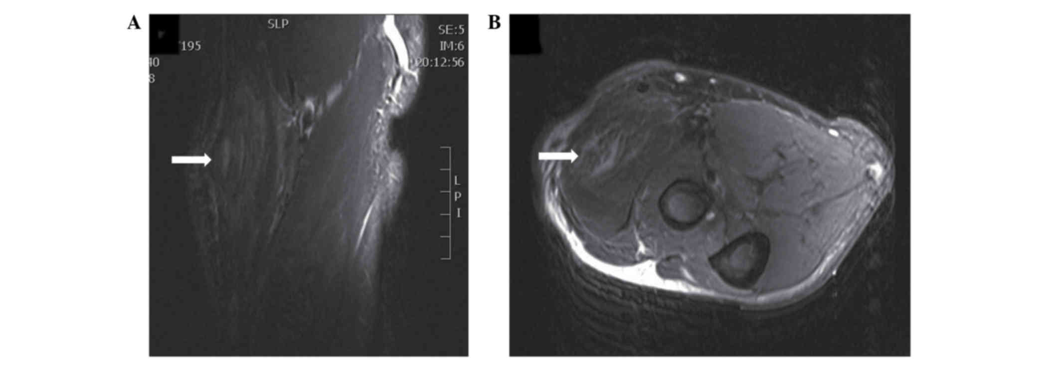
Neurological examination of the right arm was unremarkable. MRI showed a hyperintense lesion in T1- and T2-weighted images (Fig. 1). Fine-needle biopsy of the lesion revealed spindle cells and ganglion cells, suggesting proliferative myositis. Pathological examination was subsequently performed. The specimen was fixed in 10% formalin-saline solution, followed by embedding in paraffin for 24 h, sectioning at 4-µm thickness, and staining with hematoxylin and eosin. Subsequently, the sections were examined under a light microscope (DP20; Olympus Corp., Tokyo, Japan) at a magnification of ×40. Since pathological examination demonstrated that the lesion was benign and the function of the arm was not affected, a watch-and-wait strategy was recommended. However, the patient insisted on surgical resection of the mass.

Figure 1.

T2-weighted magnetic resonance images with fat suppression. (A) Coronal and (B) axial views. Arrows denote the lesion in the right brachioradialis.

Intraoperatively, the mass was discovered to be incorporated with surrounding muscular tissues. Complete resection of the mass was performed with dissection of the surrounding normal tissues.

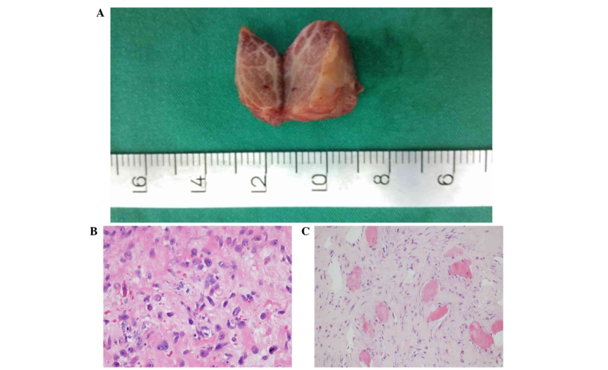
Gross pathological examination showed that the resected mass was pink-gray on the transverse section surface (Fig. 2A). Microscopic examination revealed ganglion-like giant basophilic cells and spindle cells (Fig. 2B and C). A diagnosis of proliferative myositis was confirmed pathologically. Upon follow-up 13 months after surgery, the patient exhibited a well-healed surgical incision and good elbow joint function, without any swelling or tenderness. No recurrence of the mass was detected. The patient refused a follow-up MRI examination.

Figure 2.

Pathological examination of the resected proliferative myositis. (A) The mass was pink-gray on the transverse section surface with a solid texture, integrated with the surrounding muscle. (B) Ganglion-like giant basophilic cells were detected and (C) muscle bundles were divided by the spindle cells, in a checkerboard-like pattern, as detected by hematoxylin and eosin staining (magnification, ×40).

Ethical approval for the present study was obtained from The First Hospital of Jilin University and written informed consent was obtained from the patient.

Discussion

Proliferative myositis was first reported by Kern in 1960 (2). Several theories regarding its etiology have been proposed, such as mechanical injury and ischemia, which remain inconclusive (6–9). The mass usually grows rapidly and is able to cause local compression symptoms.

Proliferative myositis shows no specific features upon laboratory and imaging examinations; therefore, pathological examination is required for a definitive diagnosis. The resected mass is lobular and infiltrative. Its section surface is gray in color, with a solid texture. Pathologically, the proliferative mass is characterized by a checkerboard of myofibroblasts infiltrating muscle fibers on the transverse section, and ganglion-like cells (10).

Proliferative myositis is distinct from rhabdomyosarcoma, myositis ossificans, nodular fasciitis, and proliferative fasciitis (2,11,12). Differentiating between proliferative myositis and rhabdomyosarcoma can be difficult. Soft tissue sarcoma is often round in shape or nodular, lacking the normal structure of muscle fibers. In large soft tissue sarcomas, necrosis, hemorrhage, and cysts may be found (12). In addition, proliferative myositis is negative for myosin staining, but positive in rhabdomyosarcoma. Myositis ossificans exhibit characteristic ossification in the lesion, as observed on MRI or computed tomography (10). MR images of proliferative myositis have been reported in several cases, with hypo- or iso-intense T1 signals, compared with those of muscle and homogeneous enhancement. T2-weighted MR images typically demonstrate a hyperintense soft-tissue mass (1,2,5).

A watch-and-wait strategy is preferred for proliferative myositis, due to its benign nature and the potential for spontaneous resolution. Although the mass grows rapidly in its initial phase, it typically stabilizes after a few weeks. No malignancy or metastasis of proliferative myositis has ever been reported (9). If the mass is causing compression symptoms or affecting the patient's daily life, surgical resection can be performed.

In conclusion, proliferative myositis is a rare, self-limiting, benign disease that has not been described in the right brachioradialis until now. Its diagnosis can be difficult and, in many cases, diagnosis is not confirmed until after surgical resection. Fine-needle biopsy is helpful in the diagnosis of proliferative myositis, thus avoiding unnecessary surgical trauma and costs.

References

1 

Mulier S, Stas M, Delabie J, Lateur L, Gysen M, Dal Cin P, Robberecht C and De Wever I: Proliferative myositis in a child. Skeletal Radiol. 28:703–709. 1999. View Article : Google Scholar : PubMed/NCBI

2 

Wlachovska B, Abraham B, Deux JF, Sibony M, Marsault C and Le Breton C: Proliferative myositis in a patient with AIDS. Skeletal Radiol. 33:237–240. 2004. View Article : Google Scholar : PubMed/NCBI

3 

Kern WH: Proliferative myositis; a pseudosarcomatous reaction to injury: A report of seven cases. Arch Pathol. 69:209–216. 1960.PubMed/NCBI

4 

Enzinger FM and Dulcey F: Proliferative myositis: Report of thirty-three cases. Cancer. 20:2213–2223. 1967. View Article : Google Scholar : PubMed/NCBI

5 

Kent MS, Flieder DB, Port JL and Altorki NK: Proliferative myositis: a rare pseudosarcoma of the chest wall. Ann Thorac Surg. 2002.73:1296–1298. View Article : Google Scholar : PubMed/NCBI

6 

Wong NL: Fine needle aspiration cytology of pseudosarcomatous reactive proliferative lesions of soft tissue. Acta Cytol. 46:1049–1055. 2002. View Article : Google Scholar : PubMed/NCBI

7 

Wong NL and Di F: Pseudosarcomatous fasciitis and myositis diagnosisby fine-needle aspiration cytology. Am J Clin Pathol. 132:857–865. 2009. View Article : Google Scholar : PubMed/NCBI

8 

Brooks JK, Scheper MA, Kramer RE, Papadimitriou JC, Sauk JJ and Nikitakis NG: Intraoral proliferative myositis: Case report and literature review. Head Neck. 29:416–420. 2007. View Article : Google Scholar : PubMed/NCBI

9 

Fauser C, Hrig J, Niedermeyer HP and Arnold W: Proliferative myositisa rare pseudomalignant tumor of the head and neck. Arch Otolaryngol Head Neck Surg. 134:437–440. 2008. View Article : Google Scholar : PubMed/NCBI

10 

Klapsinou E, Despoina P and Dimitra D: Cytologic findings and potential pitfalls in proliferative myositis and myositis ossificans diagnosed by fine needle aspiration cytology: Report of four cases and review of the literature. Diagn Cytopathol. 40:239–244. 2012. View Article : Google Scholar : PubMed/NCBI

11 

Subramanian S and Sharma R: Can MR imaging be used to reliably differentiate proliferative myositis from myositis ossificans? Radiology. 246:9872008. View Article : Google Scholar : PubMed/NCBI

12 

Demir MK, Beser M and Akinci O: Case 118: Proliferative myositis. Radiology. 244:613–616. 2007. View Article : Google Scholar : PubMed/NCBI

Related Articles

  • Abstract
  • View
  • Download
  • Twitter
Copy and paste a formatted citation
Spandidos Publications style
Wei N, Xu WJ, Dong D and Gong YB: Proliferative myositis in the right brachioradialis: A case report. Exp Ther Med 13: 2483-2485, 2017.
APA
Wei, N., Xu, W., Dong, D., & Gong, Y. (2017). Proliferative myositis in the right brachioradialis: A case report. Experimental and Therapeutic Medicine, 13, 2483-2485. https://doi.org/10.3892/etm.2017.4269
MLA
Wei, N., Xu, W., Dong, D., Gong, Y."Proliferative myositis in the right brachioradialis: A case report". Experimental and Therapeutic Medicine 13.5 (2017): 2483-2485.
Chicago
Wei, N., Xu, W., Dong, D., Gong, Y."Proliferative myositis in the right brachioradialis: A case report". Experimental and Therapeutic Medicine 13, no. 5 (2017): 2483-2485. https://doi.org/10.3892/etm.2017.4269
Copy and paste a formatted citation
x
Spandidos Publications style
Wei N, Xu WJ, Dong D and Gong YB: Proliferative myositis in the right brachioradialis: A case report. Exp Ther Med 13: 2483-2485, 2017.
APA
Wei, N., Xu, W., Dong, D., & Gong, Y. (2017). Proliferative myositis in the right brachioradialis: A case report. Experimental and Therapeutic Medicine, 13, 2483-2485. https://doi.org/10.3892/etm.2017.4269
MLA
Wei, N., Xu, W., Dong, D., Gong, Y."Proliferative myositis in the right brachioradialis: A case report". Experimental and Therapeutic Medicine 13.5 (2017): 2483-2485.
Chicago
Wei, N., Xu, W., Dong, D., Gong, Y."Proliferative myositis in the right brachioradialis: A case report". Experimental and Therapeutic Medicine 13, no. 5 (2017): 2483-2485. https://doi.org/10.3892/etm.2017.4269
Follow us
  • Twitter
  • LinkedIn
  • Facebook
About
  • Spandidos Publications
  • Careers
  • Cookie Policy
  • Privacy Policy
How can we help?
  • Help
  • Live Chat
  • Contact
  • Email to our Support Team