Introduction
Complex congenital heart disease (CHD), which is
related to cardiovascular malformations due to abnormal embryonic
development, is one of the most common cardiovascular diseases. The
incidence of CHD is 0.4–0.8% in newborns, of which 60% succumb to
CHD during their first year (1).
Therefore, CHD is a fatal defect that lowers the life quality of
young children (2). As China has the
highest birth rate in the world (3),
early diagnosis and timely surgical treatment is extremely
important for reducing the mortality rate of CHD. Preoperative
examination of CHD is conducted by echocardiography, cardiac
catheterisation angiography, magnetic resonance imaging (MRI) and
computed tomography. Ultrasound cardiograms are convenient and
non-invasive, but the vessels outside the heart cannot be displayed
clearly (4,5). Cardiac catheterisation angiography is
invasive for patients. MRI provides a non-invasive alternative and
avoids ionizing radiation exposure, although the examination time
of MRI is long, and it is not suitable for children (6). Dual-source computed tomography (DSCT)
is a non-invasive technique with high temporal resolution and
spatial resolution that is helpful for diagnosis, postoperative
effect prediction and follow-up studies (7,8).
The present study aimed to evaluate image quality
and radiation dosage using a low-dose prospectively
electrocardiogram (ECG)-gated CT protocol for DSCT in children with
complex congenital heart disease. From January 2013 to July 2013, a
specific scanning schedule was introduced to reduce the radiation
dosage in order to limit unnecessary exposure to radiation in
children with CHD.
Materials and methods
Patients
Low-dose protocols were approved by the Ethics
Committee of Wuhan Asia Heart Hospital (Wuhan, China). During the
period from January 2013 to July 2013, 206 children were initially
diagnosed with complex congenital heart diseases, which were
confirmed by ultrasound cardiogram. To clarify the intracardiac and
extracardiac abnormities, patients underwent cardiac CT scanning
prior to surgery. These 206 cases, including 105 males and 101
females with an age range of 1–142 months, a mean age of 22.21±2.89
months, a mean height of 76.54±1.88 cm and a mean weight of
9.76±0.49 kg, were equally and randomly classified into two groups
according to the registered number. Children in group A underwent
low-dose prospective ECG-gated scanning, whereas those in group B
received retrospective ECG-gated scanning on a DSCT scanner.
Informed consent was obtained from the parents of all children
enrolled in the present study.
Scanning protocol
Patients were scanned with a DSCT scanner (Siemens
Somatom Definition Flash CT; Siemens AG, Munich, Germany). All
patients remained in sinus rhythm before examination without
receiving a β-blocker. An intravenous catheter was placed into an
antecubital fossa vein or femoral vein in each patient.
Short-acting anesthesia (propofol, 1–2 mg/kg; Frsenius Kabi
Deutschland GmbH, Germany) was administered to uncooperative
patients according to their respective weights. Patients were
provided with a lead apron to cover body parts that were not to be
scanned to ensure radiation protection. All patients received a
nonionic low-osmolality contrast agent Visipaque 320 (iodixanol; GE
Healthcare Life Sciences, Chalfont, UK) injection according to
their weight (1.5–2.5 ml/kg) at an injection velocity of 0.7–2.7
ml/sec. A contrast agent dose of 2.0 mg/kg is typically used. A
double tube high pressure syringe (Medrad, Inc., Warrendale, PA,
USA) was applied for biphasic injection: 75% contrast and 25%
saline were injected simultaneously in the first phase, and 6–10 ml
saline was injected at the same velocity in the second phase to
eliminate artifacts in the superior vena cava and right atrium
(9).
The scanning range of the heart included the area
from the thoracic inlet to 1 cm below the diaphragm, and the
scanning direction was set from superior to inferior. The following
imaging parameters were used: 80 kV automatic tube current
modulation technique (caredose); rotation time, 0.28 sec; detector
array, 128×0.6 mm; and slice thickness, 0.75 mm. Epigastric CT
scanning was carried out following heart scanning to rule out situs
inversus viscerum and anomalous pulmonary venous connection.
Scanning range was set from the diaphragm to the inferior pole of
the kidney.
Scanning methods
Different scanning methods were applied to the
children in the two groups. Retrospective ECG-gated helical was
used in group A (10), with a
continuous volume scan adjusted to an exposure window of 35–75% of
the cardiac cycle. Prospective ECG-triggered axial coronary CTA was
used in group B (11,12) in a step-and-shoot scan mode, which
adjusted the exposure time between 40–70% of the cardiac cycle.
Radiation dose volume CT dose index (CTDI vol), dose length product
(DLP) and effective dose (ED), which is calculated as ED = K × DLP,
were recorded after scanning. As previously demonstrated, the
values of the coefficient K are variable at different ages
(13).
Following post-processing of the raw data, the
optimal images were captured at both systole and diastole, with a
thickness of 0.75 mm and a convolution kernel of B26. The imaging
mode included maximum intensity projection, multi-plane
reconstruction and volume representation amongst others. Diagnosis
was compared with intraoperative findings or cardiac
catheterisation, and the coincidence, false negative rate and
misdiagnosis rates of groups A and B were subsequently
recorded.
Assessments of image quality
Objective evaluation
Regions of interest (ROIs) with a length >1 cm
were drawn on the right atrium, right ventricle, left atrium, left
ventricle, ascending aorta (1 cm below the tracheal carina),
descending aorta and main pulmonary artery during diastole. ROIs
were placed at the center of each region to confirm the uniformity
of density and improve measurement performances (14).
Subjective evaluation
Raw data were transferred to workstations for
post-processing. Two radiologists subsequently assessed the image
quality at post-processing workstations using a 5-point scale
(15): (i) 5-point, excellent; (ii)
4-point, good; (iii) 3-point, moderate; (iv) 2-point, suboptimal;
and (v) 1-point, unacceptable.
Diagnostic results
A total of 82 children in group A underwent surgery,
of which two underwent transcatheter occlusion and one underwent
right catheterisation angiography. In group B, 86 children
underwent surgery, of which two underwent transcatheter occlusion
and two underwent right catheterisation angiography. Intraoperative
or cardiac catheterisation findings were subsequently compared with
the CT results.
Statistical analysis
Statistical analysis was performed using SPSS 19.0
statistical software (IBM SPSS, Armonk, NY, USA). One-sample
Kolmogorov-Smirnov test was used to present statistical data in a
normal distribution. Student's t-test analysis was performed
to determine difference among age, height, weight tube current,
image quality, mean value of ROIs, CTDI, DLP and ED factors between
two groups. P<0.05 was considered to indicate a statistically
significant difference.
Results
All 206 children in the two groups suffered from
malformation of cardiac structure, anomalous with great vascular
and malformation of trachea and visceral organs (Table I). In total, 476 different
malformations were diagnosed by DSCT, including: Tetralogy of
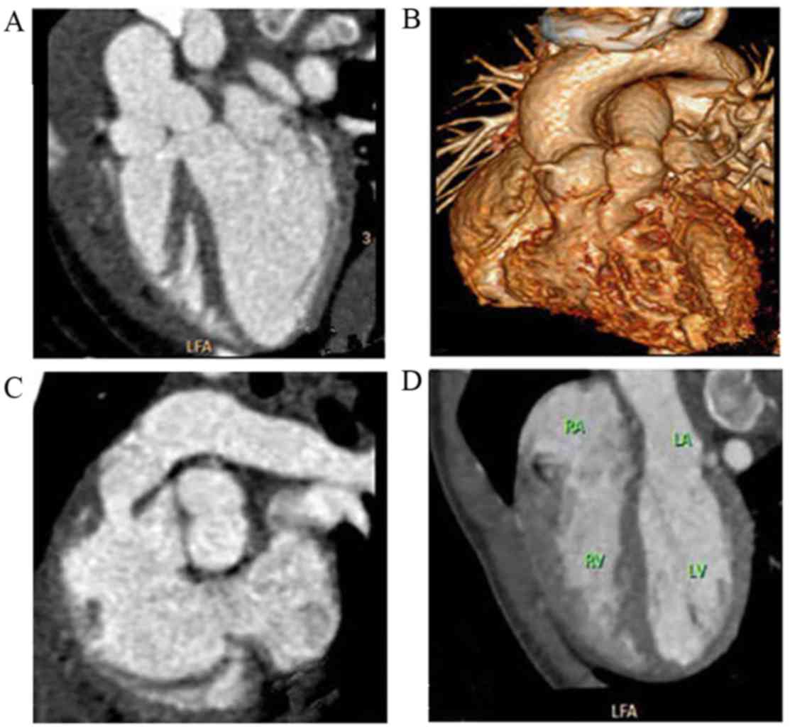
Fallot (Fig. 1), double-outlet right
ventricle (DORV), pulmonary atresia, anomalous pulmonary venous
connection, transposition of the great arteries, and coarctation of
the descending aorta (Table II).
There were no significant differences identified in the image
quality scores between the two groups (Table III).
 | Table I.Characteristics of the children in the
two groups. |
Table I.
Characteristics of the children in the
two groups.
| Group | Age (months) | Height (cm) | Weight (kg) |
|---|
| Group A | 22.2±2.9 | 76.6±1.9 | 9.76±0.49 |
| Group B | 22.9±2.7 | 77.9±1.8 | 10.16±0.56 |
| P-value | 0.851 | 0.617 | 0.459 |
| Table II.Main deformities diagnosed by
dual-source computed tomography in patients with complex congenital
heart disease in the two groups. |
Table II.
Main deformities diagnosed by
dual-source computed tomography in patients with complex congenital
heart disease in the two groups.
| Disease
deformities | Group A (n) | Group B (n) |
|---|
| Tetralogy of
Fallot | 36 | 34 |
| Double outlet right
ventricle | 18 | 13 |
| Coarctation of the
descending aorta | 7 | 8 |
| Pulmonary
atresia | 6 | 3 |
| Patent ductus
arteriosus | 5 | 6 |
| Endocardial cushions
defect | 5 | 1 |
| Anomalous pulmonary
venous connection | 3 | 10 |
| Single atrium | 3 | 2 |
| Transposition of the
great arteries | 1 | 5 |
| Single
ventricle | 1 | 2 |
| Right coronary
artery fistula to left ventricle | 1 | – |
| Right coronary
artery fistula to right ventricle | – | 2 |
| Double-chambered
right ventricle | 1 | – |
| Anomalous origin of
coronary artery from pulmonary artery | 1 | – |
| Anomalous origin of
right pulmonary artery from ascending aorta | – | 1 |
| Aneurysm of the
membranous ventricular septum | – | 1 |
| Interruption of
aortic arch | 1 | – |
| Tricuspid
atresia | – | 1 |
| Table III.Image quality scores of the two
groups. |
Table III.
Image quality scores of the two
groups.
|
| Grade |
|---|
|
|
|
|---|
|
| 5 | 4 | 3 | 2 | 1 | Image quality
scores |
|---|
| Group A | 23 | 56 | 19 | 5 | 0 | 3.94±0.08 |
| Group B | 31 | 48 | 22 | 2 | 0 | 4.05±0.08 |
| P-value | – | – | – | – | – | 0.324 |
In group A, 237 types of malformation were detected,
including: Atrial septum defects (n=25); ventricular septum defects
(n=26); patent ductus arteriosus (n=19); anomalies of the
morphological structure of the pulmonary valve (n=17); anomalous
vena caval connection (n=13); pulmonary stenosis or anomalous
origin of the arterial branch (n=10); anomalous origin of the
coronary artery (n=8); heteroplasia of the aortic arch (n=7);
patent oval foramen (n=5); bronchial stenosis or anomalies of the
morphological structure (n=4); aberrant subclavian artery (n=5);
situs viscera inversus, asplenia or polysplenia (n=3); disorders of
the mitral valve (n=2); cor triatriatum (n=1); unroofed coronary
sinus syndrome (n=1); atrioventricular discordance (n=1); and
mirror image dextrocardia (n=1).
In group B there were 239 types of malformation
detected, including: Atrial septum defects (n=29); ventricular
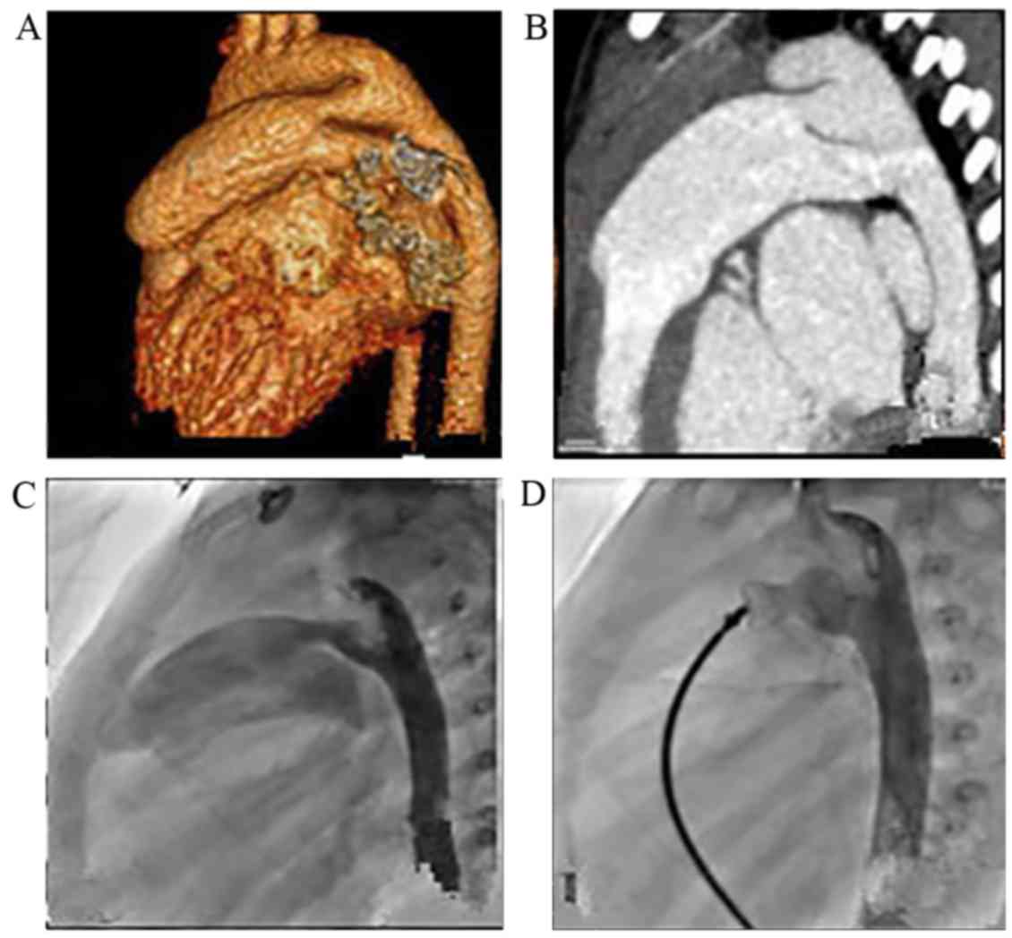
septum defects (n=32); patent ductus arteriosus (n=14) (Fig. 2); anomalies of the morphological
structure of the pulmonary valve (n=17); anomalous vena caval
connection (n=8); pulmonary stenosis (n=7); anomalous origin of the
coronary artery (n=7); heteroplasia of the aortic arch (n=9);
patent oval foramen (n=8); bronchial stenosis or anomalies of the
morphological structure (n=5); aberrant subclavian artery (n=9);
asplenia with situs viscera inversus (n=1); unroofed coronary sinus
syndrome (n=1); and hepatic carcinoma (n=1).
Among all the cases in groups A and B, there were
175 children (424 types of malformation) who underwent therapeutic
surgery or cardiac catheterisation to confirm the diagnosis of CT.
In group A, foramen ovale patent (n=16), atrial septum defects
(n=3) and muscular part of ventricular septum defect (n=1) were not
detected, and two normal ventricular septums were misdiagnosed as
having ventricular septal defects. In group B, the following were
not detected: Foramen ovale patent (n=17), atrial septum defect
(n=4), patent ductus arteriosus (n=2) and muscular part of
ventricular septum defects (n=2). Two normal ventricular septal
were misdiagnosed as being the muscular part of a ventricular
septum defect. In group A, the coincidence rate of CT and clinical
diagnoses was 89.37%, the rate of missed diagnosis was 9.66%, and
the false positive rate was 0.97%. In group B, the coincidence rate
of CT and clinical diagnoses was 88.48%, the rate of missed
diagnosis was 10.60%, and the false positive rate was 0.92%. No
significant differences were identified between the two groups. No
statistical differences in the mean values of ROI and tube current
were identified between the two groups (Table IV).
| Table IV.Mean values of ROI, tube current and
radiation dose in the two groups. |
Table IV.
Mean values of ROI, tube current and
radiation dose in the two groups.
| Group | Mean ROI (Hu) | Tube current
(mAs) | CTDI (mGy) | DLP (mGy.cm) | ED (mSv) |
|---|
| Group A | 476.2±4.96 | 80.65 | 3.24 | 47.53 | 0.93 |
| Group B | 470.9±4.32 | 84.11 | 2.27 | 27.03 | 0.53 |
| P-value | 0.755 | 0.327 | <0.001 | <0.001 | <0.001 |
Discussion
During the important stage of growth and
development, between birth and twelve years of age, children are 10
times more sensitive to radiation than adults (16); therefore, the importance of radiation
protection cannot be overemphasised. At present, the main method
used to reduce radiation dosage is auto tube current modulation and
the restriction of tube voltage (17–19). The
present study responded to the growing concern over the radiation
doses administered to children and applied a novel scanning method.
According to the two gating techniques used, children were equally
classified into groups A and B (20). In group A, retrospective ECG-gating
with an ECG-pulsing technique was applied and the exposure window
of this group was adjusted to 35–75% of the cardiac cycle; whereas
the exposure window of group B was adjusted to 40–70%. The mean ED
of group A was 0.93±0.41 mSv and the radiation dose was reduced by
75%. Therefore, the radiation dose remained inadequately controlled
due to the continuous scanning.
The mean ED of group B was 0.53±0.23 mSv, which is
markedly lower than the standard CT scanning dose by virtue of the
step-and-shoot mode (21). For
patients with situs viscera inversus, the final phase of the scan
protocol was set as a low-dose abdominal scan in order to detect
any structural abnormalities of the abdominal organs and
arteriovenous malformation. Both groups A and B underwent abdominal
scans with Siemens Care Dose technology (70 kV; 20–30 mAs), which
administers a very low radiological dose with a DLP of 4–5 mGy·cm.
DLP is the product of the CTDI vol and X-ray scan length of
subjects, ED is the X-ray dose that causes biological damage to
subjects, the K-value is the conversion coefficient of DLP and ED
[ED (mSv)=K (mSv/[mGy·cm]) × DLP (mGy·cm)] (13), and the conversion coefficient varies
based on the patient's age and body part scanned. In the present
study, according to the AAPM Report 96 (2008) (22), in chest routine scans, the K-values
were as follows: Newborn, 0.039; 1-year-old, 0.026; 5-year-old,
0.018; 10-year-old, 0.013; and adult, 0.014.
The most effective method of reducing the radiation
dosage typically comes at the cost of impairing image quality. In
the present study, the data showed that there was no significant
difference in image quality between the two groups.
By analyzing the surgical findings, one can observe
that defects <0.5 mm were easily misdiagnosed or undetected
since the resolution was >0.5 mm. In the present study, patent
foramen ovale, atrial septum defects and ventricular septum defects
<0.5 mm were misdiagnosed or undetected. This was predominantly
pronounced in cases with patent foramen ovale as this type of
defect typically appears to mimic a flap valve, only opening during
certain conditions when there is increased pressure inside the
chest; therefore, this defect is consistently detected during
exploratory surgery but remains difficult to diagnose via CT
scanning (23). In addition, two
patent ductus arteriosi were left undetected in group B, whose
sizes were <1 mm.
Typically, the three main factors influencing the
radiation dose in patients are CTDI, DLP and ED (24,25). By
comparing these factors between the two groups, significant
differences were detected. Furthermore the image quality of group B
was not significantly different to that of group A, demonstrating
that prospective ECG-gated CT with ECG-pulsing is effective at
reducing the radiation dosage and avoids the impairment of image
quality.
To optimise the scanning phase, previous studies
(20,26) have suggested that the exposure window
should be adjusted to 40% of systole as the majority of children
with CHD exhibit rapid heart rates. As such, cardiac function
cannot be evaluated without a cardiac systole during scanning
(27). In the present study,
scanning was performed during the double phase of diastole and
systole; therefore, the diameter of the defect or stricture could
be measured accurately and cardiac function was quantitatively
analysed (28–31).
Proper usage of contrast medium is a vital procedure
in cardiac angiography and bolus tracking is one of the most common
methods used to visualise vessels clearly. The volume of contrast
is tracked using an ROI at a certain level and when the CT reaches
this level the images are acquired at a rate as fast as the
contrast moves through the vessels (32). Complex CHD, however, altered the
haemodynamics to varying degrees due to the presence of
extracardiac deformities, ventriculoarterial connections,
alterations in ventricular volume and rapid heart rates. In
particular, the DORV, transposition of the great arteries,
pulmonary atresia, tricuspid atresia and other complex deformations
cause large shunting, which seriously affected the image quality
(33).
The bolus injection used in the present study has
limitations. Firstly, it may cause a banding artifact in the
superior vena cava and right atrium when normal saline is not
filling into the right atrium as soon as possible. Secondly, there
is not enough contrast medium in the left heart system, which may
result in faint development, and the poor development of the
descending aorta is one of the factors that may result in small
diameter patent ductus arteriosus or major aorta pulmonary
collateral arteries being left undetected. Thirdly, the
malformation of the atrium and the structure of the tricuspid valve
cannot be accurately identified.
Directly delayed contrast-enhanced scanning was
applied in the present study. Prior to scanning, medical history
was collected and ECG was performed, and a double tube
high-pressure syringe was subsequently applied for biphasic
injection. According to the heart rate of the patients and the type
of malformation, different integral doses, velocities, and scanning
ranges were selected. Following a delay of 20–25 sec, the scan was
processed, which lasted 2–6 sec.
The scanning method used in the present study
guaranteed the excellent demonstration of the cardiovascular
structure without artifacts and with proper usage of the contrast
medium (34), leading to the reduced
risk of renal injury. Therefore, the present study showed that
proper contrast medium combined with low kV scanning enables the
accurate diagnosis of complex CHD. Furthermore, all the patients
successfully completed the examination without any adverse
reactions.
Subsecond scanning and isotropic imaging of
64-multi-slice CT (MSCT) facilitates the clinical application of
coronary angiography. In combination with the high accuracy of
echocardiogram for the assessment of cardiac structure, 64-MSCT was
preliminarily applied in the diagnosis of complex CHD. However, due
to the restriction of time resolution, CHD patients whose heart
rates were >100 bpm required retrospective ECG-gated CT for
scanning so that the scan time was prolonged and the scan range was
limited. As a result, the radiation dosage increases, which is
harmful to children's growth and development (35).
On account of the detector array, 320-MSCT can be
used to cover the heart with a routine scan (36). Patients with slow heart rates are
able to complete the heart examination during one cardiac period,
whereas patients with faster heart rates require multiple cardiac
periods to complete the scan, and the risk of multiple exposures is
increased. For the images of patients whose heart rates exceed 100
bpm, extensive post-processing is required. As previously
described, the mean effective radiation dose of ~1.17 mSv is higher
than the mean radiation dose applied in this study (25,37).
Notably, 320-MSCT allows CT angiography examination to be performed
at high-pitch values of 3.4, and at a scanning rate of up to 43
cm/sec (38). Additionally, the
advantages include an extremely short exposure time, a low
radiation dose, and scanning is unaffected by respiratory rate.
Therefore, fast heart rates and arrhythmia remain challenging
during 320-MSCT (39). Prospective
ECG-gating is an ideal method that is applicable for patients with
rapid heart rates or arrhythmia. Advances in scientific research
concerning radiation doses have shown that, in a chest scan and
angiocardiography, the radiation dose of DSCT was only 24% of the
traditional CT, which is a notable development in overcoming the
restriction of radiation dosage (40).
High time resolution (75 msec) of DSCT overcomes the
limitations of heart rate during CT scanning, and prospective
ECG-gated CT scanning is a more suitable method for complex CHD
patients with tachycardia or tachypnoea. Moreover, the ECG-pulsing
technique is able to greatly reduce the radiation dose without
impairing image quality. Therefore, prospective ECG-gated CT
scanning with an ECG-pulsing technique, low tube voltage and
automatic tube current modulation technique may be a feasible
protocol for reducing radiation dosages.
In conclusion, ECG is the preferred method for the
diagnosis of complex CHD; however, CT scanning can provide
additional useful information that may increase the diagnosis rate
prior to surgery. DSCT has an unbeatable advantage in its scanning
pattern, which optimises the protocol according to the clinical
features of the patient and disease to effectively reduce the
radiation dosage to a level that is reasonable and achievable.
Acknowledgements
This study was supported by Wuhan Research Funded
Projects (grant no. WX13Z04).
References
|
1
|
Kocakap BD Sayin, Sanli C, Cabuk F, Koc M
and Kutsal A: Association of MTHFR A1298C polymorphism with
conotruncal heart disease. Cardiol Young. 25:1326–1331. 2015.
View Article : Google Scholar : PubMed/NCBI
|
|
2
|
Holst KA, Dearani JA, Burkhart HM,
Connolly HM, Warnes CA, Li Z and Schaff HV: Risk factors and early
outcomes of multiple reoperations in adults with congenital heart
disease. Ann Thorac Surg. 92:122–130. 2011. View Article : Google Scholar : PubMed/NCBI
|
|
3
|
Flohr TG, Leng S, Yu L, Aiimendinger T,
Bruder H, Petersilka M, Eusemann CD, Stierstorfer K, Schmidt B and
McCollough CH: Dual-source spiral CT with pitch up to 3.2 and 75 ms
temporal resolution: Image reconstruction and assessment of image
quality. Med Phys. 36:5641–5653. 2009. View Article : Google Scholar
|
|
4
|
Pushparajah K, Miller OI and Simpson JM:
3D echocardiography of the atrial septum: Anatomical features and
landmarks for the echocardiographer. JACC Cardiovasc Imaging.
3:981–984. 2010. View Article : Google Scholar : PubMed/NCBI
|
|
5
|
Song BG, Park SW, Lee SC, Choi JO, Park
SJ, Chang SA, Oh JK and Yang JH: Real-time 3D TEE for
multiperforated interatrial septum. JACC Cardiovasc Imaging.
3:11992010. View Article : Google Scholar : PubMed/NCBI
|
|
6
|
Fratz S, Chung T, Gerald F, Greil GF,
Samyn MM, Taylor AM, Buechel ER, Yoo SJ and Powell AJ: Guidelines
and protocols for cardiovascularmagnetic resonance in children and
adults withcongenital heart disease: SCMR expert consensus group on
congenital heart disease. J Cardiovasc Magn Reson. 15:12013.
View Article : Google Scholar : PubMed/NCBI
|
|
7
|
Al-Mousily F, Shifrin RY, Fricker FJ,
Feranec N, Quinn NS and Chandran A: Use of 320-detector computed
tomographic angiography for infants and young children with
congenital heart disease. Pediatr Cardiol. 32:426–432. 2011.
View Article : Google Scholar : PubMed/NCBI
|
|
8
|
Sun XH, Sun ZG, Li Z, Shi Z, Wang L, Sheng
H, Wu T and Li C: The study of dual source computed tomography
angiography in the evaluation of extracardial anomalies in patients
with congenital heart disease. J Med Imaging. 20:667–671. 2010.
|
|
9
|
Khatri S, Varma SK, Khatri P and Kumar RS:
64-slice multidetector-row computed tomographic angiocardiography
for evaluating congenital heart disease. Pediatr Cardiol.
29:755–762. 2008. View Article : Google Scholar : PubMed/NCBI
|
|
10
|
Hollingsworth CL, Yoshizumi TT, Frush DP,
Chan FP, Toncheva G, Nguyen G, Lowry CR and Hurwitz LM: Pediatric
cardiac-gated CT angiography: Assessment of radiation dose. AJR Am
J Roentgenol. 189:12–18. 2007. View Article : Google Scholar : PubMed/NCBI
|
|
11
|
Liu GR and Li YC: Dual-source definition
flash CT in diagnosis of cardiovascular and cerebrovascular
disease. People's Medical Publishing House; Beijing: 2013
|
|
12
|
Sabarudin A, Sun Z and Yusof AK: Coronary
CT angiography with single-source and dual-source CT: Comparison of
image quality and radiation dose between prospective ECG-triggered
and retrospective ECG-gated protocols. Int J Cardiol. 168:746–753.
2013. View Article : Google Scholar : PubMed/NCBI
|
|
13
|
Thomas KE and Wang B: Age-specific
effective doses for pediatric MSCT examinations at a large
children's hospital using DLP conversion coefficients: A simple
estimation method. Pediatr Radiol. 38:645–656. 2008. View Article : Google Scholar : PubMed/NCBI
|
|
14
|
Heyer CM, Mohr PS, Lemburg SP, Peters SA
and Nicolas V: Image quality and radiation exposure at pulmonary CT
angiography with 100-or 120-kVp protocol: Prospective randomized
study. Radiology. 245:577–583. 2007. View Article : Google Scholar : PubMed/NCBI
|
|
15
|
Pache G, Grohmann J, Bulla S, Arnold R,
Stiller B, Schlensak C, Langer M and Blanke P: Prospective
electrocardiography-triggered CT angiography of the great thoracic
vessels in infants and toddlers with congenital heart disease:
Feasibility and image quality. Eur J Radiol. 80:e440–e445. 2011.
View Article : Google Scholar : PubMed/NCBI
|
|
16
|
Donnelly LF: Reducing radiation dose
associated with pediatric CT by decreasing unnecessary examination.
AJR Am J Roentgenol. 184:655–657. 2005. View Article : Google Scholar : PubMed/NCBI
|
|
17
|
Zhang J, Yang M, Mo XM, Jin JY, Liu B, Li
LL and Teng GJ: Impact of different tube voltage protocols on image
quality and radiation dosage for pediatric 64-slice cardiovascular
CT angiography. Chin J Med Imaging Technol. 28:1213–1217. 2012.
|
|
18
|
Kalra MK, Maher MM, Toth TL, Schmidt B,
Westerman BL, Morgan HT and Saini S: Techniques and applications of
automatic tube current modulation for CT. Radiology. 233:649–657.
2008. View Article : Google Scholar
|
|
19
|
Park CK, Choo KS, Jeon UB, Baik SK, Kim
YW, Kim TU, Kim CW, Jeong YJ, Jeong DW and Lim SJ: Image quality
and radiation dose of 128-slice dual-source CT venography using low
kilovoltage combined with high-pitch scanning and automatic tube
current modulation. Int J Cardiovasc Imaging. 29 Suppl 1:47–51.
2013. View Article : Google Scholar : PubMed/NCBI
|
|
20
|
Nie P, Wang X, Cheng Z, Ji X, Duan Y and
Chen J: Accuracy, image quality and radiation dose comparison of
high-pitch spiral and sequential acquisition on 128-slice
dual-source CT angiography in children with congenital heart
disease. Eur Radiol. 22:2057–2066. 2012. View Article : Google Scholar : PubMed/NCBI
|
|
21
|
Klass O, Walker M, Siebach A, Stuber T,
Feuerlein S, Juchems M and Hoffmann MH: Prospectively gated axial
CT coronary angiography: Comparison of image quality and effective
radiation dose between 64- and 256-slice CT. Eur Radiol.
20:1124–1131. 2010. View Article : Google Scholar : PubMed/NCBI
|
|
22
|
Shrimpton PC, Hillier MC, Lewis MA and
Dunn M: National survey of doses from CT in the UK: 2003. Br J
Radiol. 79:968–980. 2014. View Article : Google Scholar
|
|
23
|
Yang SY: Pediatric cardiology. People's
Medical Publishing House; Beijing: 2009, View Article : Google Scholar
|
|
24
|
Xu J, Zhao H, Wang X, Bai Y, Liu L, Liu Y,
Wei M, Li J and Zheng M: Accuracy, image quality, and radiation
dose of prospectively ECG-triggered high-pitch dual-source CT
angiography in infants and children with complex coarctation of the
aorta. Acad Radiol. 21:1248–1254. 2014. View Article : Google Scholar : PubMed/NCBI
|
|
25
|
Paul JF, Rohnean A, Elfassy E and
Sigal-Cinqualbre A: Radiation dose for thoracic and coronary
step-and-shoot CT using a 128-slice dual-source machine ininfants
and small children with congenital heart disease. Pediatr Radiol.
41:244–249. 2011. View Article : Google Scholar : PubMed/NCBI
|
|
26
|
Ren H, Rong Y, Zhang YQ and Hu X:
Investigation of coronary angiography by dual-source definition
flash CT with multiple scan modes and relative radiation dose. Chin
J Radiol Med Prot. 32:329–330. 2012.
|
|
27
|
Cheng Z, Wang X, Duan Y, Wu L, Wu D, Chao
B, Liu C, Xu Z, Li H and Liang F: Low-dose prospective
ECG-triggering dual-source CT angiography in infants and children
with complex congenital heart disease: First experience. Eur
Radiol. 20:2503–2511. 2010. View Article : Google Scholar : PubMed/NCBI
|
|
28
|
Goo HW and Yang DH: Coronary artery
visibility in free-breathing young children with congenital heart
disease on cardiac 64-slice CT Dual-source ECG-triggered sequential
scan vs. single-source non-ECG-synchronized spiral scan. Pediatr
Radiol. 40:1670–1680. 2010. View Article : Google Scholar : PubMed/NCBI
|
|
29
|
Ben Saad M, Rohnean A, Sigal-Cinqualbre A,
Adler G and Paul JF: Evaluation of image quality and radiation dose
of thoracic and coronary dual-source CT in 110 infants with
congenital heart disease. Pediatr Radiol. 39:668–676. 2009.
View Article : Google Scholar : PubMed/NCBI
|
|
30
|
Oda S, Utsunomiya D, Funama Y, Awai K,
Katahira K, Nakaura T, Yanaga Y, Namimoto T and Yamashita Y: A low
tube voltage technique reduces the radiatiun dose at retrospective
ECG-gated cardiac computed tomography for anatomical and functional
analyses. Acad Radio. 18:991–999. 2011. View Article : Google Scholar
|
|
31
|
Hoe J and Toh KH: First experience with
320-row multidetector CT coronary angiography scanning with
prospective electrocardiogram gating to reduce radiation dose. J
Cardiovasc Comput Tomogr. 3:257–611. 2009. View Article : Google Scholar : PubMed/NCBI
|
|
32
|
Szucs-Farkas Z, Schibler F, Cullmann J,
Torrente JC, Patak MA, Raible S, Hoppe H, Wyttenbach R, Vock P and
Schindera ST: Diagnostic accuracy of pulmonau CT angiograph at low
tube voltage: Intraindividual comparison of a normal-dose protocol
at 120 kVp and a low-dose protocol at 80 kVp using reduced amount
of contrast medium in a simulation study. AJR Am J Roentgenol.
197:W852–W859. 2011. View Article : Google Scholar : PubMed/NCBI
|
|
33
|
Sable C, Foster E, Uzark K, Bjornsen K,
Canobbio MM, Connolly HM, Graham TP, Gurvitz MZ, Kovacs A, Meadows
AK, et al: Best practices in managing transition to adulthood
foradolescents with congenital heart disease: The transition
process and medical and psychosocial issues: A scientific statement
from the American Heart Association. Circulation. 123:1454–1485.
2011. View Article : Google Scholar : PubMed/NCBI
|
|
34
|
Rybicki FJ, Otero HJ, Steigner ML,
Vorobiof G, Nallamshetty L, Mitsouras D, Ersoy H, Mather RT, Judy
PF, Cai T, et al: Initial evaluation of coronary images from
320-detector row computed tomography. Int J Cardiovase Imaging.
24:535–546. 2008. View Article : Google Scholar
|
|
35
|
Han BK, Rigsby CK, Hlavacek A, Leipsic J,
Nicol ED, Siegel MJ, Bardo D, Abbara S, Ghoshhajra B, Lesser JR, et
al: Computed tomography imaging in patients with congenital heart
disease Part I: Rationale and utility. An Expert Consensus Document
of the Society of Cardiovascular Computed Tomography (SCCT):
Endorsed by the Society of Pediatric Radiology (SPR) and the. and
the North American Society of Cardiac Imaging (NASCI). J Cardiovasc
Comput Tomogr. 9:475–492. 2015. View Article : Google Scholar : PubMed/NCBI
|
|
36
|
Du J, Jiang T, Zhou J, Lv B, Zhang ZQ and
Chen W: Application of 320-row detector dynamic volume CT in
infants with complicated congenital heart diseases. Chin J Med
Imaging Technol. 27:1174–1177. 2011.
|
|
37
|
Tognolini A, Arellano CS, Marfori W,
Heidari G, Sayre JW, Krishnam MS and Ruehm SG: Comprehensive
low-dose imaging of carotid and coronary arteries with a
single-injection dualsource CT angiography protocol. Clinical
Radiol. 69:246–253. 2014. View Article : Google Scholar
|
|
38
|
Li J, Huan Y, Zhao HL, Wang Y, Liu Y, Wei
M, Shi M and Zheng M: Comparison of prospective
electrocardiography-gating high-pitch mode and without
electrocardiography-synchronization high-pitch mode acquisition for
the image quality and radiation doses of the aortic using
dual-source CT. Chin J Radiol. 47:301–304. 2013.
|
|
39
|
de Broucker T, Pontana F, Santangelo T,
Faivre JB, Tacelli N, Delannoy-Deken V, Duhamel A, Remy J and
Rémy-Jardin M: Single- and dual-source chest CT protocols: Levels
of radiation dose in routine clinical practice. Diagn Interv
Imaging. 93:852–858. 2012. View Article : Google Scholar : PubMed/NCBI
|
|
40
|
Weustink AC, Neefjes LA, Kyrzopoulos S,
van Straten M, Eu R Neoh, Meijboom WB, van Mieghem CA, Capuano E,
Dijkshoorn ML, Cademartiri F, et al: Impact of heart rate frequency
and variabilitv on radiation exposure, image quality, and
diagnostic performance in dual-source spiral CT coronary
angiography. Radiology. 253:672–680. 2009. View Article : Google Scholar : PubMed/NCBI
|