Introduction
Papillary thyroid cancer (PTC) is the most common
type of well-differentiated thyroid cancer (1). Primary lymph node metastasis (LNM)
occurs in approximately 30–40% of cases, and microscopic
involvement of the lymph nodes may occur in as high as 80–90%
(2). LNM is an important risk
indicator of locoregional recurrence, and regional LNM could be the
second most important independent prognostic indicator (3–5).
Identifying the characteristics of PTCs for invasion and
metastasis, is helpful for clinicians to assesse the risk of
recurrent disease and to provide a personalized therapy for each
patient. Patients with PTC have good prognosis after successfully
personalized management, with the reported 10-year survival rate
recorded at 93% (6). Recent
researches have focused on how to reduce excess therapy and screen
high-risk patients (7,8). Fine needle aspiration (FNA) is an
important diagnostic tool in the evaluation of thyroid nodules. The
2015 ATA guidelines strongly recommend FNA for nodules that appear
suspicious during ultrasound (US) (9). However, FNA cannot provide accurate
evaluation of tumor features for invasion and metastasis. Even
though the expression of genes in PTCs involved in tumor
angiogenesis and metastasis has been investigated recently
(10,11), the main constraint on its clinical
application is the high cost. It is necessary to establish a more
promising diagnostic modality concerning optimization of
cost-effectiveness and perioperative prediction of lymph nodes
clearance.
LNM in PTCs usually occurs initially in the central
cervical lymph node compartment ahead of the lateral compartment,
in spite of their occasional skip to the lateral compartment,
leaving the central compartment tumor-free (12,13). US
has been widely used for preoperative staging in patients with PTC
to determine the necessary extent of surgery. Yet, LNM in the
central neck may not appear abnormal on preoperative US detection,
the ability of preoperative US to identify central neck LNM is
limited (14). Therefore, a growing
number of researchers are advocating prophylactic central lymph
node dissection (CLND) for PTC patients staging cN0 (AJCC/UICC TNM,
7th edition, as proposed by the College of American Pathologists)
based on a preoperative US or upon cervical palpation (15,16).
However, controversy whether prophylactic CLND has major impact on
disease-specific survival still exists (17). Prophylactic CLND might benefit
greatly if PTC patients with high risk of LNM can be screened
preoperatively. Tumor angiogenesis or neovascularization is
accepted as an indicator of tumor growth and metastasis (18–20). A
recent study demonstrated higher blood supply is associated with
regional LNM of PTC (21), while
studies of microvascular density (MVD) have generated a conflicting
hypothesis that recurrent thyroid cancers expressed less
intratumoral microvessels (22).
Given that by providing visualization of the macro- and
microvascularization of the tumor, contrast-enhanced US (CEUS) can
evaluate thyroid tumors qualitatively and quantitatively. Though
some studies have evaluated the value of CEUS in the diagnosis of
thyroid malignant nodules (23,24),
there are few studies available assessing CEUS to help
risk-stratify PTC patients with LNM. In this study we investigated
the clinical implications of CEUS in PTCs, and examined the
relation between enhancements of PTC and the risk of LNM.
Patients and methods
Patients
The present study's design and protocol were
approved by the Ethics Committee of the Third People's Hospital of
Chengdu (Sichuan, China) and all study subjects signed an informed
consent. A retrospective analysis of prospectively collected data
was performed, from May 2014 to November 2016, based on 42 patients
candidate to surgery for suspicious PTCs. Subjects with the
following US-based detection criteria were included: suspicious
PTCs including sonographic findings: solid hypoechoic, long/short
ratio <2, poorly defined margin, and microcalcifications. The
exclusion criteria were: i) a nodule with a fluid content >50%
of volume; ii) the presence of a coarse calcification inside a
nodule; iii) insufficient normal thyroid tissue around the target
nodule; iv) any condition of hyperthyroidism; v) pregnancy; vi)
heart failure and vii) severe pulmonary hypertension (25). The study population that was enrolled
for investigation comprised a total of 42 individuals, including 33
women and 9 men (mean age, 49.1 years; range, 22–73 years; SD ±
13.5 years). The individuals were advised that any surgical
procedure would not be influenced by participation in the present
study.
CEUS
Once enrolled in the study, the individuals
underwent CEUS with conventional US by 2 experienced radiologists 3
days before any surgical intervention, using an IU Elite scanner
(Phillips Medical Systems, Bothell, WA, USA), equipped with an
L12-5 transducer. Initially, the 3 dimensions of the nodules were
measured by US, and the largest diameter was recorded as the nodule
size. CEUS was performed to examine the mass or suspicious region.
Under the US contrast mode the mechanical index (MI) ranged between
0.06 and 0.08, and the focal point was adjusted to the middle or
the lower edge of the thyroid nodules. Double-contrast mode was
used to ensure the probe was in parallel with the long axis of the
thyroid gland as much as possible. If there were multifocal
lesions, the most suspicious lesion was taken for analysis. The
contrast medium was obtained by adding 5 ml of physiological saline
to SonoVue (Bracco SpA, Milan, Italy). Once the largest plane of
the lesion in long axis was selected, a contrast bolus of 2.4 ml
was injected manually via a 20-gauge intravenous cannula placed in
an antecubital vein, and 5 ml of normal saline followed as a flush.
Meanwhile, the timer on the US system was started, and the imaging
plane was kept as stable as possible. Each contrast imaging
acquisition lasted ≥3 min after bolus injection and was digitally
stored. The imaging data obtained from CEUS were continuously
stored. All individuals were monitored for adverse events until 20
min after the procedure. The offline analysis was performed with
dedicated software (Q-LAB®; Philips Medical Systems, The
Netherlands). In each case, based upon CEUS, signal intensity
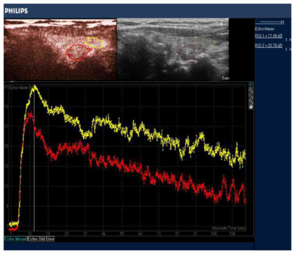
changes of the nodule were measured in decibels (dB). A region of
interest (ROI) was traced out along the edge of the nodule, and
another ROI of similar size on the peripheral thyroid parenchyma
(Fig. 1). Quantitative perfusion
parameters were obtained using the ROI method and time-intensity
curves, including baseline signal intensity (BI, dB), Time of
enhancement beginning (TEB, sec), time to peak (TP, sec), peak
accelerating time (PAT, sec), peak intensity (PI, dB), mean
intensity (MI, dB), intensity increase velocity (IIV, dB/sec), and
intensity decrease velocity (IDV, dB/sec).
US-guided FNA
Based on CEUS and US imaging, preoperative US-guided
FNA was performed to confirm the cytological diagnosis in 42 cases.
The FNA procedure was carried out using a standard 21 gauge needle.
Sampling typically targets the solid component of the lesion. If
there was more than one nodule, a sample was taken from the nodule
with suspicious or atypical US characteristics. The
cytopathological system used in the present study was the Bethesda
system.
Surgery
The surgical approach at our hospital was as
follows: i) Total thyroidectomy (TT) with prophylactic CLND in
cases of PTCs with bilateral nodularity and without suspected
central neck and/or lateral cervical LNM on US; ii) TT with neck
dissection of involved compartments in cases of PTCs with any of
following features: suspected central neck and/or lateral cervical
LNM on US, cervical LNM confirmed by positive frozen section,
extrathyroidal extension, tumor >4 cm; iii) lobectomy +
isthmusectomy (LI) with prophylactic CLND in cases of PTCs without
any of following features: contralateral lesions, suspected central
neck and/or lateral cervical LNM on US, cervical LNM confirmed by
frozen section, extrathyroidal extension.
Demographic data on patient clinical features (sex,
age at diagnosis) as well as histological characteristics of tumors
and neck lymph samples (size of tumors, lesion locations,
extrathyroidal extension) were recorded. All thyroid and lymph node
specimens were evaluated by the same pathologist (with 10 years
working experience).
MVD measurements
Measurements of MVD were performed independently by
the same one pathologist mentioned above, who was blinded to the US
findings. Formalin-fixed paraffin-embedded tissues were segmented
at 3–4 µm thick sections using a semi-automatic microtome.
Consecutive sections were placed on slides pre-coated with egg
albumin for routine hematoxylin and eosin staining and on slides
coated with poly-L-lysine for immunohistochemical staining for CD34
(a mouse antihuman CD34 monoclonal antibody (W-0117; Dako
Corporation, Carpinteria, USA) was used. Completed according to the
method by Weidner et al (26), regions with the highest vessel
density (‘hot spots’) were located by scanning the tissue sections
under ×10-power microscope. Three different fields were randomly
chosen within each hot spot, and individual microvessels were
manually outlined using freehand draw option in Image Pro Plus
software 7.0 (Media Cybernetics, Inc. Singapore, Singapore) and
counted under ×40-power power. Images of these selected fields
along with the marked microvessels were captured. The mean average
scores were tabulated and used for statistical analysis.
Statistical analysis
The SPSS 22.0 software package (IBM, Armonk, NY,
USA) was used for statistical analysis. Values are expressed as the
mean ± SD. Categorical variables were analyzed by Fisher's exact
test. Continuous variables were compared between groups using the
t-test and analysis of variance. Spearman correlation analysis was
used to analyze correlation between quantitative variables and LNM.
Diagnostic performance was estimated by ROC analysis. A P<0.05
was statistically significant.
Results
FNA biopsy yielded positive and non-diagnostic
results in 39 and 3 patients, respectively, of these 3 patients.
Two of the 3 un-diagnostic exhibited extremely weak enhancements on
CEUS, while the other a homogeneous hyper-enhancement. Both the
enhancement patterns were suggestive of benign, which was
ultimately confirmed by surgery. CEUS misdiagnosed 2 PTCs as benign
nodules, and 1 subacute thyroiditis as PTC. The sensitivity,
specificity and accuracy of CEUS for PTCs suspected by conventional
US was 97.4, 75.0, and 95.2%, respectively.
A total of 39 cases were confirmed as PTC by
surgery. They were divided into two groups: LNM+ group
(PTCs with LNM, 27 cases) and LNM− group (PTCs without
LNM, 12 cases). Table I demonstrates
that compared with LNM− group, patients in
LNM+ group had a significantly younger age (P<0.05)
and larger mean tumor size (P<0.05). It was also shown that
patients ≤45 years old (P<0.05), tumors >10 mm in size
(P<0.05) and capsular infiltration (P<0.05) were more
commonly seen in LNM+ group.
 | Table I.Comparison of clinicopathological
characteristics between LMN+ group and
LMN−group. |
Table I.
Comparison of clinicopathological
characteristics between LMN+ group and
LMN−group.
| Variables | LMN+
group (n=27) (%) | LMN−
group (n=12) (%) | P-value |
|---|
| Age (years) |
|
| 0.012a |
|
≤45 | 21 (77.8) | 4 (33.3) |
|
|
>45 | 6 (22.2) | 8 (66.7) |
|
| Mean age
(years) | 41.5±16.1 | 49.3±15.8 | 0.047a |
| Sex |
|
| 0.690 |
|
Male | 7 (25.9) | 2 (16.7) |
|
|
Female | 20 (74.1) | 10 (83.3) |
|
| Tumor size
(mm) |
|
| 0.041a |
|
≤10 | 8 (29.6) | 8 (66.7) |
|
|
>10 | 19 (70.4) | 4 (33.3) |
|
| Mean size (mm) | 28.1±10.7 | 10.5±3.6 | 0.039a |
| Right lobe
lesion | 13 (48.1) | 7 (58.3) | 0.631 |
| Left lobe
lesion | 6 (22.2) | 2 (16.7) |
|
| Multiplicity and/or
bilaterality | 8 (29.7) | 3 (25.0) |
|
| Capsular
infiltration (%) | 17 (62.9) | 3 (25.0) | 0.041a |
| Location of the
malignancy |
|
| 0.720 |
| Upper
third | 9 (33.3) | 5 (41.6) |
|
| Mid and
lower third | 18 (66.7) | 7 (58.4) |
|
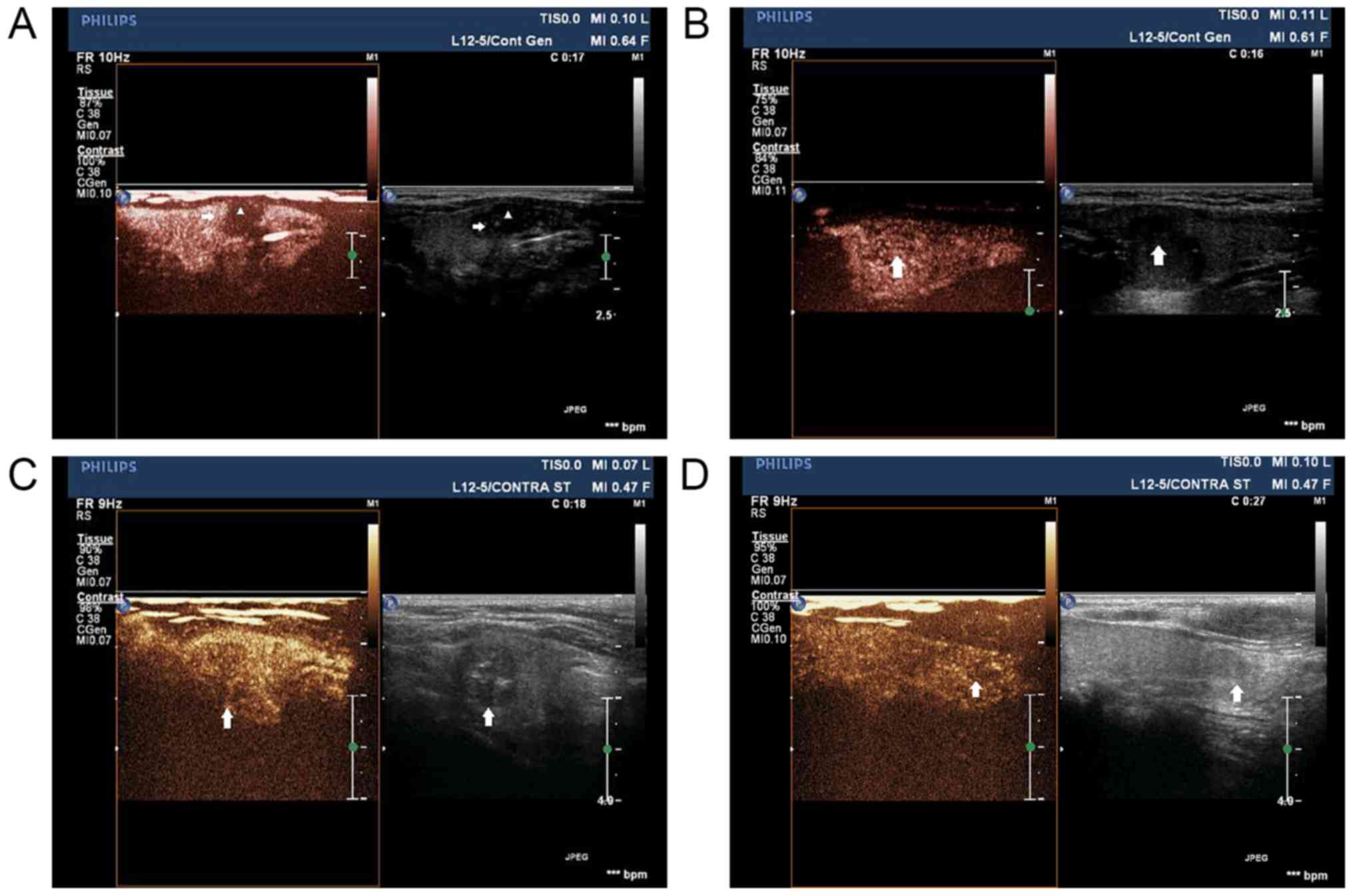
CEUS enhancements in early ascending phases was
classified into the following two new patterns for the first time
(Fig. 2). The first pattern was
early partial hyper-enhancement, presenting either complex
enhancement (characterized by periphery hyperperfused with
relatively central hypoperfused; Fig.
2A) or pervasive heterogeneous hyper-enhancement (Fig. 2B). The sencond pattern, non
hyper-enhancement, including centripetal hypo-enhancement (Fig. 2C) and iso-enhancement (Fig. 2D), both of which were heterogeneous.
It was demostrated that, from late ascending to descending phases,
PTC lesions mainly presented hypo-enhancement, no stastical
differences were found between the two goups. Table II demonstrates that, the ratio of
the first enhancement pattern was significantly higher in
LNM+ group than that in LNM− group
(P<0.01), while the second enhancement pattern was
proportionately lower in LNM− group than that in
LNM+ group (P<0.01). Time-intensity curves analysis
indicated that MI, IIV and mean intensity ratio of
intratumoral/peripheral thyroid parenchyma (MIR) were significantly
higher in LNM+ group than that in LNM− group
(all P<0.05), suggesting higher and faster enhancements were
more common during early ascending period in LNM+ group.
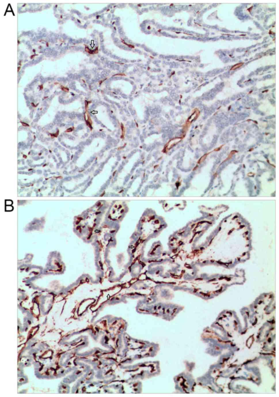
In addition, the measurements of MVD by CD34 expression detection
demonstrated that, both intratumoral MVD and MVD ratio of
intratumoral/peripheral thyroid parenchyma (MVDR) were higher in
LNM+ group than that in LNM− group
(P<0.01) (Fig. 3). As regards the
Spearman correlation analysis, MI (r=0.11, P=0.48), IIV
(r=0.09, P=0.35), MVDR (r=0.57, P<0.05) and MIR
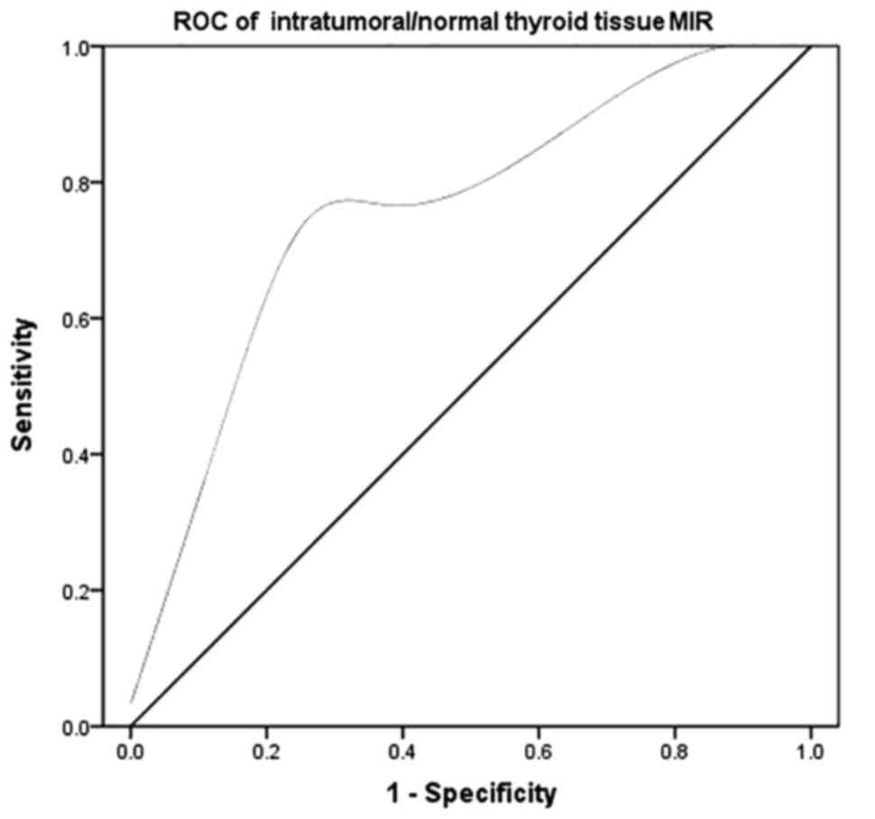
(r=0.41, P<0.05) was positively correlated with LNM. The
ROC analysis (AUC = 0.74, P<0.05) revealed the MIR of 0.86 was
the optimal threshold of LNM in PTCs (Fig. 4).
| Table II.Comparison of CEUS enhancement
characteristics and MVD features between between LMN+
group and LMN− group. |
Table II.
Comparison of CEUS enhancement
characteristics and MVD features between between LMN+
group and LMN− group.
| Characteristic | LMN+
group (n=27) (%) | LMN−
group (n=12) (%) | P-value |
|---|
| Enhancement
type |
| Early partial
hyper-enhancement | 22 (81.5) | 3 (25.0) | 0.001a |
| Complex
enhancement | 17 | 0 |
|
|
Pervasive |
|
Hyper-enhancement | 5 | 3 |
|
| Non
hyper-enhancement | 5 (18.5) | 9 (75.0) |
|
|
Centripetal |
|
Hypo-enhancement | 5 | 8 |
|
|
Iso-enhancement | 0 | 1 |
|
| Mean intensity (MI,
dB) | 7.7±4.9 | 6.4±3.2 | 0.033b |
| Intensity increase
velocity (IIV, dB/sec) | 1.7±0.8 | 1.6±0.9 | 0.019b |
| Intensity decrease
velocity (IDV, dB/sec) | 1.3±0.5 | 1.2±0.3 | 0.221 |
| Mean intensity
ratio of Intratumoral/peripheral thyroid parenchyma (MIR) | 0.89±0.19 | 0.82±0.33 | 0.045b |
| Intratumoral
MVD | 5.5±1.8 | 5.0±1.7 | 0.017b |
| peripheral thyroid
parenchyma MVD | 11.2±1.3 | 11.6±1.1 | 0.095 |
| MVD ratio of
intratumoral/peripheral thyroid parenchyma (MVDR) | 0.42±0.16 | 0.48±0.17 | 0.021b |
Discussion
ATA 2015 recommended that, FNA is the accepted
standard tool for the evaluation of thyroid nodules, as it is
reported to be safe and accurate (9). Definitely, FNA does have limitations,
which include a significant rate of non-diagnostic results ranging
from 0.6 to 43.1% (27,28). In total, 7.1% (3/42) of undiagnosis
by FNA were shown in our study. The three cases undiagnosed by FNA
alone were diagnosed as benign via combined use of FNA with CEUS,
two cases were confirmed for nodular goiter and one for follicular
adenoma by surgery ultimately. Nodules of nodular goiter are prone
to hemorrhage appearing initially nonechogenic on US. When the
blood converts from a liquid state to a solid state, the
nonechogenic turns into solid-like hypoechogenic. When US-guided
FNA targeted the solid-like hypoechogenic lesions, it's difficult
to obtain adequate cellular content. While previous hemorrhage
exhibits extremely low enhancement, no enhancement or local
non-enhancement, CEUS may prove to be useful in distinguishing
solid-like lesions from true solidary nodules. It is well known
that, capsular or vesicular invasion cannot be detected by FNA
cytology, so the accuracy of FNA has been largely compromised for
follicular lesions, namely follicular thyroid carcinoma (FTC) and
follicular adenoma (FA). CEUS could provide information of
microvascular features in a whole profile for a follicular lesion.
It might be one of the possible reasons why the case of follicular
adenoma was diagnosed as benign on preoperative CEUS accurately. On
the other hand, CEUS misdiagnosed 2 cases of PTC with
iso-enhancement as benign nodules, and 1 case of subacute
thyroiditis with hypo-enhancement as PTC. CEUS was highly sensitive
and accurate in diagnosis of PTC in our study. Its low specificity
meant CEUS could not be recommended as a first-line investigation,
but a supplement to increase the preoperative diagnostic accuracy
of FNA.
LNM is common for PTC patients. Although there is
still controversy on the prognosis of LNM, it is universally
accepted that cervical LNM should be removed (29,30).
Central cervical lymph nodes are easily covered by the thyroid
gland, and micro-metastasis of lymph nodes is difficult to detect,
so the ability of preoperative US to identify central neck LNM is
limited (31). Besides, preoperative
US showed a medium sensitivity and a relatively high diagnostic
accuracy in detecting lateral neck LNM (32). If PTC patients at high risk of LNM
identified preoperatively, they may benifit from extended resection
and lymph nodes dissection. For patients with low risk of LNM,
conservative treatment with regular monitoring may probably help
avoid unnecessary prophylactic CLND and reduce the overall
financial costs. Several studies have explored the value of CEUS in
the diagnosis of PTC (23,31), however, the application of CEUS of
PTC lesions for predicting the risk of LNM has not been reported.
The present study compared clinical and pathological
characteristics between PTCs with and without LNM, the results
showed that LNM+ group had a relatively younger mean age
and larger mean tumor size compared to LNM− group.
Besides, LNM+ group had a higher incidence of patients
≤45 years old, tumors >10 mm in size and capsular infiltration.
These findings which were similar to previous studies (33,34),
suggest that age (≤45 years old), larger primary tumor size, and
the presence of capsular infiltration indicate an increased risk of
LNM in PTC.
Studies have shown that, PTC lesions exhibit lower
enhancement than benign thyroid nodules on CEUS. As a result of
thyroid tumor neovascularization, pathological vessels differ
significantly from normal vessels. The lack of muscle and nerve
support, vascular dysfunction and tortuosity may lead to low
enhancement of the contrast agent (35,36). The
present study, for the first time, investigated differences between
PTC patients with and without LNM using qualitative and
quantitative CEUS. In the present study, PTC lesions mainly
presented hypo-enhancement from late ascending to descending
phases, no stastical differences were found between PTCs with and
without LNM. Yet in early ascending period, enhancements were
divided into two patterns (partial hyper-enhancement pattern and
non hyper-enhancement pattern) as mentioned in the Results section.
By using the newly introduced classification, the ratio of the
first pattern in LNM+ group was significantly higher
than that in LNM− group, supporting the hypothesis that
local higher blood supply may be associated with LNM (21). It was revealed that patients in
LNM+ group had a higher and faster enhancement at early
contrast agent ascending period, and had significantly higher MVD
than that in LNM− group. PTCs with LNM have been
reported to be associated with high expression of vascular
proliferative factors, for example vascular growth factor A
(VEGF-A) (35). The lymphatic vessel
density (LVD), which is directly involved with LNM, is most likely
connected to VEGF stimulation (36,37).
Besides, VEGF and MVD have significant relationship in assessing
microvascular angiogenesis in thyroid carcinomas. Given all that,
MVD could be correlated with LNM of PTC (38). Since several studies reported that,
quantification of CEUS can be applied to assess MVD in PTC tissues
(37,38), CEUS can be recognized as a
cost-effective tool for visualizing MVD of PTC. It was investigated
that higher local concentrations of microbubbles was observed in
PTC lesions with LNM than that without LNM, consistent with the
fact that LNM might rely on local rich blood supply. For PTC
patients of this study, patial hyper-enhancement on CEUS was
speculated to be associated with increased risk of LNM. MIR on
quantitative CEUS was positive correlated with LNM in PTC, instead
of other quantitative parameters such as MI, IIV, and IDV. The MIR
of 0.86 was the optimal threshold of LNM in PTCs. Further
researches on a larger population is needed to identify the
threshold accurately and help risk-stratify PTC patients with
LNM.
Acknowledgements
We would like to thank Dr Tongtong Zhang and Dr Yu
Tang for helpful conversation, Mr Geoff Cunliff and Dr Wenli Hou
for helping with language editing.
References
|
1
|
Tian X, Cong M, Zhou W, Zhu J and Liu Q:
Relationship between protein expression of VEGF-C, MMP-2 and lymph
node metastasis in papillary thyroid cancer. J Int Med Res.
36:699–703. 2008. View Article : Google Scholar : PubMed/NCBI
|
|
2
|
Noguchi S, Murakami N, Yamashita H, Toda M
and Kawamoto H: Papillary thyroid carcinoma: Modified radical neck
dissection improves prognosis. Arch Surg. 133:276–280. 1998.
View Article : Google Scholar : PubMed/NCBI
|
|
3
|
American Thyroid Association (ATA)
Guidelines Taskforce on Thyroid Nodules and Differentiated Thyroid
Cancer, . Cooper DS, Doherty GM, Haugen BR, Kloos RT, Lee SL,
Mandel SJ, Mazzaferri EL, McIver B, Pacini F, et al: Revised
American Thyroid Association management guidelines for patients
with thyroid nodules and differentiated thyroid cancer. Thyroid.
19:1167–1214. 2009. View Article : Google Scholar : PubMed/NCBI
|
|
4
|
Gimm O, Rath FW and Dralle H: Pattern of
lymph node metastases in papillary thyroid carcinoma. Br J Surg.
85:252–254. 1998. View Article : Google Scholar : PubMed/NCBI
|
|
5
|
Machens A, Hinze R, Thomusch O and Dralle
H: Pattern of nodal metastasis for primary and reoperative thyroid
cancer. World J Surg. 26:22–28. 2002. View Article : Google Scholar : PubMed/NCBI
|
|
6
|
Hundahl SA, Fleming ID, Fremgen AM and
Menck HR: A national cancer data base report on 53,856 cases of
thyroid carcinoma treated in the U.S., 1985–1995. Cancer.
83:2638–2648. 1998. View Article : Google Scholar : PubMed/NCBI
|
|
7
|
Mercante G, Frasoldati A, Pedroni C,
Formisano D, Renna L, Piana S, Gardini G, Valcavi R and Barbieri V:
Prognostic factors affecting neck lymph node recurrence and distant
metastasis in papillary microcarcinoma of the thyroid. Results of a
study in 445 patients: Thyroid. 19:707–716. 2009.
|
|
8
|
Zhang L, Liu H, Xie Y, Xia Y, Zhang B,
Shan G and Li X: Risk factors and indication for dissection of
right paraesophageal lymph node metastasis in papillary thyroid
carcinoma. Eur J Surg Oncol. 42:81–86. 2016. View Article : Google Scholar : PubMed/NCBI
|
|
9
|
Haugen BR, Alexander EK, Bible KC, Doherty
GM, Mandel SJ, Nikiforov YE, Pacini F, Randolph GW, Sawka AM,
Schlumberger M, et al: 2015 American Thyroid Association management
guidelines for adult patients with thyroid nodules and
differentiated thyroid cancer. Thyroid. 26:1–133. 2016. View Article : Google Scholar : PubMed/NCBI
|
|
10
|
Wang N, Luo HJ, Yin GB, Dong CR, Xu M,
Chen GG and Liu ZM: Overexpression of HIF-2α, TWIST, and CXCR4 is
associated with lymph node metastasis in papillary thyroid
carcinoma. Clin Dev Immunol. 2013:5894232013. View Article : Google Scholar : PubMed/NCBI
|
|
11
|
Tang C, Yang L, Wang N, Li L, Xu M, Chen
GG and Liu ZM: High expression of GPER1, EGFR and CXCR1 is
associated with lymph node metastasis in papillary thyroid
carcinoma. Int J Clin Exp Pathol. 7:3213–3223. 2014.PubMed/NCBI
|
|
12
|
Machens A, Holzhausen HJ and Dralle H:
Skip metastases in thyroid cancer leaping the central lymph node
compartment. Arch Surg. 139:43–45. 2004. View Article : Google Scholar : PubMed/NCBI
|
|
13
|
Roh JL, Kim JM and Park CI: Lateral
cervical lymph node metastases from papillary thyroid carcinoma:
Pattern of nodal metastases and optimal strategy for neck
dissection. Ann Surg Oncol. 15:1177–1182. 2008. View Article : Google Scholar : PubMed/NCBI
|
|
14
|
Kouvaraki MA, Shapiro SE, Fornage BD,
Edeiken-Monro BS, Sherman SI, Vassilopoulou-Sellin R, Lee JE and
Evans DB: Role of preoperative ultrasonography in the surgical
management of patients with thyroid cancer. Surgery. 134:946–955.
2003. View Article : Google Scholar : PubMed/NCBI
|
|
15
|
Caliskan M, Park JH, Jeong JS, Lee CR,
Park SK, Kang SW, Jeong JJ, Chung WY and Park CS: Role of
prophylactic ipsilateral central compartment lymph node dissection
in papillary thyroid microcarcinoma. Endocr J. 59:305–311. 2012.
View Article : Google Scholar : PubMed/NCBI
|
|
16
|
Wada N, Duh QY, Sugino K, Iwasaki H,
Kameyama K, Mimura T, Ito K, Takami H and Takanashi Y: Lymph node
metastasis from 259 papillary thyroid microcarcinomas: Frequency,
pattern of occurrence and recurrence, and optimal strategy for neck
dissection. Ann Surg. 237:399–407. 2003. View Article : Google Scholar : PubMed/NCBI
|
|
17
|
White ML, Gauger PG and Doherty GM:
Central lymph node dissection in differentiated thyroid cancer.
World J Surg. 31:895–904. 2007. View Article : Google Scholar : PubMed/NCBI
|
|
18
|
Eloy C, Santos J, Soares P and
Sobrinho-Simões M: Intratumoural lymph vessel density is related to
presence of lymph node metastases and separates encapsulated from
infiltrative papillary thyroid carcinoma. Virchows Arch.
59:595–605. 2011. View Article : Google Scholar
|
|
19
|
Lee K, Park DJ, Choe G, Kim HH, Kim WH and
Lee HS: Increased intratumoral lymphatic vessel density correlates
with lymph node metastasis in early gastric carcinoma. Ann Surg
Oncol. 17:73–80. 2010. View Article : Google Scholar : PubMed/NCBI
|
|
20
|
Folkman J, Merler E, Abernathy C and
Williams G: Isolation of a tumor factor responsible for
angiogenesis. J Exp Med. 133:275–288. 1971. View Article : Google Scholar : PubMed/NCBI
|
|
21
|
Zhan WW, Zhou P, Zhou JQ, Xu SY and Chen
KM: Differences in sonographic features of papillary thyroid
carcinoma between neck lymph node metastatic and nonmetastatic
groups. J Ultrasound Med. 31:915–920. 2012. View Article : Google Scholar : PubMed/NCBI
|
|
22
|
Hakala T, Sand J, Kellokumpu-Lehtinen PL,
Huhtala H, Leinonen R and Kholová I: Recurrent thyroid cancers have
more peritumoural lymphatic vasculature than nonrecurrent thyroid
cancers. Eur J Clin Invest. 44:825–832. 2014. View Article : Google Scholar : PubMed/NCBI
|
|
23
|
Chen M, Zhang KQ, Xu YF, Zhang SM, Cao Y
and Sun WQ: Shear wave elastography and contrast-enhanced
ultrasonography in the diagnosis of thyroid malignant nodules. Mol
Clin Oncol. 5:724–730. 2016. View Article : Google Scholar : PubMed/NCBI
|
|
24
|
Piscaglia F, Nolsøe C, Dietrich CF,
Cosgrove DO, Gilja OH, Nielsen Bachmann M, Albrecht T, Barozzi L,
Bertolotto M, Catalano O, et al: The EFSUMB guidelines and
recommendations on the clinical practice of contrast enhanced
ultrasound (CEUS): Update 2011 on non-hepatic applications.
Ultraschall med. 33:33–59. 2012. View Article : Google Scholar : PubMed/NCBI
|
|
25
|
Rosário PW, de Faria S, Bicalho L, Alves
MF, Borges MA, Purisch S, Padrão EL, Rezende LL and Barroso AL:
Ultrasonographic differentiation between metastatic and benign
lymph nodes in patients with papillary thyroid carcinoma. J
Ultrasound Med. 24:1385–1389. 2005. View Article : Google Scholar : PubMed/NCBI
|
|
26
|
Weidner N, Semple JP, Welch WR and Folkman
J: Tumor angiogenesis and metastasis-correlation in invasive breast
carcinoma. N Engl J Med. 324:1–8. 1991. View Article : Google Scholar : PubMed/NCBI
|
|
27
|
Ceresini G, Corcione L, Morganti S, Milli
B, Bertone L, Prampolini R, Petrazzoli S, Saccani M, Ceda GP and
Valenti G: Ultrasound-guided fine-needle capillary biopsy of
thyroid nodules, coupled with on-site cytologic review, improves
results. Thyroid. 14:385–389. 2004. View Article : Google Scholar : PubMed/NCBI
|
|
28
|
Tabaqchali MA, Hanson JM, Johnson SJ,
Wadehra V, Lennard TW and Proud G: Thyroid aspiration cytology in
Newcastle: A six year cytology/histology correlation study. Ann R
Coll Surg Engl. 82:149–155. 2000.PubMed/NCBI
|
|
29
|
Hay ID, Thompson GB, Grant CS, Bergstralh
EJ, Dvorak CE, Gorman CA, Maurer MS, McIver B, Mullan BP, Oberg AL,
et al: Papillary thyroid carcinoma managed at the Mayo Clinic
during six decades (1940–1999): Temporal trends in initial therapy
and long-term outcome in 2444 consecutively treated patients. World
J Surg. 26:879–885. 2002. View Article : Google Scholar : PubMed/NCBI
|
|
30
|
Yamashita H, Noguchi S, Murakami N,
Kawamoto H and Watanabe S: Extracapsular invasion of lymph node
metastasis is an indicator of distant metastasis and poor prognosis
in patients with thyroid papillary carcinoma. Cancer. 80:2268–2272.
1997. View Article : Google Scholar : PubMed/NCBI
|
|
31
|
Xiang D, Hong Y, Zhang B, Huang P, Li G,
Wang P and Li Z: Contrast-enhanced ultrasound (CEUS) facilitated US
in detecting lateral neck lymph node metastasis of thyroid cancer
patients: Diagnosis value and enhancement patterns of malignant
lymph nodes. Eur Radiol. 24:2513–2519. 2014. View Article : Google Scholar : PubMed/NCBI
|
|
32
|
Na DK, Choi YJ, Choi SH, Kook SH and Park
HJ: Evaluation of cervical lymph node metastasis in thyroid cancer
patients using real-time CT-navigated ultrasonography: Preliminary
study. Ultrasonography. 34:39–44. 2015. View Article : Google Scholar : PubMed/NCBI
|
|
33
|
Lang BH, Lo CY, Chan WF, Lam KY and Wan
KY: Staging systems for papillary thyroid carcinoma: A review and
comparison. Ann Surg. 245:366–378. 2007. View Article : Google Scholar : PubMed/NCBI
|
|
34
|
Qu N, Zhang L, Ji QH, Chen JY, Zhu YX, Cao
YM and Shen Q: Risk factors for central compartment lymph node
metastasis in papillary thyroid microcarcinoma: A meta-analysis.
World J Surg. 39:2459–2470. 2015. View Article : Google Scholar : PubMed/NCBI
|
|
35
|
Meng XY, Zhang Q, Li Q, Lin S and Li J:
Immunohistochemical levels of cyclo-oxygenase-2, matrix
metalloproteinase-9 and vascular endothelial growth factor in
papillary thyroid carcinoma and their clinicopathological
correlations. J Int Med Res. 42:619–627. 2014. View Article : Google Scholar : PubMed/NCBI
|
|
36
|
Sun XF and Zhang H: Clinicopathological
significance of stromal variables: Angiogenesis, lymphangiogenesis,
inflammatory infiltration, MMP and PINCH in colorectal carcinomas.
Mol Cancer. 5:432006. View Article : Google Scholar : PubMed/NCBI
|
|
37
|
Dufort S, Sancey L, Hurbin A, Foillard S,
Boturyn D, Dumy P and Coll JL: Targeted delivery of a proapoptotic
peptide to tumors in vivo. J Drug Target. 19:582–588. 2011.
View Article : Google Scholar : PubMed/NCBI
|
|
38
|
Yu XM, Lo CY, Lam AK, Leung P and Luk JM:
Serum vascular endothelial growth factor C correlates with lymph
node metastases and high-risk tumor profiles in papillary thyroid
carcinoma. Ann Surg. 247:483–489. 2008. View Article : Google Scholar : PubMed/NCBI
|