Introduction
Atherosclerosis, which results in a significant
number of deaths worldwide annually, is becoming a serious burden
in almost all countries (1,2). As a chronic immune-inflammatory
disease, macrophages in the arterial wall engulf oxidized low
density lipoprotein (ox-LDL) and undergo apoptosis, which
contributes to the formation of foam cells. The assembly of foam
cells develops into atherosclerotic plaque and accelerates
atherosclerosis development (3,4). During
this process, ox-LDL-load macrophage apoptosis and lipid uptake
play the key role. Relieving the ox-LDL-induced cholesterol
metabolism obstacle in macrophages may play a vital role in
alleviating atherosclerosis.
Metformin, a widely used anti-T2D (type 2 diabetes)
drug (5), exerts anti-hyperglycemic
effects by inhibiting gluconeogenesis in the liver, strengthening
glucose uptake in muscles and suppressing glucose absorption from
the small intestine (6,7). Several researchers have reported that
metformin can slow or reverse the progress of the atherosclerotic
plaque in patients with diabetes and impaired glucose tolerance
(IGT) (8). Moreover, metformin also
inhibited lipid accumulation and cholesterol biosynthesis in
macrophages (9,10). However, the detailed effect of
metformin on macrophages warrants further investigation.
In the present study, we further explored the
function of metformin on ox-LDL-induced macrophage apoptosis and
the possible mechanism, including endoplasmic reticulum (ER)
stress, mitochondrial membrane potential depolarization, cytochrome
c (cyto-c) release, and lipid uptake.
Materials and methods
Cell culture
The THP-1 cell line was purchased from American Type
Culture Collection (Manassas, VA, USA) and was maintained in
RPMI-1640 culture medium containing 10% FBS, 100 U/ml penicillin
and 100 µg/ml streptomycin (all from Gibco; Thermo Fisher
Scientific, Inc., Waltham, MA, USA). Cells were passaged
approximately twice per week to maintain logarithmic growth and
were cultured at 37°C and 5% CO2 in a humidified
incubator. Macrophages were obtained as described (11). Briefly, THP1 cells (2×10⁵
cells/ml) were cultured in a medium with 100 nM
phorbol-12-myristate 13-acetate (PMA; Sigma-Aldrich; Merck KGaA,
Darmstadt, Germany) for 3 days. Next, the PMA-containing media was
discarded, and cells were cultured in fresh RPMI-1640 (10% FBS, 100
U/ml penicillin and 100 µg/ml streptomycin) for another 24 h to
obtain macrophages for later experiments.
Western blotting
Protein extraction and immunoblotting were performed
as follows: In brief, cells were lysed, and the supernatant was
collected. Concentrations of proteins was determined by the BCA
assay kit (Beyotime Institute of Biotechnology, Shanghai, China).
An equal amount of proteins (40 µg) were resolved and run on a 10%
polyacrylamide SDS gels and transferred onto
polyvinylidenedifluoride membranes (EMD Millipore, Billerica, MA,
USA). Immunoblotting was performed using relevant antibodies
respectively. Immuno-detection was accomplished using a mouse
anti-rabbit or goat anti-mouse secondary antibody and later
visualized using ECL detection (Pierce; Thermo Fisher Scientific,
Inc.). The GAPDH protein was used as endogenous control. Primary
antibodies used were as follows: Rabbit anti-Bak antibody (1:1,000;
Abcam, Cambridge, MA, USA); rabbit anti-Bax antibody (1:1,000;
Abcam); rabbit; rabbit anti-Bad antibody (1:1,000; Abcam); rabbit
anti-Bcl-2 antibody (1:1,000; Abcam); mouse anti-GAPDH antibody
(1:2,000; Nuoyang, Beijing, China); rabbit anti-cleaved PARP
antibody (1:1,000; Abcam); rabbit anti-glucose-regulated protein 78
(GRP78) antibody (1:1,000; Abcam); rabbit anti-PDI antibody
(1:1,000; Abcam); rabbit anti-PERK antibody (1:1,000; Abcam);
rabbit anti-CHOP antibody (1:1,000; Abcam); rabbit anti-p-eIFα
antibody (1:1,000; Abcam); rabbit anti-CD36 antibody (1:1,000;
Abcam); rabbit anti-SRA antibody (1:1,000; EMD Millipore); rabbit
anti-LOX-1 antibody (1:1,000; Abcam); rabbit anti-β-catenin
antibody (1:1,000; Cell Signaling Technology, Inc., Danvers, MA,
USA); rabbit anti-PPAR-γ antibody (1:1,000; Cell Signaling
Technology, Inc.); rabbit anti-AP-1 antibody (1:1,000; Abcam);
rabbit anti-lamin B antibody (1:2,000; Abcam).
Nuclear protein isolation
Nuclear protein extraction was performed using the
nuclear and cytoplasmic protein extraction kit (Beyotime Institute
of Biotechnology,) according to the manufacturer's directions. In
brief, cells were harvested and resuspended using buffer A on ice,
mixed with buffer B and certrifugated. Cell debris was collected
and later lysed using buffer C for 10 min on ice. After
certrifugated, supernatant was collected as nuclear protein
extraction. Western blots were performed as previous description
with Lamin B used as a loading control for the analysis of nuclear
protein expression, while GAPDH was used as a loading control for
the analysis of total protein expression.
Flow cytometry analysis
The flow cytometry detection was performed using the
FITC Annexin V Apoptosis Detection kit I (BD Biosciences, Franklin
Lakes, NJ, USA) according to the manufacturer's directions.
Briefly, the harvested cells were washed twice with cold PBS and
later resuspended in 1X binding buffer at a concentration of
1×10⁶ cells/ml. Next, 100 µl of solution
(1×10⁵ cells) were transferred to a culture tube, and 5
µl of FITC Annexin V and 5 µl of PI were added. Tubes were gently
vortexed and incubated for 15 min at room temperature in the dark,
and 400 µl of 1X binding buffer was added to each tube. Cells were
analyzed using CellQuest software (BD Biosciences).
Immunofluorescence staining
Immunofluorescence staining was performed as
follows: First, cells were fixed in 4% paraformaldehyde for 30 min
at room temperature. Then, cells were permeabilized with 0.2%
Triton X-100 for 10 min. After blocking in 2% BSA for 30 min, cells
were incubated with primary antibodies against CD36 and SRA
overnight at 4°C. Later, after incubating for 1 h at 37°C with the
appropriate fluorescent-conjugated secondary antibody, cells were
counterstained with 4′,6-diamidino-2-phenylindole (DAPI; Roche
Diagnostics, Basel, Switzerland) and examined with a fluorescence
microscope (Nikon TE300; Nikon Corporation, Tokyo, Japan). All
images were collected with the same settings for software and
hardware.
Mitochondrial membrane potential (MMP)
analysis
Mitochondrial membrane potential was measured by
5,5′,6,6′-Tetrachloro-1,1′,3,3′tetraethylbenzimidazolylcarbocyanine
iodide (JC-1; Sigma-Aldrich; Merck KGaA) staining. Cells were
incubated with 2 µM JC-1 at 37°C for 10 min. After washing three
times with PBS, cells were visualized using a fluorescence
microscope (Axio Observer Z1, CCD camera; Carl Zeiss AG,
Oberkochen, Germany).
Lipid uptake detection
Cells were washed with RPMI-1640 (Gibco; Thermo
Fisher Scientific, Inc.) twice and incubated with dil-ox-LDL (10
µg/ml; Yiyuan Biotech Inc., Guangzhou, China) at 37°C for 30 min in
the dark according to the manufacturer's instruction. Cells were
washed with PBS for total of 15 min in the dark. Cells were
observed and photos were taken using an Olympus fluorescence
microscope (Olympus Corporation, Tokyo, Japan).
Statistical analysis
All the experiments were repeated three times. Data
were presented as the mean ± standard deviation. Statistically
significant differences among three or more groups, statistical
analysis was performed using one-way analysis of variance (ANOVA).
P<0.05 was considered to indicate a statistically significant
difference.
Results
Metformin suppresses ox-LDL
loaded-macrophage apoptosis
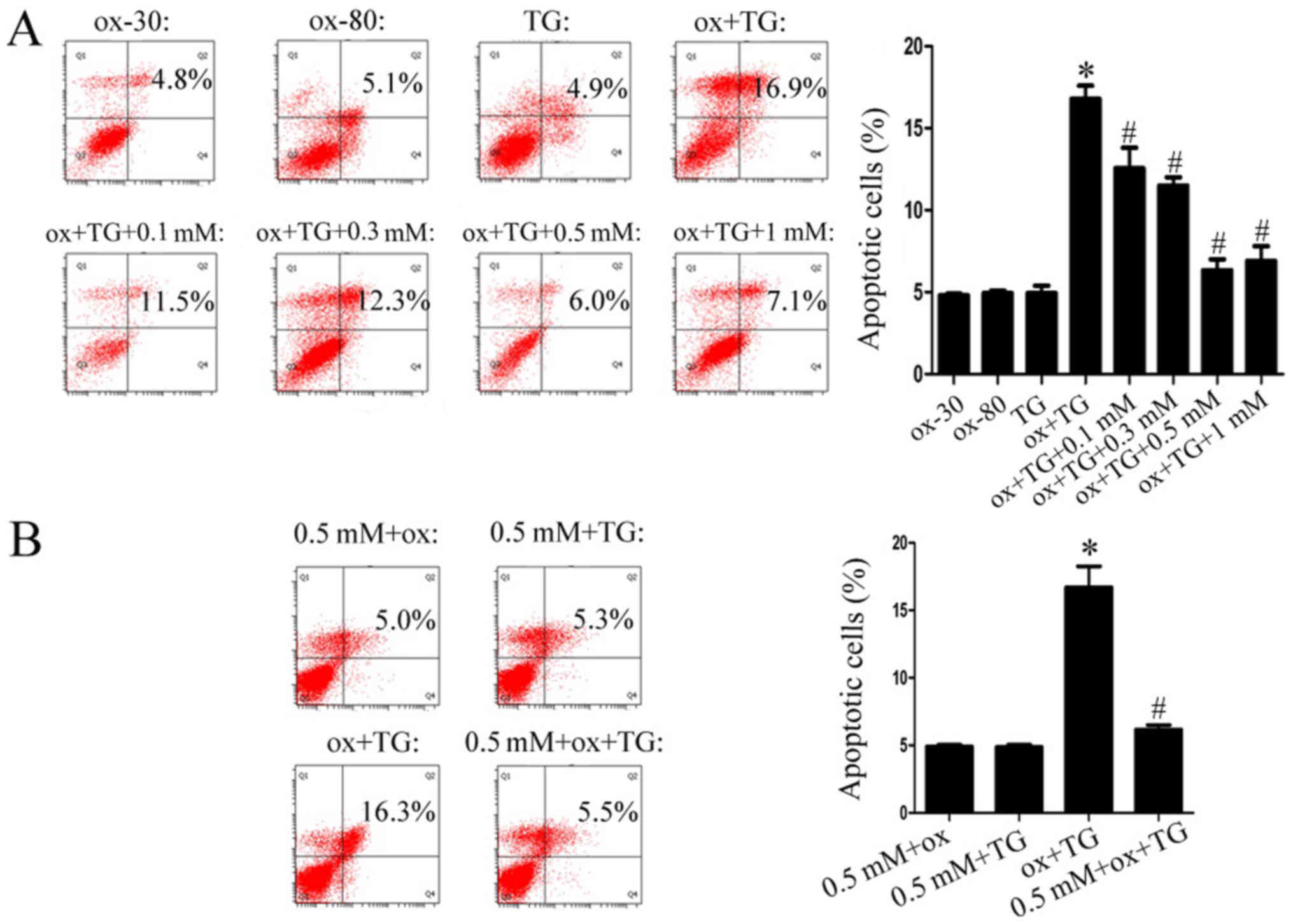
As reported by previous studies (12,13),
ox-LDL did not influence macrophage apoptosis alone. Combined with
the ER stress activator-thapsigargin (TG), ox-LDL prominently
induced macrophage apoptosis (Fig.
1A) (14,15). When macrophages were treated with
metformin excessively with the indicated concentrations (0.1, 0.3,
0.5, 1 mM) for 24 h, the percentage of apoptotic cells was
decreased from 16.9 to 6.0% (Fig.
1A). After pretreatment with metformin for 24 h, macrophages
were later stimulated with ox-LDL and TG. The proportion of
apoptotic cells was also reduced (Fig.
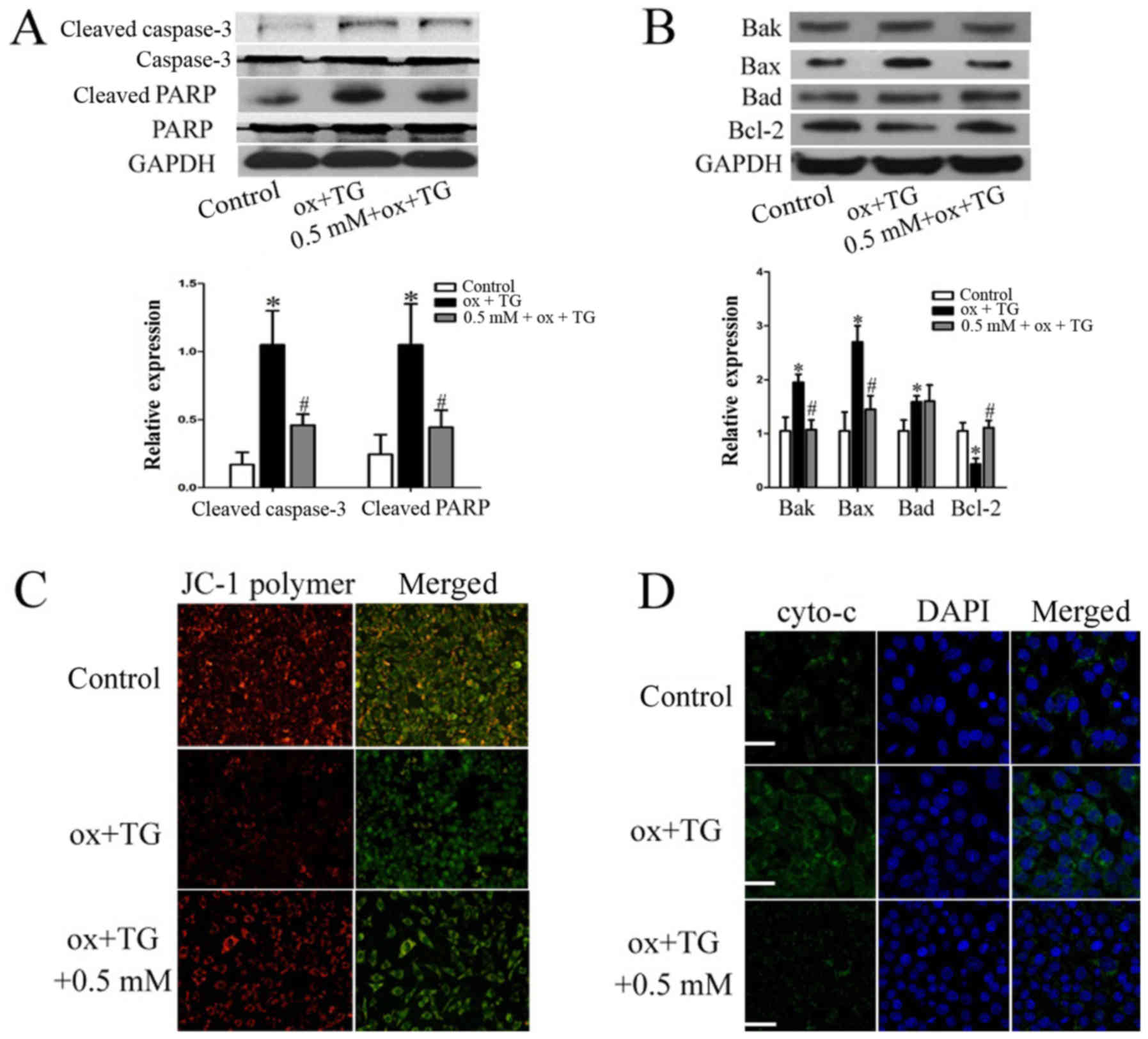
1B). Next, PARP cleavage, triggered by caspase-3 activation
during apoptotic processes, and apoptosis-related Bcl-2 family
proteins were also detected when macrophages were treated with
ox-LDL TG and metformin (0.5 mM). As shown in Fig. 2A, there was no difference in the
expression of full-length caspase-3 and PARP among these groups as
expected. But, PARP cleavage and caspase-3 activation were induced
by ox-LDL and TG while metformin attenuated it. Levels of the
pro-apoptotic proteins Bax and Bak were increased and the
anti-apoptotic proteins Bcl-2 and Bad were decreased when
macrophages were treated with ox-LDL and TG. However, metformin
reversed the upregulation of Bak and Bax and the down-regulation of
Bcl-2 (Fig. 2B). Next, we also
detected mitochondrial membrane potential alteration and cyto-c
distribution. As shown in Fig. 1,
metformin alleviated ox-LDL-induced mitochondrial membrane
depolarization (Fig. 2C) and cyto-c
release (Fig. 2D). Taken together,
these results implicated that metformin suppresses mitochondrial
pathway-mediated macrophage apoptosis triggered by ox-LDL and
TG.
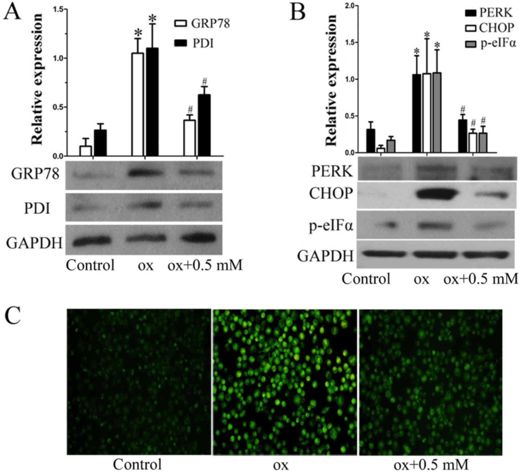
Metformin attenuate sox-LDL-induced ER
stress and ROS generation in macrophages
Endoplasmic reticulum stress triggered by ox-LDL
contributed to ROS generation and subsequent macrophage apoptosis,
which had been reported by Tracie A (16). Whether metformin influenced
ox-LDL-induced ER stress and subsequent ROS generation was unknown.
The upregulation of ER chaperones, including GRP78 and protein
disulfide isomerase (PDI), is a pivotal marker of ER stress. As
shown in Fig. 3A, both GRP78 and PDI
were induced by ox-LDL, while metformin partly canceled this
promotion. In ER stress events, PERK separates from GRP78
deactivated eIF2α through phosphorylation, which terminates protein
synthesis and subsequently activates the downstream proapoptotic
protein CHOP (17,18). This phenomenon was also detected in
ox-LDL-treated macrophages (Fig. 3).
Metformin addition simultaneously reversed the ox-LDL induced
upregulation of PERK, p-eIF2α, and CHOP (Fig. 3B). Moreover, ROS generation was also
suppressed by metformin under ox-LDL treatment (Fig. 3C). In conclusion, metformin can
inhibit ox-LDL-induced ER stress and ROS generation, leading to the
decrease of macrophage apoptosis.
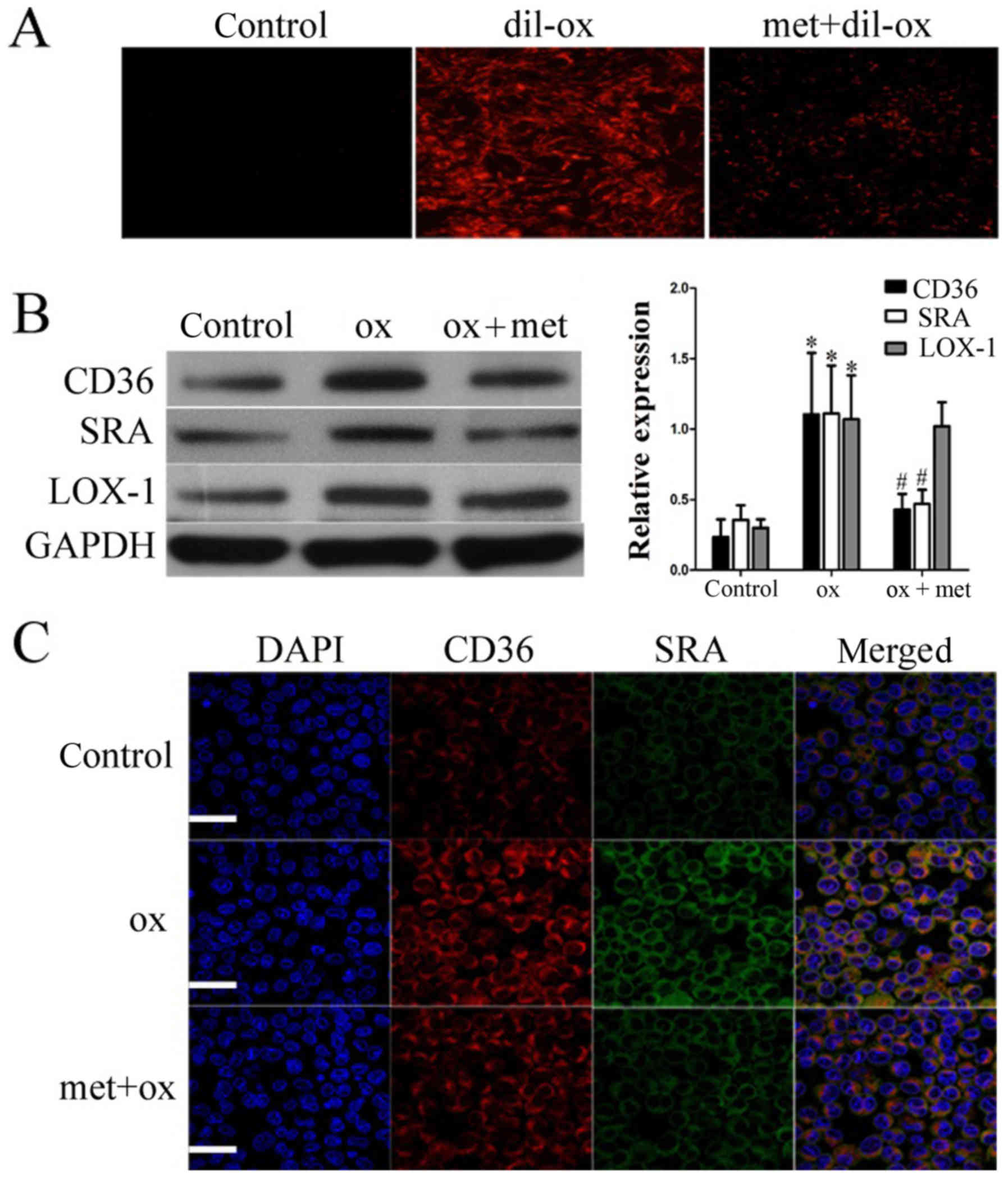
Metformin inhibits lipid uptake
through reducing scavenger receptor expression
Lipid accumulation continually in macrophages
contributes to several biological disorders, including ER stress
and mitochondrial dysfunction, resulting in apoptosis (19,20).
Consistent with Song's study (21),
pretreatment of macrophages with metformin for 24 h suppressed
lipid uptake (Fig. 4A). Scavenger
receptors, including SRA, CD36, and LOX-1, had been reported to
mediate lipid uptake, primarily in macrophages (22,23). As
shown in Fig. 4B and C, metformin
treatment decreased ox-LDL-induced CD36 and SRA expression but had
no significant effect on LOX-1 expression. Above all, metformin
attenuated lipid uptake by inhibiting the expression of CD36 and
SRA.
Metformin inhibits CD36 and SRA
expression through suppressing β-catenin and AP-1 respectively
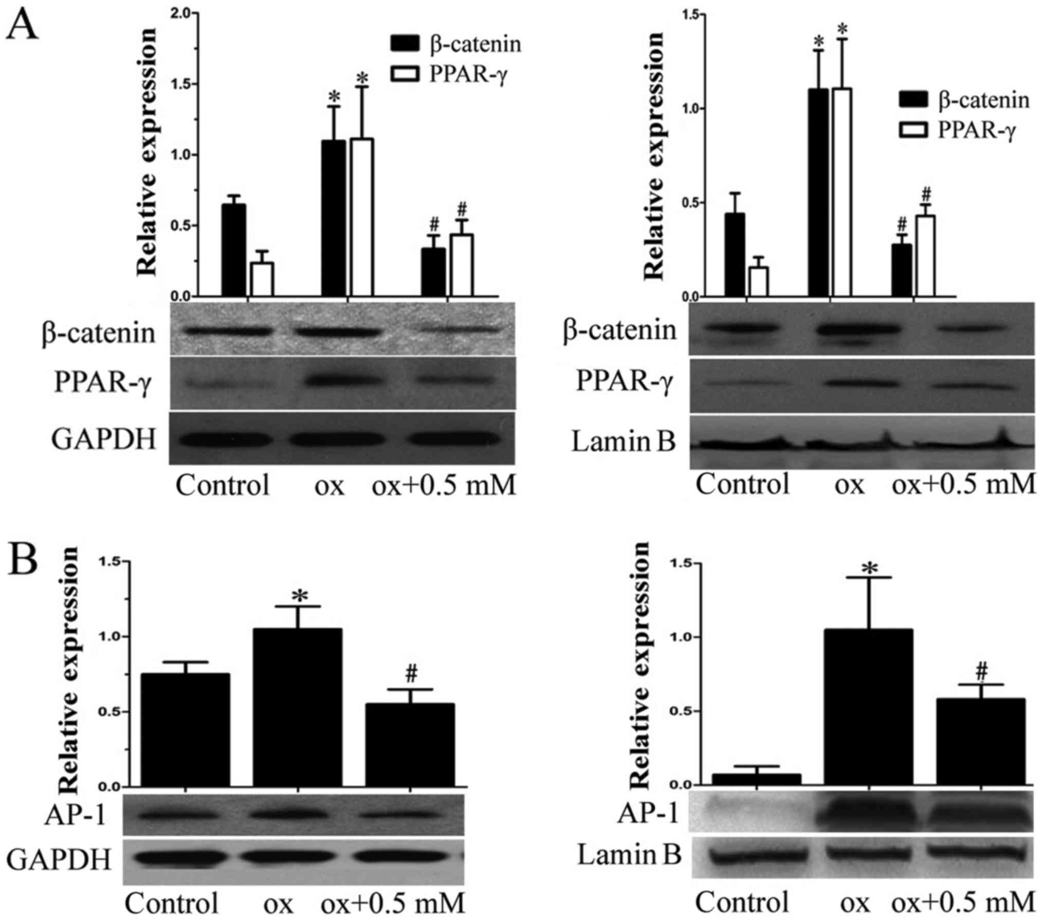
In our previous study, we identified that β-catenin
increased CD36 transcription through cooperating with PPAR-γ in
macrophages (24). Thus, we
investigated whether decreased expression of CD36 under metformin
treatment was caused by the inhibition of β-catenin and PPAR-γ. To
test our hypothesis, we tested the total and nuclear content of
β-catenin and PPAR-γ respectively. As shown in Fig. 5A, metformin indeed suppressed
ox-LDL-induced β-catenin and PPAR-γ upregulation. Simultaneously,
nuclear content of β-catenin and PPAR-γ induced by ox-LDL was also
inhibited by metformin. Previous studies had demonstrated that
activating protein-1 (AP-1) binding element located between −67 and
−50 bp relative to the transcriptional start site was critical for
macrophage SRA activity (25,26).
Therefore, we speculated whether metformin inhibited SRA expression
through controlling AP-1 expression. As shown in Fig. 5B, the increased expression of total
or nuclear AP-1 stimulated by ox-LDL in macrophages was indeed
partly canceled by metformin. Thus, we concluded that metformin
inhibited CD36 and SRA expression through suppressing β-catenin and
PPAR-γ and AP-1 expression, respectively.
Discussion
In recent years, many studies have found that
metformin has many excess effects on blood vessels besides its
hypoglycemic effect (27–29). Clinical researchers have also
identified that metformin was the only hypoglycemic drug which
could lower the risk of cardio-cerebrovascular complications of
T2DM patients (30,31). Regarding the relationship between
metformin and atherosclerosis, studies determined that metformin
could suppress diabetes-related atherosclerosis via inhibiting
Drp1-mediated mitochondrial fission (32). Furthermore, metformin could
ameliorate the pro-inflammatory state in patients with carotid
artery atherosclerosis (33). With
the deepening of the research on metformin, many studies have
identified its anti-inflammation effects. Song et al
observed that metformin reduced lipid accumulation in macrophages
by inhibiting FOXO1-mediated transcription of fatty acid-binding
protein 4 (21). Koren-Gluzer et
al demonstrated that metformin inhibits macrophage cholesterol
biosynthesis rate through attenuated oxidative stress (34). In addition, metformin could inhibit
monocyte-to-macrophage differentiation via AMPK-mediated inhibition
of STAT3 activation, which may play an important role in
atherosclerosis (35). Since
macrophages play a vital role in the progress of atherosclerosis,
we assume that metformin attenuated atherosclerosis may be related
to the inhibition of lipid uptake in macrophages.
In this study, we further investigate the mechanism
underlying the inhibition of lipid uptake in macrophages by
metformin. By detecting scavenger receptor expression, we found
that metformin treatment suppressed CD36 and SRA expression in
macrophages with ox-LDL. Combined with previous works, canonical
WNT pathway and AP-1 were observed to be involved in
metformin-mediated reduction of CD36 and SRA in macrophages with
ox-LDL stimulation (24,26). Since several studies had confirmed
the beneficial effect of metformin on slowing atherosclerosis
development (36,37), our study is the first to demonstrate
the detailed mechanism involved in atherosclerosis alleviation by
metformin, which provides the theoretical basis for applying
metformin for atherosclerosis treatment. In summary, we identified
that metformin could suppress ox-LDL loaded-macrophage apoptosis by
alleviating mitochondrial membrane depolarization, cyto-c release,
ER stress and ROS generation. Additionally, metformin could inhibit
lipid uptake through down-regulation of SRA and CD36 by suppressing
β-catenin and NF-kB.
References
|
1
|
Hansson GK and Hermansson A: The immune
system in atherosclerosis. Nat Immunol. 12:204–212. 2011.
View Article : Google Scholar : PubMed/NCBI
|
|
2
|
Yan H, Wang S, Li Z, Zhao W, Wang Z, Sun
Z, Pan Y and Zhu J: Upregulation of miRNA-155 expression by OxLDL
in dendritic cells involves JAK1/2 kinase and transcription factors
YY1 and MYB. Int J Mol Med. 37:1371–1378. 2016. View Article : Google Scholar : PubMed/NCBI
|
|
3
|
Moore KJ and Tabas I: Macrophages in the
pathogenesis of atherosclerosis. Cell. 145:341–355. 2011.
View Article : Google Scholar : PubMed/NCBI
|
|
4
|
Libby P, Ridker PM and Hansson GK:
Progress and challenges in translating the biology of
atherosclerosis. Nature. 473:317–325. 2011. View Article : Google Scholar : PubMed/NCBI
|
|
5
|
Bailey CJ and Turner RC: Metformin. N Engl
J Med. 334:574–579. 1996. View Article : Google Scholar : PubMed/NCBI
|
|
6
|
Hundal RS, Krssak M, Dufour S, Laurent D,
Lebon V, Chandramouli V, Inzucchi SE, Schumann WC, Petersen KF,
Landau BR and Shulman GI: Mechanism by which metformin reduces
glucose production in type 2 diabetes. Diabetes. 49:2063–2069.
2000. View Article : Google Scholar : PubMed/NCBI
|
|
7
|
Hundal RS and Inzucchi SE: Metformin-New
understandings, new uses. Drugs. 63:1879–1894. 2003. View Article : Google Scholar : PubMed/NCBI
|
|
8
|
Fitch K, Abbara S, Lee H, Stavrou E, Sacks
R, Michel T, Hemphill L, Torriani M and Grinspoon S: Effects of
lifestyle modification and metformin on atherosclerotic indices
among HIV-infected patients with the metabolic syndrome. AIDS.
26:587–597. 2012. View Article : Google Scholar : PubMed/NCBI
|
|
9
|
Song J, Ren PP, Zhang L, Wang XL, Chen L
and Shen YH: Metformin reduces lipid accumulation in macrophages by
inhibiting FOXO1-mediated transcription of fatty acid-binding
protein 4. Biochem Biophys Res Commun. 393:89–94. 2010. View Article : Google Scholar : PubMed/NCBI
|
|
10
|
Koren-Gluzer M, Aviram M and Hayek T:
Metformin inhibits macrophage cholesterol biosynthesis rate:
Possible role for metformin-induced oxidative stress. Biochem
Biophys Res Commun. 439:396–400. 2013. View Article : Google Scholar : PubMed/NCBI
|
|
11
|
Daigneault M, Preston JA, Marriott HM,
Whyte MK and Dockrell DH: The identification of markers of
macrophage differentiation in PMA-stimulated THP-1 cells and
monocyte-derived macrophages. PLoS One. 5:e86682010. View Article : Google Scholar : PubMed/NCBI
|
|
12
|
Wang Q, Ji J, Hao S, Zhang M, Li K and
Qiao T: Iron together with lipid downregulates protein levels of
ceruloplasmin in macrophages associated with rapid foam cell
formation. J Atheroscler Thromb. 23:1201–1211. 2016. View Article : Google Scholar : PubMed/NCBI
|
|
13
|
Yan H, Li Y, Peng X, Huang D, Gui L and
Huang B: Resistance of mitochondrial DNA-depleted cells against
oxidized low-density lipoprotein-induced macrophage pyroptosis. Mol
Med Rep. 13:4393–4399. 2016. View Article : Google Scholar : PubMed/NCBI
|
|
14
|
Goeritzer M, Schlager S, Radovic B,
Madreiter CT, Rainer S, Thomas G, Lord CC, Sacks J, Brown AL, Vujic
N, et al: Deletion of CGI-58 or adipose triglyceride lipase
differently affects macrophage function and atherosclerosis. J
Lipid Res. 55:2562–2575. 2014. View Article : Google Scholar : PubMed/NCBI
|
|
15
|
Aflaki E, Doddapattar P, Radović B,
Povoden S, Kolb D, Vujić N, Wegscheider M, Koefeler H, Hornemann T,
Graier WF, et al: C16 ceramide is crucial for
triacylglycerol-induced apoptosis in macrophages. Cell Death Dis.
3:e2802012. View Article : Google Scholar : PubMed/NCBI
|
|
16
|
Seimon TA, Nadolski MJ, Liao X, Magallon
J, Nguyen M, Feric NT, Koschinsky ML, Harkewicz R, Witztum JL,
Tsimikas S, et al: Atherogenic lipids and lipoproteins trigger
CD36-TLR2-dependent apoptosis in macrophages undergoing endoplasmic
reticulum stress. Cell Metab. 12:467–482. 2010. View Article : Google Scholar : PubMed/NCBI
|
|
17
|
Cnop M, Igoillo-Esteve M, Cunha DA,
Ladrière L and Eizirik DL: An update on lipotoxic endoplasmic
reticulum stress in pancreatic beta-cells. Biochem Soc Trans.
36:909–915. 2008. View Article : Google Scholar : PubMed/NCBI
|
|
18
|
Ron D and Walter P: Signal integration in
the endoplasmic reticulum unfolded protein response. Nat Rev Mol
Cell Biol. 8:519–529. 2007. View
Article : Google Scholar : PubMed/NCBI
|
|
19
|
Yao S, Zong C, Zhang Y, Sang H, Yang M,
Jiao P, Fang Y, Yang N, Song G and Qin S: Activating transcription
factor 6 mediates oxidized LDL-induced cholesterol accumulation and
apoptosis in macrophages by up-regulating CHOP expression. J
Atheroscler Thromb. 20:94–107. 2013. View Article : Google Scholar : PubMed/NCBI
|
|
20
|
Sekiya M, Yamamuro D, Ohshiro T, Honda A,
Takahashi M, Kumagai M, Sakai K, Nagashima S, Tomoda H, Igarashi M,
et al: Absence of Nceh1 augments 25-hydroxycholesterol-induced ER
stress and apoptosis in macrophages. J Lipid Res. 55:2082–2092.
2014. View Article : Google Scholar : PubMed/NCBI
|
|
21
|
Song J, Ren P, Zhang L, Wang XL, Chen L
and Shen YH: Metformin reduces lipid accumulation in macrophages by
inhibiting FOXO1-mediated transcription of fatty acid-binding
protein 4. Biochem Biophys Res Commun. 393:89–94. 2010. View Article : Google Scholar : PubMed/NCBI
|
|
22
|
Murphy JE, Tedbury PR, Homer-Vanniasinkam
S, Walker JH and Ponnambalam S: Biochemistry and cell biology of
mammalian scavenger receptors. Atherosclerosis. 182:1–15. 2005.
View Article : Google Scholar : PubMed/NCBI
|
|
23
|
Yan H, Wang S, Li Z, Sun Z, Zan J, Zhao W,
Pan Y, Wang Z, Wu M and Zhu J: Rspo2 suppresses CD36-mediated
apoptosis in oxidized low density lipoprotein-induced macrophages.
Mol Med Rep. 14:2945–2952. 2016. View Article : Google Scholar : PubMed/NCBI
|
|
24
|
Wang S, Sun Z, Zhang X, Li Z, Wu M, Zhao
W, Wang H, Chen T, Yan H and Zhu J: Wnt1 positively regulates CD36
expression via TCF4 and PPAR-γ in macrophages. Cell Physiol
Biochem. 35:1289–1302. 2015. View Article : Google Scholar : PubMed/NCBI
|
|
25
|
Zhao W, Sun Z, Wang S, Li Z and Zheng L:
Wnt1 participates in inflammation induced by lipopolysaccharide
through upregulating scavenger receptor A and NF-kB. Inflammation.
38:1700–1706. 2015. View Article : Google Scholar : PubMed/NCBI
|
|
26
|
Mietus-Snyder M, Glass CK and Pitas RE:
Transcriptional activation of scavenger receptor expression in
human smooth muscle cells requires AP-1/c-Jun and C/EBPbeta: Both
AP-1 binding and JNK activation are induced by phorbol esters and
oxidative stress. Arterioscler Thromb Vasc Biol. 18:1440–1449.
1998. View Article : Google Scholar : PubMed/NCBI
|
|
27
|
Gillessen S, Gilson C, James N, Adler A,
Sydes MR and Clarke N; STAMPEDE Trial Management Group, :
Repurposing metformin as therapy for prostate cancer within the
STAMPEDE trial platform. Eur Urol. 70:906–908. 2016. View Article : Google Scholar : PubMed/NCBI
|
|
28
|
Cameron AR, Morrison VL, Levin D, Mohan M,
Forteath C, Beall C, McNeilly AD, Balfour DJ, Savinko T, Wong AK,
et al: Anti-inflammatory effects of metformin irrespective of
diabetes status. Circ Res. 119:652–665. 2016. View Article : Google Scholar : PubMed/NCBI
|
|
29
|
Ramakrishna G, Sen B, Trehanpati N and
Sarin SK: Repurposing of metformin in liver injury: The JNK
conundrum. J Hepatol. 64:749–750. 2016. View Article : Google Scholar : PubMed/NCBI
|
|
30
|
Gerstein HC, Miller ME, Ismail-Beigi F,
Largay J, McDonald C, Lochnan HA and Booth GL; ACCORD Study Group,
: Effects of intensive glycaemic control on ischaemic heart
disease: Analysis of data from the randomised, controlled ACCORD
trial. Lancet. 384:1936–1941. 2014. View Article : Google Scholar : PubMed/NCBI
|
|
31
|
ACCORD Study Group, . Nine-year effects of
3.7 years of intensive glycemic control on cardiovascular outcomes.
Diabetes Care. 39:701–708. 2016. View Article : Google Scholar : PubMed/NCBI
|
|
32
|
Wang Q, Zhang M, Torres G, Wu S, Ouyang C,
Xie Z and Zou MH: Metformin suppresses diabetes-accelerated
atherosclerosis via the inhibition of Drp1-mediated mitochondrial
fission. Diabetes. 66:193–205. 2017. View Article : Google Scholar : PubMed/NCBI
|
|
33
|
Xu W, Deng YY, Yang L, Zhao S, Liu J, Zhao
Z, Wang L, Maharjan P, Gao S, Tian Y, et al: Metformin ameliorates
the proinflammatory state in patients with carotid artery
atherosclerosis through sirtuin 1 induction. Transl Res.
166:451–458. 2015. View Article : Google Scholar : PubMed/NCBI
|
|
34
|
Koren-Gluzer M, Aviram M and Hayek T:
Metformin inhibits macrophage cholesterol biosynthesis rate:
Possible role for metformin-induced oxidative stress. Biochem
Biophys Res Commun. 439:396–400. 2013. View Article : Google Scholar : PubMed/NCBI
|
|
35
|
Vasamsetti SB, Karnewar S, Kanugula AK,
Thatipalli AR, Kumar JM and Kotamraju S: Metformin inhibits
monocyte-to-macrophage differentiation via AMPK-mediated inhibition
of STAT3 activation: Potential role in atherosclerosis. Diabetes.
64:2028–2041. 2015. View Article : Google Scholar : PubMed/NCBI
|
|
36
|
Liu X, Mei T, Chen W and Ye S: Comparison
of antidiabetic medications during the treatment ofatherosclerosis
in T2DM patients. Mediators Inflamm. 2017:50327082017. View Article : Google Scholar : PubMed/NCBI
|
|
37
|
Luo F, Guo Y, Ruan GY, Long JK, Zheng XL,
Xia Q, Zhao SP, Peng DQ, Fang ZF and Li XP: Combined use of
metformin and atorvastatin attenuates atherosclerosis in rabbits
fed a high-cholesterol diet. Sci Rep. 7:21692017. View Article : Google Scholar : PubMed/NCBI
|