Introduction
Tongue squamous cell carcinoma (TSCC) is the most
common malignancy in the oral cavity, accounting for about
one-third of all oral cancers (1).
The morbidity and typical mortality rates of TSCC has been
increasing rapidly during the past five years (2,3). Being
an aggressive subtype of oral cancer, TSCC is characterized by high
frequency of local invasion and occult lymph node metastasis.
Consequently, the long-term survival rate continue to be low, with
the overall five-year survival rate at the level of less than 50%
(4). Therefore, identification of
novel biomarkers and therapeutic targets against TSCC is necessary
and urgent to illustrate the clinical behavior of TSCC and to
personalize therapy.
The heterochromatin proteins 1 (HP1) family, known
as chromatin binding proteins, directly bind to the promoter region
of the methylated H3K9 (the methyl groups of histone H3 at lysine
9) to participate in the heterochromatin silence of gene expression
(5). Recent studies indicated that
H3K9 methylation may play key roles in tongue cancer tumorigenesis
(6). HP1γ, encoded by the CBX3, is a
paralog of HP1. By binding methylated H3K9, CBX3/HP1γ can recruit
lots of cofactors which perform various biological functions
including RNA alternative splicing, DNA damage response,
transcription elongation, cell growth and differentiation (7). Moreover, CBX3/HP1γ is also located in
euchromatin areas, which suggest that it may be also associated wih
transcriptional activation (8).
Previous studies proved that CBX3/HP1γ was abnormally expressed in
multiple cancers such as prostate, colon and lung cancer (9–11), and
was always an unfavorable prognostic factor. Thus, it seems that
CBX3/HP1γ plays a potential role as a novel cancer biomarker and a
putative oncogene. However, the expression level of CBX3/HP1γ in
TSCC and its clinical pathologic parameters remains unclear.
Herein, we sought to investigate the expression of
CBX3/HP1γ in primary human TSCC specimens and identify potential
relationship between its expression and clinicopathological
features as well as patients' survival.
Materials and methods
Oncomine database analysis
To obtain the outline of the CBX3/HP1γ expression
pattern, we explored the CBX3 mRNA levels of human TSCC in Oncomine
database (http://www.oncomine.org), which is
publicly available.
Patients and specimen collection
A cohort of 126 patients with primary TSCC treated
in Sun Yat-sen University Cancer Center between January 2009 and
December 2010 were enrolled retrospectively. The enrollment
criteria included: i) primary TSCC without any chemotherapy or
radiotherapy before surgery; ii) patients underwent radical tumor
resection and neck dissection and iii) complete information
available about clinical, pathological and follow-up data. The
formalin-fixed paraffin-embedded samples were collected from the
archives of the hospital, with adjacent non-tumor tissues from the
same patients as a control group. Cancer stage was defined
according to the TNM staging based on American Joint Committee on
Cancer (AJCC), 7th edition (12). In
order to verify the diagnosis, histological grade and stage, all
samples were reviewed by a pathologist. This study was under the
approval of the Clinical Research Ethics Committee of Sun Yat-sen
University Cancer Center. Informed consent was obtained from all
patients.
Immunohistochemistry (IHC)
IHC studies were performed on formalin-fixed,
paraffin-embedded specimens following routine procedure. In brief,
3 µm-thick tissue sections were deparaffnized in xylene and
hydrated in graded alcohol ranging from 100, 95, and 80 to 70%.
Endogenous peroxidase activity was blocked with 3% hydrogen
peroxide for 10 min, and then washed with phosphate-buffered saline
(PBS, pH 7.3). Antigen retrival was completed by boiling in citric
acid buffer (10 mM, pH 6.0) for 15 min. In order to block
nonspecific binding, slides were incubated with 10% normal goat
serum for 15 min at room temperature. These sections were in
further incubation of a rabbit anti-CBX3 antibody (diluted 1:400 in
PBS; Wuhan Sanying Biotechnology, Wuhan, China) overnight at 4°C,
following with incubation of secondary anti-rabbit antibody at a
concentration of 1:100 at 37°C for 30 min. Subsequently, the
sections were washed with PBS, colorized by diaminobenzidine (DAB)
solution and counterstained with haematoxylin. PBS was used to
replace the primary antibody as a negative control, whereas known
IHC-positive CBX3/HP1γ staining slides of colon cancer were used as
positive control.
The immunoreactivity was scored independently by two
pathologists insensible of relevant clinicopathological data
according to the stained intensity of cancer cells and
positively-stained proportion. The estimated fraction of
positive-stained tumor cells was defined as the proportion score
(0, none; 1, 10%; 2, 11–50%; 3, 51–80%; 4, >80%), whereas the
estimated staining intensity was demonstrated as the intensity
score (0, no staining; 1, weak; 2, moderate; 3, strong) with the
aggregate score ranging from 0 to 12, as described elsewhere
(13). Overexpression of CBX3/HP1γ
was defined as an aggregate score of >4. Therefore, the
immunoreactivity of each slide was categorized into three subgroups
based on the final score: negative (0); low expression (1–4); high
expression (6–12).
Statistical analyses
Comparisons of raw IHC outcomes between groups were
performed using the Wilcoxon signed-rank test. The Chi-square test
was used to evaluate the associations between clinicpathological
parameters of patients and CBX3/HP1γ expression. The overall
survival was analyzed using Kaplan-Meier method and compared with
Log-rank test. Univariate and multivariate Cox regression models
were used to estimate individual clinical and pathological
variables with patients' overall survival. P-values <0.05
(two-sided) were considered to indicate a statistically significant
difference. All statistical analyses were performed using Graphpad
Prism 5 (GraphPad Software, Inc., La Jolla, CA, USA) or SPSS v24.0
(IBM Corp., Armonk, NY, USA).
Results
mRNA expressing pattern of CBX3 on
oncomine database
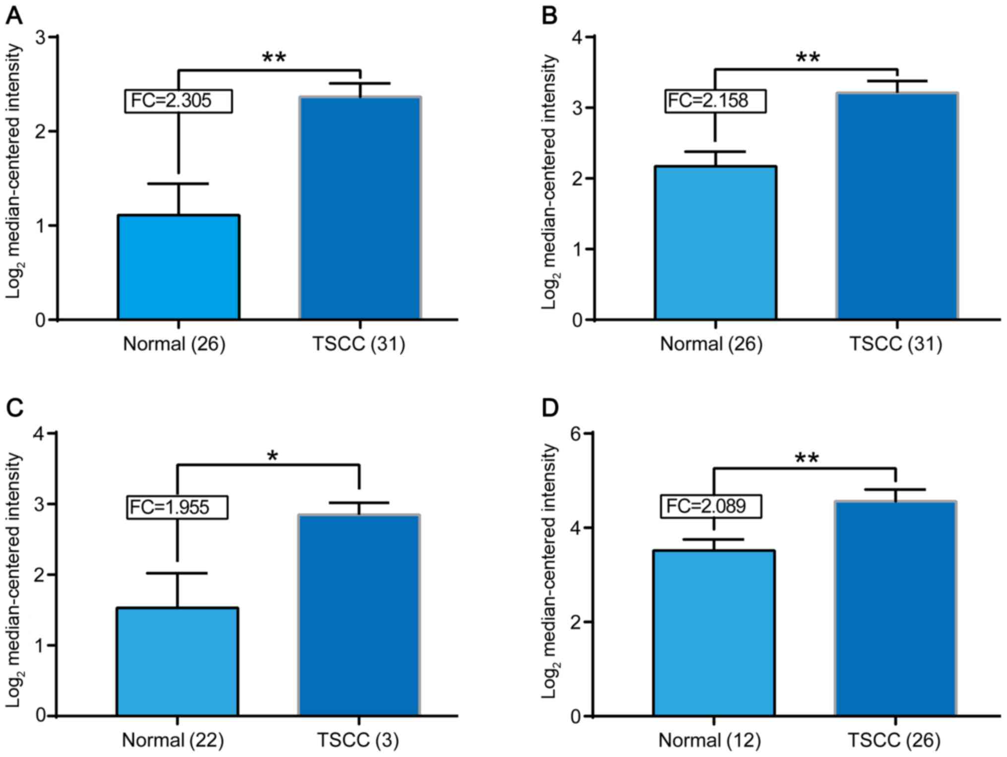
We explored the mRNA expression of CBX3 in 4
datasets on Oncomine Database (Fig.
1). CBX3 mRNA was found significantly elevated in human TSCC
tissues compared with normal tissues in datasets from Oncomine
Database [Talbot et al (14),
Estilo et al (15), Kuriakose
et al (16) and Ye et
al (17)]. We noticed that the
number of cases examined in each group was quite different. For
example, in Kuriakose's group, there are only 3 cancer cases
against 22 normal controls. However, the data from study came to a
consistent result that CBX3 mRNA significantly elevated in human
TSCC tissues.
Overexpression of CBX3/HP1γ at protein
level in TSCC tissues
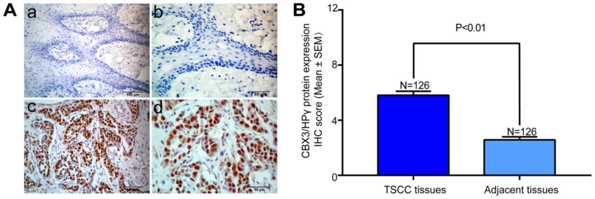
The CBX3/HP1γ protein expression of all TSCC tissues
and adjacent non-tumor tissues were evaluated by IHC. The
representative immunohistochemical staining of CBX3/HP1γ in TSCC
and adjacent tongue mucosa was shown in Fig. 2A. High CBX3/HP1γ expression was
mainly located in the nucleus areas rather than the cytoplasm in
cancer cells, whereas weak or negative staining was observed in
normal tongue epithelial cells. According to our IHC scoring
regime, CBX3/HP1γ protein abundance in these primary TSCC and
adjacent tissues was categorized, respectively. These data showed
that CBX3/HP1γ was significantly overexpressed in TSCC than in the
adjacent tissues (P<0.001; Fig.
2B).
Association between CBX3/HP1γ
expression and clinicopathological characteristics
The association between CBX3/HP1γ expression and
clinical pathological characteristics (age, gender, smoking,
pathological grade, T stage, TNM stage, cervical node status) were
summarized in Table I. Briefly, 82
male and 44 female patients were enrolled with mean age 54.1 years
(25–83 years). The follow-up period ranged from 6.5 to 97.8 months
with average 67.6 months. The results showed that cervical node
metastasis (P=0.010), clinical stage (P=0.025) were related to high
CBX3/HP1γ expression (Table I).
There were no significant correlations between CBX3/HP1γ expression
with patients' age, gender, smoking state, tumor size and
pathological grade (Chi square test).
 | Table I.Associations between CBX3/HP1γ
expression and multiple clinicopathological parameters in primary
TSCC. |
Table I.
Associations between CBX3/HP1γ
expression and multiple clinicopathological parameters in primary
TSCC.
|
|
| CBX3/HP1γ
expression |
|
|---|
|
|
|
|
|
|---|
| Variables | Cases | Lowa | High | χ2
P-value |
|---|
| Age |
|
|
| 0.182 |
|
<60 | 81 | 28 | 53 |
|
| ≥60 | 45 | 21 | 24 |
|
| Gender |
|
|
| 0.273 |
| Male | 80 | 34 | 46 |
|
|
Female | 46 | 15 | 31 |
|
| Smoking |
|
|
| 0.745 |
|
Yes | 64 | 24 | 40 |
|
| No | 62 | 25 | 37 |
|
| Tumor size |
|
|
| 0.843 |
|
T1–2 | 107 | 42 | 65 |
|
|
T3-4 | 19 | 7 | 12 |
|
| Pathological
grade |
|
|
| 0.824 |
| G1 | 99 | 38 | 61 |
|
|
G2-3 | 27 | 11 | 16 |
|
| Cervical node
metastasis |
|
|
| 0.010 |
| N- | 89 | 41 | 48 |
|
| N+ | 37 | 8 | 29 |
|
| Clinical stage |
|
|
| 0.025 |
|
I–II | 80 | 37 | 43 |
|
|
III–IV | 46 | 12 | 34 |
|
Association between CBX3/HP1γ
expression and overall survival of TSCC patients
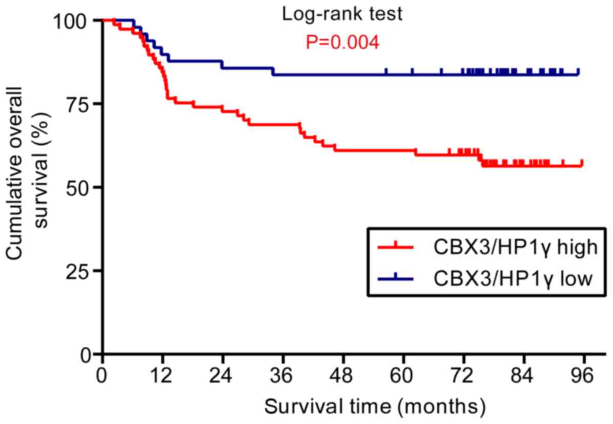
The correlation between CBX3/HP1γ expression and
patients' survival was analyzed to uncover the potential prognostic
value of CBX3/HP1γ expression. Up to the last follow-up, 85 of 126
(67.5%) patients were alive, 41 (32.5%) patients died of cancer
recurrence or metastases. The Kaplan-Meier survival analyses showed
that high CBX3/HP1γ expression indicated unfavorable outcomes. As
shown in Fig. 3, patients with low
CBX3/HP1γ expression had significantly better overall survival than
those with high expression (Log-rank test, P=0.004).
We conducted the univariate and multivariate
survival analyses to further evaluate the clinical significance of
CBX3/HP1γ expression as a prognostic predictor for TSCC patients.
The univariate survival analysis revealed that CBX3/HP1γ expression
status, cervical node metastasis and clinical stage were
significantly associated with overall survival, while other
clinicopathological parameters had not reached the statistical
significance. Furthermore, multivariate survival analysis was also
performed to exclude the confounding factors. Both the CBX3/HP1γ
expression status (HR=2.461; 95% CI 1.128–5.370; P=0.024) and
clinical stage (HR=3.663; 95% CI 1.322–10.146; P=0.013) were
identified as an independent prognostic factor for the overall
survival of patients with TSCC in this Cox regression model
(Table II).
| Table II.Cox proportional hazard model
analysis of variables affecting survival in patients with TSCC. |
Table II.
Cox proportional hazard model
analysis of variables affecting survival in patients with TSCC.
|
| Univariate survival
analysis | Multivariate
survival analysis |
|---|
|
|
|
|
|---|
| Variables | Hazard ratio | 95% CI | P-value | Hazard ratio | 95% CI | P-value |
|---|
| Age (<60 vs.
≥60) | 1.468 | 0.749–2.878 | 0.263 |
|
|
|
| Gender (male vs.
female) | 1.947 | 0.954–3.973 | 0.067 |
|
|
|
| Smoking (yes vs.
no) | 1.427 | 0.767–2.657 | 0.262 |
|
|
|
| Tumor size (T1-2
vs. T3-4) | 1.610 | 0.744–3.488 | 0.227 |
|
|
|
| Pathological grade
(G1 vs. G2-3) | 0.802 | 0.393–1.636 | 0.544 |
|
|
|
| Cervical node
metastasis (N- vs. N+) | 3.431 | 1.855–6.345 | 0.000a | 1.054 | 0.397–2.802 | 0.916 |
| Clinical stage
(I–II vs. III–IV) | 4.257 | 2.249–8.057 | 0.000a | 3.663 | 1.322–10.146 | 0.013a |
| CBX3/HP1γ
expression (lowa vs. high) | 2.967 | 1.370–6.426 | 0.006a | 2.461 | 1.128–5.370 | 0.024a |
Discussion
Being a key hallmark of human cancer, aberrant
epigenetic alternations including histone modification are
significantly involved in multiple stages of oncogenesis (18). HP1, a well-known marker of histone
modification for transcriptionally silenced heterochromatin
(19), is reported to have various
cellular functions including heterochromatin formation, chromatin
remodeling, transcription regulation, DNA damage repairing and
cancer progression (20–22). Previous studies have suggested that
the CBX3/HP1γ, one paralog of HP1, might be an oncogene driving
tumorigenesis and a novel diagnostic and prognostic biomarker in
multiple cancers (11,23–25).
Herein we investigated the expression pattern of CBX3/HP1γ in
primary TSCC and its clinical pathological relevance and prognostic
significance. We found that CBX3/HP1γ was aberrantly overexpressed
in TSCC tissues than in normal tissues and its overexpression was
significantly related to aggressive clinical pathological
parameters and an unfavorable prognosis. This result was consistent
with the trend in other cancers (11,25).
Tumorigenesis of TSCC is a multistep process driven
by numerous abnormal gene alterations, such as oncogenes activation
and tumor suppressor inactivation (26). Particularly, inactivation of tumor
suppressor genes modulated by epigenetics is a major player in this
multistep carcinogenesis and progression of tongue cancer (27,28).
Among them, H3K9/HP1 is one of the key epigenetic silencing
pathways for gene regulation (29).
As an isoforms of HP1, CBX3/HP1γ has been proved to have various
biological functions including RNA alternative splicing, DNA damage
response, transcription elongation, cell growth and differentiation
(7). Notably, highly elevated
expression had been found in non-small cell lung cancer, prostate
cancer and colorectal cancer (9,11,25) etc.
In line with these previous finding, our study showed that
CBX3/HP1γ was significantly overexpressed in human TSCC, thus
indicating that the CBX3/HP1γ might be fostering tumorigenesis in
multiple tissues including tongue. To the best of our knowledge,
this might be the first study to uncover the abnormal expression
pattern of CBX3/HP1γ in TSCC. However, the number of patients
enrolled in this study was limited. Therefore, more cases from
different institutions are needed to definitively confirm the
expression pattern of CBX3/HP1γ as well as its diagnostic value in
TSCC.
There is increasing evidence corroborating the
notion that elevated expression of CBX3/HP1γ is significantly
associated with aggressiveness in various types of cancers
(13). For example, elevated
CBX3/HP1γ is significantly associated with lymph node metastasis
and clinical stages in non-small cell lung cancer or with high
recurrence rate in prostate cancer (9,11).
Similarly, our data showed that overexpression of CBX3/HP1γ was
significantly associated with cervical lymph nodes metastasis and
clinical stages in TSCC. Moreover, previous studies have
demonstrated that CBX3/HP1γ can cooperate with MMP3 (matrix
metalloproteinase 3), a well-known secretory endopeptidase that
degrades extracellular matrices, in transcriptional regulation by
serving as a nuclear MMP3-associated protein (NuMAP) (30). Taken together, we propose that
CBX3/HP1γ might be an efficient biomarker for cervical node
metastasis in TSCC. It will be interesting to explore the
expression level of CBX3/HP1γ in the metastatic lesions and its
clinical significance in further studies.
During the past decades the long term survival rate
of TSCC remains disappointing despite the progress in diagnosis and
treatment, suggesting that an accurate prognostic marker is highly
desired and beneficial in the clinical practice (31). Previous researches showed that
CBX3/HP1γ expression was associated with patients' survival and was
identified as an independent prognostic predictor for patients with
colorectal cancer, non-small cell lung cancer (11,25). For
example, Slezak J et al (9)
found that high expression of CBX3/HP1γ positively correlated with
overexpression of Ki67 and predicted unfavorable prognosis in
patients with prostate cancer. In agreement with these findings,
our results indicated that patients with low CBX3/HP1γ expression
had significantly longer survival comparing to those with high
CBX3/HP1γ. Furthermore, univariate analysis identified CBX3/HP1γ
expression as an independent prognostic factor affecting survival
of patients with TSCC. Thus, the expression status of CBX3/HP1γ
might offer valuable information for predicting patients' prognosis
and effective follow-up management.
Increasing evidence had uncovered that CBX3/HP1γ was
critically involved in tumorigenesis by promoting cell
proliferation via regulating the expression of P21 or cyclin
dependent kinases 6 (CDK6) during cell cycle (7,25). In
addition, CBX3/HP1γ was demonstrated to have novel function in the
epigenetic regulation of both cell differentiation and cancer
development in various types of cancers (10). In particular, Michael Su et al
found that reducing the levels of Cbx3/HP1γ could enhance
tumor-killing capacity on CD8+ T cells (32). Taken these findings together, we
suppose that Cbx3/HP1γ serve as not only a novel cancer biomarker
of high value in diagnosis and prognosis, but also an effective
target for gene therapy against various human cancers. Therefore,
further researches are solely needed to uncover the roles and
mechanisms of Cbx3/HP1γ during tongue tumorigenesis.
In conclusion, Cbx3/HP1γ is aberrantly overexpressed
in TSCC tissues. CBX3/HP1γ expression is an independent prognostic
factor of patients with TSCC, suggesting that Cbx3/HP1γ may be a
candidate prognostic marker for TSCC patients. More studies are
warranted to gain the insight into the mechanism of CBX3/HP1γ
playing during the tumorigenesis and progression in TSCC.
Acknowledgements
The authors would like to thank Professor WuGuo Deng
and Mr. DingBo Shi of The State Key Laboratory of Oncology in South
China (Guangzhou, China) for their help during the experiment.
Funding
No funding was received.
Availability of data and materials
All data generated or analyzed during this study are
included in this published article.
Authors' contributions
AY designed the study. XS collected the patients'
samples and performed the statistical analysis. HZ and XF performed
the experiments and were major contributors in writing the
manuscript. All authors read and approved the final manuscript.
Ethics approval and consent to
participate
The experimental protocol was established, according
to the ethical guidelines of the Helsinki Declaration and was
approved by the Human Ethics Committee of Sun Yan-sen University
Cancer Centre. Written informed consent was obtained from
individual or guardian participants.
Consent for publication
Not applicable.
Competing interests
The authors declare that they have no competing
interests.
References
|
1
|
Siegel RL, Miller KD and Jemal A: Cancer
statistics, 2017. CA Cancer J Clin. 67:7–30. 2017. View Article : Google Scholar : PubMed/NCBI
|
|
2
|
Chi AC, Day TA and Neville BW: Oral cavity
and oropharyngeal squamous cell carcinoma-an update. CA Cancer J
Clin. 65:401–421. 2015. View Article : Google Scholar : PubMed/NCBI
|
|
3
|
Duz MB, Karatas OF, Guzel E, Turgut NF,
Yilmaz M, Creighton CJ and Ozen M: Identification of miR-139-5p as
a saliva biomarker for tongue squamous cell carcinoma: A pilot
study. Cell Oncol (Dordr). 39:187–193. 2016. View Article : Google Scholar : PubMed/NCBI
|
|
4
|
Li Z, Wang Y, Qiu J, Li Q, Yuan C, Zhang
W, Wang D, Ye J, Jiang H, Yang J and Cheng J: The polycomb group
protein EZH2 is a novel therapeutic target in tongue cancer.
Oncotarget. 4:2532–2549. 2013. View Article : Google Scholar : PubMed/NCBI
|
|
5
|
Canzio D, Larson A and Narlikar GJ:
Mechanisms of functional promiscuity by HP1 proteins. Trends Cell
Biol. 24:377–386. 2014. View Article : Google Scholar : PubMed/NCBI
|
|
6
|
Yuan C, Li Z, Qi B, Zhang W, Cheng J and
Wang Y: High expression of the histone demethylase LSD1 associates
with cancer cell proliferation and unfavorable prognosis in tongue
cancer. J Oral Pathol Med. 44:159–165. 2015. View Article : Google Scholar : PubMed/NCBI
|
|
7
|
Fan Y, Li H, Liang X and Xiang Z: CBX3
promotes colon cancer cell proliferation by CDK6 kinase-independent
function during cell cycle. Oncotarget. 8:19934–19946.
2017.PubMed/NCBI
|
|
8
|
Minc E, Courvalin JC and Buendia B:
HP1gamma associates with euchromatin and heterochromatin in
mammalian nuclei and chromosomes. Cytogenet Cell Genet. 90:279–284.
2000. View Article : Google Scholar : PubMed/NCBI
|
|
9
|
Slezak J, Truong M, Huang W and Jarrard D:
HP1γ expression is elevated in prostate cancer and is superior to
Gleason score as a predictor of biochemical recurrence after
radical prostatectomy. BMC Cancer. 13:1482013. View Article : Google Scholar : PubMed/NCBI
|
|
10
|
Takanashi M, Oikawa K, Fujita K, Kudo M,
Kinoshita M and Kuroda M: Heterochromatin protein 1gamma
epigenetically regulates cell differentiation and exhibits
potential as a therapeutic target for various types of cance. Am J
Pathol. 174:309–316. 2009. View Article : Google Scholar : PubMed/NCBI
|
|
11
|
Zhou J, Bi H, Zhan P, Chang C, Xu C, Huang
X, Yu L, Yao X and Yan J: Overexpression of HP1γ is associated with
poor prognosis in non-small cell lung cancer cell through promoting
cell survival. Tumour Biol. 35:9777–9785. 2014. View Article : Google Scholar : PubMed/NCBI
|
|
12
|
Edge SB and Compton CC: The American joint
committee on cancer: The 7th edition of the AJCC cancer staging
manual and the future of TNM. Ann Surg Oncol. 17:1471–1474. 2010.
View Article : Google Scholar : PubMed/NCBI
|
|
13
|
Zheng X, Jia B, Lin X, Han J, Qiu X, Chu
H, Sun X, Hu W, Pan J, Chen J and Zhao J: FRMD4A: A potential
therapeutic target for the treatment of tongue squamous cell
carcinoma. Int J Mol Med. 38:1443–1449. 2016. View Article : Google Scholar : PubMed/NCBI
|
|
14
|
Talbot SG, Estilo C, Maghami E, Sarkaria
IS, Pham DK, O-charoenrat P, Socci ND, Ngai I, Carlson D, Ghossein
R, et al: Gene expression profiling allows distinction between
primary and metastatic squamous cell carcinomas in the lung. Cancer
Res. 65:3063–3071. 2005. View Article : Google Scholar : PubMed/NCBI
|
|
15
|
Estilo CL, O-charoenrat P, Talbot S, Socci
ND, Carlson DL, Ghossein R, Williams T, Yonekawa Y, Ramanathan Y,
Boyle JO, et al: Oral tongue cancer gene expression profiling:
Identification of novel potential prognosticators by
oligonucleotide microarray analysis. BMC Cancer. 9:112009.
View Article : Google Scholar : PubMed/NCBI
|
|
16
|
Kuriakose MA, Chen WT, He ZM, Sikora AG,
Zhang P, Zhang ZY, Qiu WL, Hsu DF, McMunn-Coffran C, Brown SM, et
al: Selection and validation of differentially expressed genes in
head and neck cancer. Cell Mol Life Sci. 61:1372–1383. 2004.
View Article : Google Scholar : PubMed/NCBI
|
|
17
|
Ye H, Yu T, Temam S, Ziober BL, Wang J,
Schwartz JL, Mao L, Wong DT and Zhou X: Transcriptomic dissection
of tongue squamous cell carcinoma. BMC Genomics. 9:692008.
View Article : Google Scholar : PubMed/NCBI
|
|
18
|
Suvà ML, Riggi N and Bernstein BE:
Epigenetic reprogramming in cancer. Science. 339:1567–1570. 2013.
View Article : Google Scholar : PubMed/NCBI
|
|
19
|
Zeng W, Ball AR Jr and Yokomori K: HP1:
Heterochromatin binding proteins working the genome. Epigenetics.
5:287–292. 2010. View Article : Google Scholar : PubMed/NCBI
|
|
20
|
Maison C and Almouzni G: HP1 and the
dynamics of heterochromatin maintenance. Nat Rev Mol Cell Biol.
5:296–304. 2004. View
Article : Google Scholar : PubMed/NCBI
|
|
21
|
Bártová E, Malyšková B, Komůrková D,
Legartová S, Suchánková J, Krejčí J and Kozubek S: Function of
heterochromatin protein 1 during DNA repair. Protoplasma.
254:1233–1240. 2017. View Article : Google Scholar : PubMed/NCBI
|
|
22
|
Dialynas GK, Vitalini MW and Wallrath LL:
Linking heterochromatin protein 1 (HP1) to cancer progression.
Mutat Res. 647:13–20. 2008. View Article : Google Scholar : PubMed/NCBI
|
|
23
|
Saini V, Hose CD, Monks A, Nagashima K,
Han B, Newton DL, Millione A, Shah J, Hollingshead MG, Hite KM, et
al: Identification of CBX3 and ABCA5 as putative biomarkers for
tumor stem cells in osteosarcoma. PLoS One. 7:e414012012.
View Article : Google Scholar : PubMed/NCBI
|
|
24
|
Lomberk G, Bensi D, Christensen T,
Salisbury J and Urrutia R: HP1 γ has a significant impact on cell
cycle progression through the regulation of G2/M, centrosome
biology and, chromosomal stability in pancreatic cancer cells.
Pancreas. 35:4122007. View Article : Google Scholar
|
|
25
|
Liu M, Huang FF, Zhang D, Ju J, Wu XB,
Wang Y, Wang Y, Wu Y, Nie M, Li Z, et al: Heterochromatin protein
HP1γ promotes colorectal cancer progression and is regulated by
miR-30a. Cancer Res. 75:4593–4604. 2015. View Article : Google Scholar : PubMed/NCBI
|
|
26
|
Scully C and Bagan J: Oral squamous cell
carcinoma overview. Oral Oncol. 45:301–308. 2009. View Article : Google Scholar : PubMed/NCBI
|
|
27
|
González-Ramírez I, Soto-Reyes E,
Sánchez-Pérez Y, Herrera LA and García-Cuellar C: Histones and long
non-coding RNAs: The new insights of epigenetic deregulation
involved in oral cancer. Oral Oncol. 50:691–695. 2014. View Article : Google Scholar : PubMed/NCBI
|
|
28
|
Mascolo M, Siano M, Ilardi G, Russo D,
Merolla F, De Rosa G and Staibano S: Epigenetic disregulation in
oral cancer. Int J Mol Sci. 13:2331–2353. 2012. View Article : Google Scholar : PubMed/NCBI
|
|
29
|
Nestorov P, Tardat M and Peters AH:
H3K9/HP1 and Polycomb: two key epigenetic silencing pathways for
gene regulation and embryo development. Curr Top Dev Biol.
104:243–291. 2013. View Article : Google Scholar : PubMed/NCBI
|
|
30
|
Eguchi T, Calderwood SK, Takigawa M,
Kubota S and Kozaki KI: Intracellular MMP3 promotes HSP gene
expression in collaboration with chromobox proteins. J Cell
Biochem. 118:43–51. 2017. View Article : Google Scholar : PubMed/NCBI
|
|
31
|
Hu H, Wang Y, Li Z, Zhu Y, Zhang W, Wang
D, Lin T, Yang J, Wang Y and Cheng J: Overexpression of suppressor
of zest 12 is associated with cervical node metastasis and
unfavorable prognosis in tongue squamous cell carcinoma. Cancer
Cell Int. 17:262017. View Article : Google Scholar : PubMed/NCBI
|
|
32
|
Sun M, Ha N, Pham DH, Frederick M, Sharma
B, Naruse C, Asano M, Pipkin ME, George RE and Thai TH: Cbx3/HP1γ
deficiency confers enhanced tumor-killing capacity on CD8+ T cells.
Sci Rep. 7:428882017. View Article : Google Scholar : PubMed/NCBI
|